1
Comparative Study between the Therapeutic
Effects of Surgical Myocardial Revascularization
and Coronary Angioplasty in Equivalent Ischemic
Situations: Analysis Through Myocardial
Scintigraphy with
99m
Tc-Sestamibi
Anellys E. L. C. Moreira, Whady A. Hueb, Paulo Rogério Soares, José Cláudio Meneghetti,
Maria Clementina P. Jorge, William A. Chalela, Eulogio E. Martinez Filho,
Sérgio Almeida de Oliveira, Fábio Biscegli Jatene, José Antônio Franchini Ramires
Instituto do Coração do Hospital das Clínicas - FMUSP - São Paulo, SP - Brazil
Mailing address: Anellys Emilia Lourenço da Costa Moreira Rua Cincinato Braga, 102 - 01333-010 - São Paulo, SP - Brazil E-mail: [email protected]
Sent for publishing on 11/17/2004 Accepted on 12/03/2004
Approximately 60% of the patients with multivessel coronary artery disease and indication for myocardial revascularization can, technically, be treated both through surgical myocardial revascu-larization (SMR) and through percutaneous coronary transluminal angioplasty (CTA). However, the diversity of coronary lesions, the diameter of ill-taken arteries, the extension of myocardial ischemia and the ill-taking of left ventricular function makes difficult the choice among the methods, which emphasize the need of per-formance of comparative studies among them.
Despite all benefits achieved by these two therapeutic models, randomized and controlled clinical studies involving SMR and CTA in patients with multivessel coronary artery disease could not es-tablish a consensus concerning the procedure with long term better clinical results, with many clinical, angiographic and prog-nostic conditions related to them being under questioning1-9.
The world literature is rich in data that demonstrate that the presence of exercise-induced perfusion myocardial changes provide more accurate information than those obtained through anatomic data interpreted through cinecoronariography: angiographically similar coronary lesions can have different functional meanings10,11.
Therefore, the main determining factor of the evolution of coronary artery disease would be represented by the functional meaning of the lesion (repercussion on myocardial perfusion), and the identi-fication of ischemia would make possible to intervene in the natu-ral history of the disease: maybe, the desirable complete myocardial revascularization (absence of residual coronary lesion greater than 70% in any large epicardial ramus after CTA or in accordance to the number of distal anastomoses performed through SMR in vessels regarded as ill) would be excessively valued to the detriment of functionally suitable myocardial revascularization8-10,12,13.
So, the residual level of myocardial ischemia after SMR or CTA could explain and correlate the results found in many studies with the characteristics of the patients submitted to each proce-dure. However, there is a scarce of information concerning the effect of those procedures of revascularization on myocardial is-chemic load.
So, this study aims at analyzing and quantifying previous (moment 1 – M1) and later (moment 2 – M2; 6 months)
myocar-Objective
To assess the myocardial ischemic load to previous and after myocardial revascularization.
Methods
Ninety-six randomized patients, carriers of multivessel coronary artery disease, stable angina, preserved left ventricular function, and exercise-induced myocardial ischemia treated with revascu-larization (SMR) or coronary angioplasty (TCA). Myocardial scinti-graphy with 99mTc-Sestamibi was performed prior to and 6 months after myocardial revascularization.
Results
The SMR determined a significant greater index of complete revascularization (p=0.001), an increase in the number of ma-ximum ergometric tests (p=0.001) and reduction in the number of positive ergometric tests with exercise angina (p=0.018). Both procedures provided an important improvement in the functional class of angina (p=0.001), an increase in the average value of double peak product (p=0.009), and the time of exer-cise tolerance (p<0.001), besides the reduction in the average value of the summed of exercise score (p<0.001) and the dif-ference of the summed of scores (p<0.001) in both groups.
Conclusion
TCA and SMR did not differ significantly concerning the reduction of myocardial ischemic load 6 months after the pro-cedure. The myocardial revascularization was more complete with the SMR than the TCA, but it did not represent a significant factor for the reduction myocardial ischemic load.
Key words
2
Scintigraphy with Tc-Sestamibi
dial ischemia to SMR or CTA (parameters provided through clinical follow-up and myocardial scintigraphy with 99mTc-Sestamibi) and
identifying the variation in the level of myocardial ischemic load determined by the two types of myocardial revascularization under equivalent situations (assessing the real benefit of the complete myocardial revascularization).
Methods
This study assessed and followed a subgroup of patients parti-cipating in a unicentric, randomized, prospective, multidisciplinary study involving patients with age between 35 and 75 years old, carriers of angiographically documented multivessel coronary artery disease (lesions ≥ 50% located in two or more different epicardial vessels and territories) and of clinically stable angina or objective evidence of myocardial ischemia. The carriers of the following diseases were not included: lesion in the left coronary artery trunk or more than two chronic coronary occlusions; associated heart diseases (congenital, valvar, myocardial, pericardial); left ventricular fraction of ejection (LVFE) < 35%; left ventricular aneurysm; pre-vious myocardial revascularization (SMR of CTA); acute myocardial infarction in the last four weeks; cerebrovascular accident or tran-sient ischemic attack in the last 6 months; life expectancy limited by the presence of associated severe disease (neoplasia, renal or hepatic failure, hematological disease or blood dyscrasias).
The patients had their cinecoronariography (performed up to 2 months prior to the procedure) analyzed by a clinical cardiologist, two interventionist cardiologists and a cardiovascular surgeon, who agreed among themselves concerning the possibility of treatment of the patients through any of the techniques for myocardial re-vascularization, in accordance to clinical and angiographic condi-tions, which were already established in consensus14-16. That study
was approved by the Scientific and Ethical Commissions of the institution where it was performed and the patients agreed on their participation, by signing the post-informed consent term and only then they were randomized for SMR or CTA.
Consecutively to randomization, the patients with suitable condi-tions for the performance of ergometric test (ET) on a treadmill, were submitted to the myocardial scintigraphy: when there was a present evidence of myocardial ischemia (reversible captation de-fects at scintigraphy), the patient was admitted to a follow-up in that study. The procedure of myocardial revascularization was perfor-med with a maximum interval of four weeks after the examination. The inexistence of any event during the first six months after myocardial revascularization that could somehow change the status of native coronary arteries or coronary grafts, as well as disable the performance of ergometric test on treadmill, was an indispen-sable condition for the continuance in the study.
The patients had, whenever possible, the following medications discontinued: betablockers and antagonists of calcium channels suspended in the four days prior to the exam, and the prolonged action nitrates, 24 hours before the test. The patients were sub-mitted to the test in treadmill according to the protocol of Ellestad, with electrocardiographic monitoring using the 12 classic deriva-tions associated to the orthogonal system of Frank, with the ob-jective of reaching the maximum heart rate (HRmax=220 - (age x 0.65)) or physical exhaustion. The criteria considered for the interruption of the test were: accented increase of diastolic blood
pressure (DBP ≥ 120 mmHg in normotensive or 140 mmHg in hypertensive), sustained drop of systolic blood pressure (SBP), accented increase of SBP ≥ 260 mmHg, appearance of angina, dizziness or pre-syncope, dyspnea disproportional to the intensity of exercise, depression of segment ST > 2 mm, increase of seg-ment ST ≥ 2 mm in a non-infarcted area, sustained ventricular arrhythmia, sustained supraventricular tachycardia, atrioventricular blocking of 2nd and 3rd degree. The exercise test was considered
as inefficient when it was interrupted with HR with a lower value than 80% of maximum HR. The following variables were analyzed: presence of angina, unlevelling of segment ST, maximum double product (DP), maximum myocardial O2 consumption (maximum VO2), exercise capacity (assessed through TEM) and exercise to-lerance time (ETT)17.
The images of myocardial scintigraphy were obtained through tomographic technique (SPECT - Single Photon Emission Com-puted Tomography), synchronized with electrocardiographic signal (gated), which allows for the simultaneous assessment of myo-cardial perfusion and quantitative parameter of ventricular function, by using the 99mTc-Sestamibi (2-methoxy-isobutyl-isonitrile marked
with technetium-99m) as myocardial perfusion marker.
Under resting conditions, 740 MBq of 99mTc-Sestamibi were
endovenously administrated to the patient, and the acquisition of images was performed 60 to 90 minutes after the injection. Near the end of ergometric test, 740 MBq de 99mTc-Sestamibi were
injected, and the patient was stimulated to continue exercising for at least 1 minute, if possible. After the recovering stage data were recorded, the patient was placed under the gamma-chamber detector for obtention of images.
The acquisition of images was performed in a non-circular orbit, started from DAO 45o projection to PAO 45o projection,
thus completing 180o scan for tomographic images, at 64 frames
in a 64 x 64 matrix (1 detection every 2.8o with duration of 30s),
using 140 keV energy windows23. No coefficient of spreading
at-tenuation or correction was used. The images were pre-filtered with a two-dimensional Butterworth filter with order 5, cutoff frequency of 0.6 Nyquist, and reconstructed through iterative me-thod (5 iterations) in a Sun computer, Ultra 60 model, through the AUTO SPECT + Instill 5.0 software, according to the existing recommendations18,19. The analysis of fraction of ejection was
performed through an AUTO QUANT 4.21 software.
For the quantification of ischemic myocardium, a semi-quan-titative assessment was performed of a 17-segment model (short axle and long vertical axle were divided in 6 in the apical, middle and basal sections of the short axle and in 2 apical segments in the long vertical section), retrospectively analyzed by at least 3 experienced observers, without any knowledge on clinical, ergo-metric and cinecoronariographic19. Each follow-up had its perfusion
image analyzed and quantified through the 5-point semi-quantita-tive score system: 0 (normal), 1 (discreet reduction in captation), 2 (moderate reduction in captation), 3 (important reduction in captation of radioisotope), 4 (apparent absence of detectable cap-tation in the follow-up).
The exercise score summed (ESS) index was obtained by sum-ming the scores of 17 follow-ups of exercise image20. The resting
3
Scintigraphy with Tc-Sestamibi the score summed (DSS) index was calculated through the
sub-traction between the RSS and ESS indexes.
SMR was performed using a standardized surgical technique, being the surgeon stimulated to intervene in all approached coronary arteries, including those with lesion ≥ 50% and chronically oc-cluded, by using venous grafts or, whenever possible, arterial con-duits, such internal thoracic artery (ITA), radial artery (RA) and epigastric artery (EA).
CTA was performed in accordance to conventional protocol, with all catheter-based technological options available (balloon-catheter, coronary stent, laser, directional atherectomy, rotational atherectomy)16. The interventionist cardiologist was stimulated
to intervene in all arteries that could be contributing to myocardial ischemia and/or showed lesion ≥ 70%. The result was considered as successful when the residual lesion obtained was lower than 50% with TIMI 3 (Thrombolysis In Myocardial Infarction) flow in the absence of greater complications at in-hospital stage (AMI, urgency SMR and death).
The follow-up was bimonthly during the first six months after the intervention, consisting of clinical and electrocardiographic exams, as well as record of events in the period. In the 6th month
after the procedure, the patients were submitted to a new myo-cardial scintigraphy with 99mTc-Sestamibi, for comparative
func-tional assessment.
The variable analysis was descriptive, being the quantitative variables analyzed through the observation of minimum and maxi-mum values, the calculation of means and standard deviations, and the qualitative variables were analyzed through the calculation of absolute frequencies and percentages. For the analysis of mean equality hypothesis between the two groups, the test t of Student was used, and for the comparison between the proportions, the chi-square test or exact test of Fisher was used. The behavior of the studied groups, considering the conditions shown, was studied through the variance analysis technique with repeated measure-ments. The significance level used for the tests was 5%.
Results
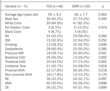
From October 1998 to August 2000, 123 patients were in-cluded in this study, in a consecutive way, and 27 were exin-cluded for the following reasons: death (8), refusal in being submitted to the procedure (5), myocardial infarction after CTA (1), cerebro-vascular accident (1), new procedure for myocardial recerebro-vasculari- revasculari-zation in the period (4), refusal in being submitted to a new myo-cardial scintigraphy (4), other associated diseases (4). Both groups did not show significant differences concerning the variables, being observed a predominance of coronary disease with bilateral onset in the group randomized for CTA and trilateral onset in the group randomized for SMR (p=0.002) (tab. I).
Angina was classified from I to IV, according to functional classification of Canadian Cardiovascular Society. The patients submitted to SMR and CTA were grouped, concerning coronary symptomatology (angina), in 3 subgroups: asymptomatic, functional class I and II angina carriers, and functional class III and IV angina carriers. The presence and severity of angina did not show significant difference at M1 in both groups (p=0.110), as well as at M2 (p=0.184) (tab. II). When each group was analyzed in particular, both showed an important improvement in angina functional class
(p=0.001), without significant difference among the subgroups concerning the functional class (tab. III).
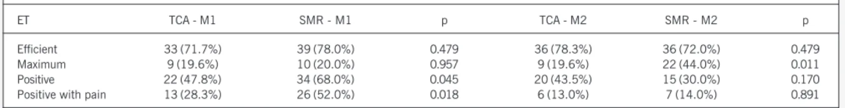
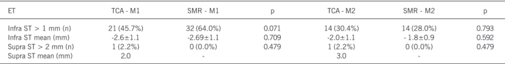
There was a significant increase in the number of maximum ergometric tests from M1 to M2, when the SMR-submitted group (p=0.01) was assessed, as well as a significant decrease in this number of positive ergometric tests from M1 to M2, when the SMR-submitted groups (p=0.045) were assessed, and positive tests with pain from M1 to M2 when the SMR-submitted group (p=0.018) was assessed (tab. IV). The SMR-submitted group showed a significant increase in HR under rest when M1 and M2 were compared (p=0.019) (tab. V). There was a significant in-crease in the mean of peak HR reached at M1 when compared to the peak HR at M2 in both groups (p=0.007) (tab. V). A significant decrease in the average values of SBP under rest from M1 to M2 in the two groups (p<0.001) (tab. V). Both groups showed a fewer number of patients with infra-unlevelling of segment ST at the exercise peak at M2, without significant difference, as well as a reduction of the infra-unlevelling mean (tab. VI). A sig-nificant increase of peak DP from M1 to M2 in both groups (p=0.009) (tab. VII). The CTA-submitted group showed MET
sig-Table I - Distribution of clinical and angiographic characteristics of patient groups
Variable (n - %) TCA (n=46) SMR (n=50) p
Average Age (years old) 59 ± 9.2 60 ± 7.7 0.563 Male Sex 30 (65.2%) 37 (74.0%) 0.349 White Color 39 (84.8%) 41 (82.0%) -Far-Eastern Color 3 (6.5%) 5 (10.0%) 0.919 Black Color 4 (8.7%) 4 (8.0%) -SH 24 (52.2%) 29 (58.0%) 0.566 DM 15 (32.6%) 22 (44.0%) 0.252 Smoking 13 (28.3%) 16 (32.0%) 0.690 Dyslipidemia 28 (60.9%) 25 (50.0%) 0.285 Previous AMI 18 (39.1%) 20 (40.0%) 0.931 Biarterial CAD 26 (56.5%) 13 (26.0%) 0.002 Triarterial CAD 20 (43.5%) 37 (74.0%) 0.002 Collateral flow 21 (45.7%) 24 (48.0%) 0.818 Proximal AIVA 22 (17.8%) 36 (26.3%) 0.218 Non-proximal AIVA 18 (17.8%) 14 (10.2%) 0.170 RC 36 (32.2%) 44 (32.1%) 0.997 AIVA 40 (35.6%) 50 (36.5%) 0.898 CX 36 (32.2%) 43 (31.4%) 0.899
TCA- coronary angioplasty; SMR- surgical myocardial revascularization; SH- systemic hypertension; DM- diabetes mellitus; AMI- acute myocardial infarction; CAD- coronary artery disease; AIVA- anterior interventricular artery; CX- circumflex artery; RC- right coronary artery.
Table II - Analysis of angina symptoms: a subdivision regarding the type of revascularization and moment of assessment
Classification TCA SMR p
M1
Asymptomatic 2 (4.4%) 7 (14.0%)
FC I / II Angina 35 (76.1%) 29 (58.0%) 0.110 FC III/IV Angina 9 (19.6%) 14 (28.0%)
M2
Asymptomatic 36 (78.3%) 45 (90.0%)
FC I / II Angina 8 (17.4%) 5 (10.0%) 0.184 FC III/IV Angina 2 (4.4%) 0 (0.0%)
4
Scintigraphy with Tc-Sestamibi
credibility to the comparison between the results from SMR and CTA and their influence on the myocardial ischemic load, making possible the extension of the findings in the study to the number of patients with coronary artery disease and the carriers of those same characteristics.
The results shown in this study refer to the analysis of the exams undertaken by 100% of the studied population, as that protocol included the performance of the ergometric test before the procedure for myocardial revascularization, as well as six months after the intervention, and the impossibility of its accom-plishment, in any of the opportunities, determined the exclusion of the patient from the study. The valuation of that fact is neces-sary, as it highlights the flaw of big randomized studies, idealized to compare the results from SMR and CTA in patients with mul-tivessel coronary artery disease, who performed ergometric test, in variable percentages of its participants, by recording, in a scanty way in the literature, details from the results obtained.
The presence of diabetes mellitus in a considerable part of the CTA-submitted population (32.6%), did not determine lower results when compared to those found in the SMR-submitted subgroup. Some peculiarities of the population included in this study, as the prevalence of male sex, the white race, and average age not so old, as well as the short follow-up time after the procedure (6 months), may have contributes to neutralize the possible negative influence of diabetes on CTA-submitted patients with coronary artery disease, as observed in BARI study at the end of 5 years of follow-up21.
Despite the prevalence of coronary artery disease with side onset in the SMR-submitted group (p=0.002) and a greater index of complete myocardial revascularization determined by SMR when compared to CTA, it was not effectively observed a greater benefit to that group. The analysis of the presence of angina symptoms prior to the procedures for myocardial revascularization showed that both groups of patients showed statistically significant im-provement (p=0.001) and similar concerning the functional class of angina, a finding that repeats in the analysis carried out six months after myocardial revascularization, confirming that the index of complete myocardial revascularization obtained through SMR did not determine a greater reduction of the symptoms in that subgroup of patients. Those data allow for questioning the relevance in reaching the complete myocardial revascularization. From the clinical point of view, none of the methods of myo-cardial revascularization was better in the follow-up carried out six months after the intervention, a disagreeing report on the results observed through randomized studies, which showed the superiority of SMR for the reduction of angina symptoms. The explanation for that fact lies in the design of this research, which differs substantially from the follow-up protocols of big studies, in
Table IV - Results from ergometric test: subdivision regarding the type of revascularization and moment of assessment
ET TCA - M1 SMR - M1 p TCA - M2 SMR - M2 p
Efficient 33 (71.7%) 39 (78.0%) 0.479 36 (78.3%) 36 (72.0%) 0.479 Maximum 9 (19.6%) 10 (20.0%) 0.957 9 (19.6%) 22 (44.0%) 0.011 Positive 22 (47.8%) 34 (68.0%) 0.045 20 (43.5%) 15 (30.0%) 0.170 Positive with pain 13 (28.3%) 26 (52.0%) 0.018 6 (13.0%) 7 (14.0%) 0.891
ET- ergometric test; TCA- coronary angioplasty; SMR- surgical myocardial revascularization; M - moment. Table III - Clinical classification of the patients: subdivision regarding
the type of revascularization and moment of assessment
Moment p
TCA
Asymptomatic FC I/II Angina FC III/IV Angina M1 2 (4.4%) 35 (76.1%) 9 (19.6%) 0.001 M2 36 (78.3%) 8 (17.4%) 2 (4.4%)
SMR
Asymptomatic FC I/II Angina FC III/IV Angina M1 7 (14.0%) 29 (58.0%) 14 (28.0%) 0.001 M2 45 (90.0%) 5 (10.0%) 0 (0.0%)
TCA - coronary angioplasty; SMR - surgical myocardial revascularization; M1 - moment; FC - functional class.
nificantly higher means than those in the SMR-submitted group in both moments (p=0.039) (tab. VII). There was a significant increase in the values of MET from M1 to M2 in the two groups (p<0.001), as well as a significant increase of values of VO2max from M1 to M2 in both groups (p<0.001) and the values of ETT from M1 to M2 in the two groups (p<0.001) (tab. VII).
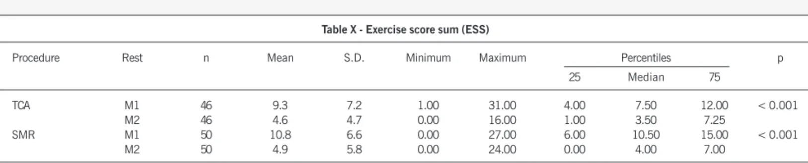
A significant decrease was observed in the values of rest LVFE from M1 to M2 in both groups (p=0.025) (tab. VIII). Both groups showed a significant decrease of ESS between M1 and M2 (p< 0.001 for both), as well as of DSS between M1 and M2 (p< 0.001 for both groups) (tabs. IX, X and XI).
SMR-submitted patients showed a complete myocardial re-vascularization index in 78.0% of the cases, in a contraposition to 39.1% of CTA-submitted patients (p=0.001). In the group of patients randomized to SMR 2.3 coronary grafts per patient were performed, being 34.5% venous and 65.5% arterial (56.0% in-volving left ATI anastomosis ATI - AIVA, 4.3% the epigastric artery, 5.2% radial artery). The success rate in CTA was 97.7%, and in 70.5% of the a coronary stent was implanted (1.4 coronary stent/ patient). In the CTA-submitted patients, 88 vessels were approa-ched (a rate of 1.9 dilated vessel/patient). There was no difference concerning the approached vessels (proximal AIVA in 17 cases, non-proximal AIVA in 26 cases, RC in 24 cases and CX in 21 cases). Regarding the type of coronary lesion approached, there was a prevalence of type B (2.3% type A, 34.1% type B1, 46.6% type B2 and 17.0% type C).
Discussion
5
Scintigraphy with Tc-Sestamibi
a singular point. This work prioritized the analysis of myocardial ischemic load under equivalent conditions and the follow-up of patients in similar clinical situations after the procedure. So, the absence of events that could be related to coronary restenosis or the occlusion of grafts in the follow-up period of six months after the procedure, or that determine the realization of cinecoronario-graphy and a later intervention during that period, was, a sine qua non condition for their permanence in it. That particularity of the study allowed for the analysis of the reflex of the procedure for myocardial revascularization in ischemic load without interfe-rence of other events. Besides, the current use of new strategies and interventionist (implantation of 70.5% pf coronary stent in the CTA-submitted group) and surgical (implantation of 65.5% of arterial grafts in the SMR-submitted group) techniques can also be positively influenced the results.
The important increase in the number of SMR-submitted pa-tients who had maximum ergometric tests six months after the procedure and the reduction in the number of tests regarded as positive for myocardial ischemia and positive for myocardial is-chemia with angina can be consequent to the higher index of complete myocardial revascularization determined through SMR. Most of patients with multivessel coronary artery disease in-creased the tolerance towards exercise after myocardial revascu-larization, and that in the presence of complete myocardial revas-cularization it was observed better indexes of maximum ergometric test, maximum heart rate, maximum double product, with lower number of positive ergometric tests and exercise-induced angina. The analysis of left ventricular fraction of ejection is the functional parameter that solely represents the best prognostic indicator for the assessment of coronary artery disease. We can infer that, in this study, both procedures for myocardial revascularization did not interfere negatively in the prognostic of that subgroup of patients22.
Among the randomized studies, we found few references
con-cerning the performance of myocardial scintigraphy, and that was performed at different moments of the evolution, which makes any comparisons difficult6,23.
An important aspect to be considered is the coexistence of coronary lesion and the functional meaning of such condition to determine myocardial ischemia. Many studies report a greater in-dex of complete anatomic myocardial revascularization determined through SMR in comparison with the index determined through CTA, with the functional revascularization index similar in both methods24-26. Such fact is confirmed in this study through the
semi-quantitative analysis of myocardial ischemia under rest (RSS), which did not show significant changes in the quantification in both sub-groups at the two moments, as well as through the semi-quantitative analysis of myocardial ischemia under exercise (ESS) that showed a statistically significant change of quantification at the M2 moment in relation to M1 moment in both subgroups, without difference between them. So, the difference between myocardial ischemia under rest and under exercise (DSS) showed a significant reduction of quantification at M2 moment in relation to M1 moment in both subgroups, without difference between them.
In this study we can ascertain that the lower rate of complete anatomic revascularization in CTA was not able to interfere nega-tively in the result of myocardial ischemia, which translated into similar and proper functional revascularization, as the reduction of myocardial ischemia and improvement in symptomatology took place in a statistically significant way.
Although the initial purpose of this study included equivalent revascularization, the anatomic extension of myocardial revascu-larization in the CTA-randomized subgroup was lower than in the SMR-randomized subgroup, as much freedom was given to the interventionist cardiologist and the cardiovascular surgeon to the approach to ill-taken coronary arteries. That fact reinforces the assumption that the interventionist cardiologist usually approaches the angiographically and clinically significant coronary lesions, whereas the cardiovascular surgeon approaches all technically possible lesions, aiming at a complete revascularization.
Despite the criticisms when important occlusions or coronary lesions persist after CTA, the complete revascularization seems to influence the prognostic only when related to surgical treatment27-29.
Recent studies have shown that incomplete myocardial revasculari-zation after CTA determined an increase in the need of a new inter-vention (early risk of new CTA and SMR 2.5 times greater) and recurrence of angina for a follow-up period of 9 years, but it did not interfere in the rates of death and AMI Q12,13,28-30. Those findings
support the strategy of CTA with incomplete myocardial revasculari-zation when it is not possible to perform a complete revascularirevasculari-zation or when it was not previously planned. So, the incomplete anatomic myocardial revascularization, but functionally suitable, can determine a similar evolution as the complete anatomic revascularization’s24.
Table VI - Analysis of segment ST in ergometric test: subdivision regarding type of myocardial revascularization and moment of assessment
ET TCA - M1 SMR - M1 p TCA - M2 SMR - M2 p
Infra ST > 1 mm (n) 21 (45.7%) 32 (64.0%) 0.071 14 (30.4%) 14 (28.0%) 0.793 Infra ST mean (mm) -2.6±1.1 -2.69±1.1 0.709 -2.0±1.1 - 1.8±0.9 0.592 Supra ST > 2 mm (n) 1 (2.2%) 0 (0.0%) 0.479 1 (2.2%) 0 (0.0%) 0.479
Supra ST mean (mm) 2.0 - 3.0
-ET- ergometric test; TCA- percutaneous myocardial revascularization; SMR- surgical myocardial revascularization; M1- moment 1; M2- moment 2. Table V - Variables in ergometric test: subdivision regarding the type of
revascularization and moment of assessment
Variables TCA - M1 SMR - M1 TCA - M2 SMR - M2
HR under Rest 78.8±15.6 72.6±13.2 76.5±16.2 78.9±17.9 (b.p.m.)
Peak HR 134.3±21.3 128.2±23.8 137.1±17.8 139.7±20.9 (b.p.m.)
SBP under Rest 149.9±23.1 153.3±25.3 139.4±17.6 139.4±15.9 (mmHg)
Peak SBP 189.1±27.8 187.9±26.5 197.6±29.7 189.3±24.8 (mmHg)
6
Scintigraphy with Tc-Sestamibi
in 88% to 98% of the cases, a revascularization rate from 2.2 to 2.6 vessels/patient, ITA used in 37% to 93% of the cases3,8,9,31,32.
The distribution of ill-taken coronary arteries, as well as the onset proportion of the proximal third of AIVA, was similar between the two subgroups in this study. That fact could be relevant as that studies showed that carriers of multivessel coronary artery disease with obstruction in the proximal third of AIVA showed a longer survival when submitted to SMR34,35.
For involving a population with heterogeneous characteristics, complex coronary lesions, lesions type B2 and C, the multivessel CTA may compromise the result of the procedure, especially if associated to diabetes mellitus, chronic coronary occlusion or high level of coronary lesion or tortuousity36,37. In this study,
al-though the group of CTA-submitted patients showed a prevalence of complex coronary lesions (34.1% type B1, 46.6% type B2 and 17% type C), a high rate of immediate success after CTA (97.7%) was obtained, a fact that can maybe be explained through the use of stent in 70.5% of the cases.
Those observations suggest the need for reassessment of the concept of complete myocardial revascularization and the valuation of the concept of functional myocardial revascularization. So, in a near future, we believe that the repercussion of coronary lesions treated on perfused myocardium (myocardial ischemia) will be responsible for the characterization of complete revascularization when, then, this concept will not be any longer determined from anatomic inferences obtained through spatial distribution of ill-taken and non-approached coronary arteries. As a reflex from the analysis of myocardial ischemic load (symptoms and exercise-induced myocardial ischemia) previous and after (6 months) the surgical or percutaneous myocardial revascularization in equivalent situations, this study allows for reaching the following conclusions: 1) the surgical and percutaneous myocardial revascularization de-termined a significant and similar reduction of myocardial ischemic load quantified 6 months after the intervention, and no statistically significant difference was not observed between the procedures; 2) the surgical myocardial revascularization determined a higher In this study, the patients randomized to CTA had 88 from
112 ill-taken vessels, approached, with the implantation of coronary stent in 70.5% of the cases, with a rate of 1.4 stents/patient, and 97.7% of success, similar findings to those found through big randomized studies2,3,8,9,31-33.
When the group randomized for SMR was analyzed, 116 coro-nary arteries were revascularized (137 ill-taken), reaching a rate of 2.3 grafts/patient, and the ITA was implanted in 56.0% of the cases. Those indexes do not differ either from those reported in the big randomized studies, and the complete revascularization was obtained
Table VII - Functional variables in ergometric test: subdivision regarding the type of revascularization and moment
Variable TCA SMR
DP under rest at M1 11,876.9±3,260.2 11,190.7±2,998.8 DP under rest at M2 10,782.1±3,190.6 11,001.9±2,864.6 Peak DP at M1 25,636.8±6,294.8 24,247.3±6,117.9 Peak DP at M2 27,086.63±5,467.9 26,477.7±5,221.1 WME at M1 9.33±2.1 8.4±2.1 WME at M2 10.6±2.7 9.8±1.9 VO2max at M1 32.6±7.3 29.7±6.9 VO2max at M2 36.7±8.9 34.3±6.8 ETT at M1 (seconds) 419.5±119.1 376.8±108.6 ETT at M2 (seconds) 486.4±163.5 444.8±111.3
DP- double product; WME- work metabolic equivalent; VO2max- maximum consumption of oxygen; ETT- exercise tolerance time; TCA- coronary angioplasty; SMR- surgical myocardial revascularization; M- moment.
Table VIII - Left ventricular fraction of ejection (GATED SPECT): subdivision regarding the type of myocardial revascularization
and moment of assessment
LVFE TCA SMR
Rest at M1 56.2±11.8 55.0±9.9 Rest at M2 55.8±12.8 51.1±11.1 Exercise at M1 52.9±13.0 49.2±10.3 Exercise at M2 52.8±11.9 49.8±12.5
LVFE- left ventricular fraction of ejection; TCA- coronary angioplasty; SMR- surgical myocardial revascularization; M- moment.
Table IX - Rest score sum (RSS)
Procedure Rest n Mean S.D. Minimum Maximum Percentiles p 25 Median 75
TCA M1 46 1.8 2.6 0.0 12.0 0.0 0.0 3.0 0.439 M2 46 1.6 2.6 0.0 12.0 0.0 0.0 3.0
SMR M1 50 2.8 4.0 0.0 18.0 0.0 1.5 4.3 0.393 M2 50 2.6 3.8 0.0 19.0 0.0 1.0 4.0
S.D.- standard deviation; TCA- coronary angioplasty; SMR- surgical myocardial revascularization; M- moment.
Table X - Exercise score sum (ESS)
Procedure Rest n Mean S.D. Minimum Maximum Percentiles p 25 Median 75
TCA M1 46 9.3 7.2 1.00 31.00 4.00 7.50 12.00 < 0.001 M2 46 4.6 4.7 0.00 16.00 1.00 3.50 7.25
SMR M1 50 10.8 6.6 0.00 27.00 6.00 10.50 15.00 < 0.001 M2 50 4.9 5.8 0.00 24.00 0.00 4.00 7.00
7
Scintigraphy with Tc-Sestamibi
index of complete revascularization, but it did not represent a statistically significant factor in the reduction of myocardial is-chemic load 6 months after the intervention, when compared to the result found in the group of patients submitted to percutaneous myocardial revascularization.
This study began in October 1998, with a follow-up up to six months after the procedure for every patient included in it. Once the medical practice has been evolving continuously, it is relevant to consider the differences among the techniques used for myocardial revascularization in this study and the most recently developed ones, which have not been frequently used and that
can substantially influence these results (surgery without extra-corporeal flow or minimally invasive, use of inhibitors of the gly-coprotein IIb/IIIa and covered stents) in the future.
As the choice of treatment for multivessel coronary artery disea-se is a complex decision, which involves the analysis of the didisea-seadisea-se under multiple dimensions, such as evolution, current functional status of the patient (symptoms) and the perception of life quality, the patients can be favored through the combination of surgical and percutaneous techniques for myocardial revascularization, thus be-nefiting from the low morbidity rate of the percutaneous procedure and the longevity of arterial grafts (surgical procedure).
References
1. Bourassa MG, Roubin GS, Detre KM,Sopko G, Krone RJ, Attabuto MJ, Bjerre-gaad P, Bolling S, Herman MV, Frye RL and the BARI Study Group. Bypass Angio-plasty Revascularization Investigation: patient screening, selection and recruit-ment. Am J Cardiol 1995; 75(suppl C):3-8.
2. CABRI Trial Participants. First-year results of CABRI (Coronary Angioplasty versus Bypass Revascularisation Investigation). Lancet 1995; 46:1179-84. 3. Hamm CW, Reimers J, Ischinger T et al. A randomized study of coronary
angio-plasty compared with bypass surgery in patients with symptomatic multivessel co-ronary disease. N Engl J Med 1994; 331 1037-43.
4. Henderson RA, for the Randomised Intervention Treatment of Angina Trial. The Randomised Intervention Treatment of Angina (RITA) Trial protocol: a long term study of coronary angioplasty and coronary artery bypass surgery in patients with angina. Br Heart J 1989; 62:411-4.
5. King III SB, Lembo NJ, Weintraub WS et al. Emory Angioplasty versus Surgery Trial (EAST): design, recruitment and baseline description of patientes. Am J Cardiol 1995; 75: 42C-59C.
6. Rodriguez A, Boullon F, Perez-Baliño N et al. Argentine randomized trial of percu-taneous transluminal coronary angioplasty versus coronary artery bypass surgery in multivessel disease (ERACI): in-hospital results and 1-year follow-up. J Am Coll Cardiol 1993; 22:1060-7.
7. Hueb W, Soares PR, Gersh BJ et al. The medicine, angioplasty or surgery study (MASS-II): a randomized controlled clinical trial of medical therapy, coronary an-gioplasty or bypass surgery for multivessel coronary artery disease. J Am Coll Car-diol 2004; 43:1743-51.
8. Rodriguez A, Bernardi V, Navia J et al. Argentine randomized study: coronary an-gioplasty with stenting versus coronary bypass surgery in patients with multiple-vessel disease (ERACI II): 30-day and one-year follow-up results. J Am Coll Car-diol 2001; 37;51-8.
9. Serruys PW, Unger F, Souza JE et al. Comparison of coronary-artery bypass surgery and stenting for the treatment of multivessel disease. N Engl J Med 2001; 344: 117-24.
10. Alkeylan A, Miller D, Shaw LJ et al. Influence of race on the prediction of cardiac events with stress technetium-99m sestamibi tomographic imaging in patients with stable angina pectoris. Am J Cardiol 1998; 81:293-7.
11. Haronian HL, Remetz MS, Sinusas AJ et al. Myocardial risk area defined by techne-tium-99m Sestamibi imaging during percutaneous transluminal coronary angioplas-ty: comparison with coronary angiography. J Am Coll Cardiol 1993, 22:1033-43. 12. Faxon DP, Ghalilli MD, Jacobs AK et al. The degree of revascularization and
out-come after multivessel coronary angioplasty. Am Heart J 1992; 123:854-9. 13. Bourassa MG, Yeh W, Holubkov R, Sopko G, Detre KM, for the Investigators of
The NHLBI PTCA Registry. Long-term outcome of patients with incomplete vs com-plete revascularization after multivessel PTCA. A report from the NHLBI PTCA Re-gistry. Eur Heart J 1998; 19:103-11.
14. Eagle KA, Guyton RA, Davidoff R et al. ACC / AHA guidelines for coronary artery by-pass graft surgery. A report of the American College of Cardiology / American Heart As-sociation Task Force on practice guidelines (Committee to revise the 1991 guidelines for coronary artery bypass graft surgery). J Am Coll Cardiol 1999; 34:1262-347. 15. Ryan TJ, Bauman WB, Kennedy JW et al. Guidelines for percutaneous
translumi-nal coronary angioplasty. A report for the American College of Cardiology / Ameri-can Heart Association Task Force on assessment of diagnostic and therapeutic cardiovascular procedures (Commitee on Percutaneous Transluminal Coronary An-gioplasty). J Am Coll Cardiol 1993; 22:2033-54.
16. Smith Jr. SC, Dove JT, Jacobs AK et al. ACC / AHA Guidelines for percutaneous transluminal coronary angioplasty. (Revision of the 1993 PTCA Guidelines) A re-port for the American College of Cardiology / American Heart Association Task Force on Practice Guidelines (Commitee to revise the 1993 Guidelines for Percutaneous Transluminal Coronary Angioplasty). J Am Coll Cardiol 2001, 37: 2239i-lXVi. 17. Gibbons RJ, Balady GJ, Beasley JW et al. ACC / AHA Guidelines for exercise testing.
A report of the American College of Cardiology / American Heart Association Task Force on practice Guidelines (Committee on Exercise Testing). J Am Coll Cardiol 1997; 30: 260-315.
18. Committee on advanced cardiac imaging and technology, Council on Clinical Cardiol-ogy, American Heart Association; Cardiovascular Imaging Committee, American College of Cardiology, and board of Directors, Cardiovascular Council, Society of Nuclear Me-dicine. Standardization of cardiac tomographic imaging. Circulation 1992; 86:338-9. 19. Port SC, Berman D, Garcia E et al. Imaging guidelines for nuclear cardiology
proce-dures. J Nucl Cardiol 1999; 6:G47-84.
20. Berman DS, Kiat HS, Friedman JD et al. Separate acquisition rest thallium-201/ stress technetium-99m sestamibi dual-isotope myocardial perfusion single-photon emission computed tomography: a clinical validation study. J Am Coll Cardiol 1993; 22: 1455-64.
21. The BARI Investigators. Seven-year outcome in the Bypass Angioplasty Revas-cularization Investigation (BARI) by treatment and diabetic status. J Am Coll Cardiol 2000; 35:1122-9.
22. Sharir T, Germano G, Kavanagh PB et al. Incremental prognostic value of post-stress left ventricular ejection fraction and volume by gated myocardial perfusion single photon emission computed tomography. Circulation 1999; 100:1035-42. 23. Alazraki NP, Krawczynska EG, Kosinski AS et al. Prognostic value of thallium-201 single-photon emission computed tomography for patients with multivessel coro-nary artery disease after revascularization (The Emory Angioplasty versus Surgery Trial (EAST)). Am J Cardiol 1999; 84:1369-74.
24. Cowley MJ, Vandermael M, Topol EJ et al. Is traditionally defined complete revas-cularization needed for patients with multivessel disease treated by elective corona-ry angioplasty? J Am Coll Cardiol 1993; 22:1289-97.
25. Pocock SJ, Henderson RA, Rickards AF et al. Meta-analysis of randomised trials comparing coronary angioplasty with bypass surgery. Lancet 1995; 346:1184-9. Table XI - Difference of score sum (DSS)
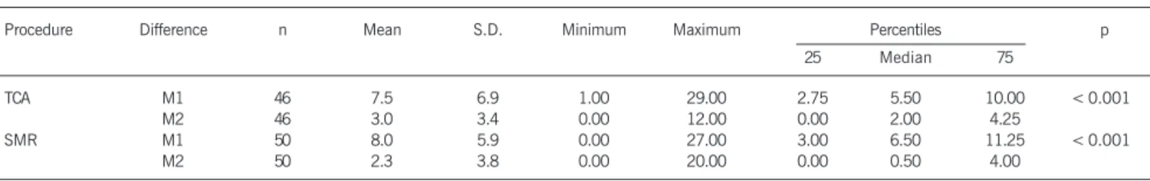
Procedure Difference n Mean S.D. Minimum Maximum Percentiles p 25 Median 75
TCA M1 46 7.5 6.9 1.00 29.00 2.75 5.50 10.00 < 0.001 M2 46 3.0 3.4 0.00 12.00 0.00 2.00 4.25
SMR M1 50 8.0 5.9 0.00 27.00 3.00 6.50 11.25 < 0.001 M2 50 2.3 3.8 0.00 20.00 0.00 0.50 4.00
8
Scintigraphy with Tc-Sestamibi
26. Sim I, Gupta M, McDonald K, Bourassa MG, Hlatky MA. A meta-analysis of ran-domized trials comparing coronary artery bypass grafting with percutaneous transluminal coronary angioplasty in multivessel coronary artery disease. Am J Cardiol 1995; 76:1025-9.
27. Bell MR, Bailey KR, Reeder GS, Lapeyre III AC, Holmes Jr DR. Percutaneous transluminal angioplasty in patients with multivessel coronary disease: how important is complete revascularization for cardiac event-free survival? J Am Coll Cardiol 1990; 16:553-62.
28. Jones EL, Craver JM, Guyton RA, Bone DK, Hatcher Jr. CR, Riechwald N. Impor-tance of complete revascularization in performance of the coronary bypass opera-tion. Am J Cardiol 1983; 51:7-12.
29. Samson M, Meester HJ, De Feyter PJ, Strauss B, Serruys PW. Successful multiple segment coronary angioplasty: effect of completeness of revascularization in sin-gle-vessel multilesions and multivessels. Am Heart J 1990; 120:1-12. 30. Bourassa MG, Kip KE, Jacobs AK et al. Is a strategy of intended incomplete
percutaneous transluminal coronary angioplasty revascularization acceptable in nondiabetic patients who are candidates for coronary artery bypass graft surgery? The Bypass Angioplasty Revascularization Investigation (BARI). J Am Coll Cardiol 1999; 33:167-36.
31. King III SB, Lembo NJ, Weintraub WS et al. A randomized trial comparing angio-plasty with coronary bypass surgery. N Engl J Med 1994; 331:1044-50. 32. RITA Trial Participants. Coronary angioplasty versus coronary artery bypass surgery: the
Randomised Intervention Treatment of Angina (RITA) trial. Lancet 1993; 341: 573-80. 33. The BARI Investigators. Comparison of coronary bypass surgery with angioplasty
in patients with multivessel disease. New Engl J Med 1996; 335:217-25. 34. Hannan EL, Racz MJ, McCallister BD et al. A comparison of three-year survival
after coronary artery bypass graft surgery and percutaneous transluminal corona-ry angioplasty. J Am Coll Cardiol 1999; 33:63-72.
35. Jones RH, Kesler K, Phillips III HR et al. Long-term survival benefits of coronary artery bypass grafting and percutaneous transluminal angioplasty in patients with coronary artery disease. J Thorac Cardiovasc Surg 1996; 111:1013-25. 36. Botas J, Stadius ML, Bourassa MG et al. Angiographic correlates of lesion
rele-vance and suitability for percutaneous transluminal coronary angioplasty and co-ronary artery bypass grafting in the Bypass Angioplasty Revascularization Investi-gation study (BARI). Am J Cardiol 1996; 77:805-14.