DOI: 10.1590/0004-282X20130240
ARTICLE
A new expansive two-open-doors laminoplasty
for multilevel cervical spondylotic myelopathy:
technical report and follow-up results
Nova técnica de laminoplastia expansiva para tratamento de mielopatia espondilotica
cervical: descrição técnica e resultados de follow-up
Aluizio Augusto Arantes Júnior1, Geraldo Alves da Silva Junior1, José Augusto Malheiros1,
Fernando Flavio Gonçalves, Marcelo Magaldi1, Erica Santiago2, Arthur Adolfo Nicolato2,
Sebastião Nataniel Silva Gusmão1
Cervical degenerative myelopathy is a common condi
tion afecting 2% of all hospital admissions according to a
number of studies1,2. he two main causes of this condition
are cervical spondylosis and ossiication of the posterior
longitudinal ligament (OPLL)3. Such conditions progress
with age and can eventually result in the impairment of mul
tilevel segments and multilevel cervical spondylotic mye lopathy (MCSM).
1Department of Neurosurgery, Clinical Hospital of the Federal University of Minas Gerais, Belo Horizonte MG , Brazil;
2Federal University of Minas Gerais - School of Medicine, Belo Horizonte MG , Brazil;
3Orthopedist at Madre Tereza Hospital, Belo Horizonte MG, Brazil.
Correspondence:Aluizio Augusto Arantes Junior; Rua dos Inconidentes 320/801; 30140-120 Belo Horizonte MG – Brasil; E-mail: [email protected]
Conflict of interest:There are no conlicts of interest to declare.
Received 27 August 2012; Received in inal form 26 October 2013; Accepted 04 November 2013.
ABSTRACT:
The laminoplasty technique was devised by Hirabayashi in 1978 for patients diagnosed with multilevel cervical spondylotic myelopathy. Objective: To describe an easy modiication of Hirabayashi’s method and present the clinical and radiological results from a ive-year follow-up study. Method and Results: Eighty patients had 5 levels of decompression (C3-C7), 3 patients had 6 levels of decompression (C2-T1) and 3 patients had 4 levels of decompression (C3-C6). Foraminotomies were performed in 23 cases (27%). Following Nurick`s scale, 76 patients (88%) improved, 9 (11%) had the same Nurick grade, and one patient worsened and was advised to undergo another surgical procedure. No deaths were observed. The mean surgery time was 122 min. Radiographic evaluation showed an increase in the mean sagittal diameter from 11.2 mm at pretreatment to 17.3 mm post surgery. There was no signiicant difference between pretreatment and post-surgery C2-C7 angles. Conclusions: This two-open-doors laminoplasty technique is safe, easy and effective and can be used as an alternative treatment for cases of multilevel cervical spondylotic myelopathy without instability.
Keywords: laminoplasty, cervical spondylotic myelopathy, ossiication of the posterior longitudinal ligament.
RESUMO:
A laminoplastia é técnica clássica descrita por Hirabayashi em 1978 para descompressão do canal cervical sem utilizar prótese. A principal indicação é o tratamento da mielopatia espondilotica cervical sem instabilidade. Objetivo: Descrever modiicação simples da técnica de laminoplastia clássica de Hirabayashi com resultados clínicos e radiográicos em 5 anos de acompanhamento. Resultados e Método: Fo-ram acompanhados 86 pacientes. Em 80, foi feita descompressão por laminoplastia em 5 níveis (C3-C7); em 3, descompressão em 6 níveis (C2-T1); em 3, descompressao em 4 níveis (C3-C6). Em 23 casos (27%), foi realizada foraminotomia associada a descompressão medular. O acompanhamento dos pacientes foi feito utilizando a escala de Nurick. Em 76 pacientes (88%) houve melhora do grau de Nurick. Não houve mortalidade associada à técnica. O tempo médio do procedimento cirúrgico foi de 122 minutos. Em relação à avaliação radiográica, houve aumento do diâmetro sagital médio do canal cervical de 11,2mm para 17,3mm. Não houve diferença estatística do ângulo C2-C7 nas ava-liações antes e após o procedimento cirúrgico. Conclusão: A nova técnica de laminoplastia descrita no presente estudo foi segura, de fácil execução, efetiva, não utiliza protese e não há instabilidade do canal cervical.
here are numerous surgical strategies for MCSM, and controversies have arisen between researchers regarding the use of an anterior or posterior approach, prophylac tic surgery, and conservative (nonsurgical) versus surgi cal treatment.
his paper describes a new technique for cervical lamino
plasties with a minimum of 5 years of follow-up in 86 patients.
Series
We prospectively analyzed patients undergoing lamino
plasties from March 2001 to March 2006 in the Hospital das Clinicas of the Federal University of Minas Gerais and the Hospital Madre Tereza, Belo Horizonte, Brazil. hese patients were followed for at least 5 years with both clinical and radio graphic evaluations.
METHODS
All of the patients ( from the Hospital das Clinicas and
the Hospital Madre Tereza) who were exhibiting clinical evi
dence of myelopathy, with or without radiculopathy, under
went complete radiographic evaluation.
Radiographic evaluation consisted of neutral anterior-posterior (AP) imaging, conventional x-ray imaging, lateral
imaging, lexion/extension x-ray imaging, computed tomog
raphy (CT) and magnetic resonance imaging (MRI).
Patients with MCSM and radiographic signs of instabil
ity4,5 or deformity were excluded and directed towards an
other surgical strategy. We considered cases of cervical insta
bility with any antero-posterior deviation >3 mm, a relative translation of a vertebra in a sagittal plane >3.5 mm, or angu
lation of one vertebra to another >11°.
he patients with cervical multilevel stenosis (cervical spine canal diameter <12 mm) without instability or even with
some clinical impairment, such as those of grade 2 or 3 accord
ing to the physical status classiication system of the American Society of Anesthesiologists (ASA), were enrolled in this study.
he Nurick scale6 was used to clinically evaluate and ob
jectively compare the preoperative data and the postopera
tive follow-up data.
Follow-up was conducted over a minimum of ive years and consisted of both clinical and radiographic evaluations. Postoperative follow-up data were recorded at 1, 3, 6, 12, 18, 24, 36, 48 and 60 months.
An independent examiner carried out the preoperative
assessment and recorded the results.
his study was approved by the ethics board of the Federal University of Minas Gerais.
Technical report
After a longitudinal median incision and a subperios
teal dissection, the cervical spine is exposed, as well as the
laminae and spinous processes. Care must be taken taken to preserve the paraspinal muscles where they join the spinous process of C2.
he next step is to decide how to perform the arthrodesis and on which side the laminae will be opened. his decision
is made individually according to both clinical and radiologi
cal indings in the preoperative evaluation. he procedure al
lows an alternated foraminotomy (alternated at successive levels) for radicular decompression.
he interspinous, interlaminar and yellow ligaments must be cut between vertebrae C2 and C3, C7 and T1 and C4 and C5, respectively, to construct two blocks of laminae. Inside each block, the yellow ligament must be preserved.
To create the opening for the laminae, a high-speed drill and a 2 mm Kerrison rongeur are used to cut a complete path through the lateral part of the laminae on one side of C3-C4 and another complete path through C5-C6 on the opposite side. he external cortical bone layers of both closed sides
must also be drilled, creating a greenstick fracture and open
ing the doors (Figures 1 and 2). his technique allows C5 fo
raminotomy on both sides.
Instead of one open door, as described by Hirabayashi, the goal of this procedure is to obtain two open doors that will be united in the middle (between C4 and C5) by sutures. herefore, two doors and three arthrodesis points are cons-tructed at C4-C5 and at both lateral parts of the laminae (on
opposite sides).
Depending on the clinical and radiographic indings, the
decompression can be extended to include C7 or C2 by par
tial laminectomy.
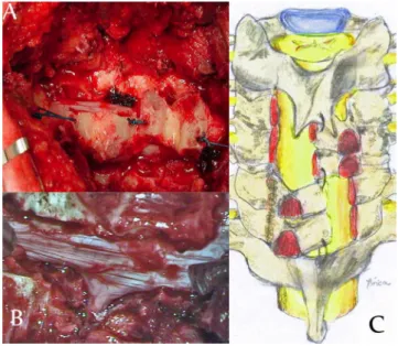
Figure 1. A) surgical view of the two parts of the construction
he blocks are held by sutures between the C2 spinous process and the C3 lamina, between the C4 and C5 laminae, and between the C6 lamina and C7 spinous process. he irst
and most important suture is between C4 and C5. his su
ture lines the extremities of both laminae (C4-C5) such that arthrodesis can be performed in the center and middle of the construction. A bone graft is taken from the spinous process and inserted into the excavation created in the lateral part of the laminae (Figure 1).
During postoperative recuperation, muscle relaxants are administered and a Philadelphia cervical collar is used for 30 days in order to provide immobilization and comfort for the
patient. After one month, the patients are driven to physio
therapy for the purpose of cervical muscle rehabilitation.
RESULTS
From March 2001 to March 2006, 86 patients underwent laminoplasties. Of these patients, 64 were male (74%), and 22 were female (26%). he mean age was 63 years.
In 80 patients (92%), the laminoplasties were performed between vertebrae C3 and C7; in 3 patients (4%), they were performed between C2 and C6; and in 3 patients (4%) they were performed between C2 and T1. Foraminotomies were performed in 23 cases (27%).
Following the Nurick scale, 76 patients (88%) improved, 9 (11%) had the same Nurick grade, and one patient worsened and was scheduled for another surgical procedure. No deaths were observed (Tables 1 and 2).
he mean surgery time was 122 min. Radiographic
eva lua tion showed an increase in the mean sagittal diam
eter from 11.2 mm pretreatment to 17.3 mm post surgery (Table 3). here was no signiicant diference between the C2-C7 angle before treatment and post surgery. No cases of kyphosis or progressive spine instability were observed (Table 4).
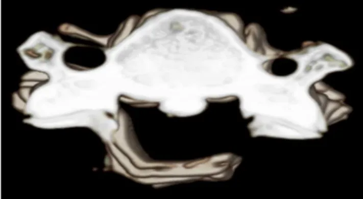
Figure 2. Three-dimensional CT scan showing a greenstick
fracture (right) after the opening of the lamina on the left side.
Table 1. Results following surgery, based on the Nurick scale.
Nurick grade Before surgery After surgery
n % n %
0 4 4.7 6 7.0
1 2 2.3 14 16.3
2 0 0.0 58 67.4
3 5 5.8 7 8.1
4 57 66.3 1 1.2
5 18 20.9 0 0.0
Median 4 2
P value for marginal homogeneity test <0.001.
Table 2. Results based on improvement after surgery.
Results n %
Improvement* 76 88
1 grade 7 8
2 or more grades 89 80
0 grades (the same grade as preoperatively) 9 11
Worse 1 1
Death 0 0
*Based on the Nurick scale
Table 3. Comparison between the sagittal diameter before
and after laminoplasty (n=86).
Sagittal diameter Before surgery (mm) After surgery (mm)
Mean 13.3 19.4
Median 13.0 19.0
Standard deviation 3.4 3.2
Minimum 7.0 14.0
Maximum 22.0 30.0
*p-value for the Wilcoxon signed rank test <0.001.
Table 4. C2-C7 angle comparison: before and after
laminoplasty (n=86).
C2-C7 angle Before surgery After surgery
Mean 17.5 17.9
Median 20.0 20.0
Standard deviation 6.7 6.4
Minimum 5.0 5.0
Maximum 36.0 35.0
*p-value for the Wilcoxon signed rank test=0.002.
hirty days post surgery (with good outcomes after treat
ment with analgesics and anti-inlammatory drugs), cervi
calgia was reported in 8 patients (9%). One patient required reoperation because of a local hematoma. Another patient
required reoperation after clinical worsening caused by frac
ture and migration of the lamina on one side of the lamino
DISCUSSION
here are numerous surgical strategies for treating multi
level cervical spondylotic myelopathy (MCSM), and there is
no standard procedure. Controversies over the superiority of
anterior or posterior approaches remain, and no consensus
has been reached. he laminoplasty is one option among the wide range of posterior approaches.
he anterior approach is performed by multilevel cervi
cal discectomy with or without corpectomy and fusion. he main advantage of this approach is the possibility of direct spinal decompression through the removal of osteophytes, extruded discs, or thin longitudinal ligaments. his techni-que is safe for up to three segments7, after which negative
results, such as pseudoarthrosis, graft migration and instru
mentation failure8,9, occur.
he classic posterior approach for MCSM is the laminec
tomy. Despite impressive decompression results, this techni
que has been associated with post-surgery spine instability
and kyphosis11, post-surgery compression by ibrous tissue,
and diminished clinical results12.
he laminoplasty technique is a variant of the laminec
tomy, which was irst described by Hirabayashi13 in 1978
with the purpose of decompressing the spine and reducing the complications resulting from the classic laminectomy. Multiple variations on the technique have been created, but
all of these methods share the same idea of cervical expan
sion with a protective dorsal element14,15.
Wada et al.16 comparatively analyzed the corpectomy and
the laminoplasty as treatments for MCSM. here was no difer
ence in the functional outcomes of these groups, but pseudoar
throsis was reported in 26% of the patients in the corpectomy
group. Herkowitz17 compared anterior fusion with laminecto
mies and laminoplasties in 45 patients with 2 years of follow-up. Excellent to good results were reported for 92% of patients in the anterior group compared with 86% in the laminoplasty
group and 66% in the laminectomy group. No signiicant dif
ference in outcomes was found between the anterior fusion and laminoplasty groups, but pseudoarthrosis was reported in 37% of the patients in the anterior fusion group. hese results are consistent with other published series18,19.
Another option is to combine the laminectomy and the pos
terior cervical fusion in order to avoid kyphosis related to the loss of the posterior column resulting from the laminectomy.
Heller et al.20 analyzed patients undergoing laminectomies and
instrumented posterior fusion with laminoplasties, and they ob
served no diferences in functional outcomes, although a greater number of complications were found in the laminectomy group.
A review of the literature reveals no diference in the func
tional outcomes of the laminoplasty variants21-24. Conversely, a
great number of these variants are technically diicult to per
form, while others are cumbersome in their use of bone grafting
and require a prohibitive number of suture points. Other disad
vantages in certain types of laminoplasties result from the re
quired use of instrumentation to maintain the open door and from the fact that the foraminotomy is possible on only one side. Our results were descriptive but not comparative. he refore, our main goal was to describe our technique and not compare the diferent kinds of technique available in the literature.
In order to avoid these shortcomings, we have proposed an easier laminoplasty technique involving just three su
ture points, requiring no instrumentation to maintain the
open door, and making foraminal decompression possi
ble on both sides. Using this procedure, the door remained opened throughout the ive years of follow-up on account of three arthrodesis points that were created: two later
al points (one on either side of the construction) and one
point in the middle of the construction. his technique in
creased the median sagittal diameter from 11.2 mm to 17.3 mm. Compared with other series21-24, our technique
caused the greatest expansion in the medium sagittal diame-ter, as determined by radiographic evaluation (Figure 3). Other published series have reported similar results: Wang
et al.3 reported an increase from 9.8 mm to 16.6 mm; O`Brien
et al.23 reported an increase from 8.2 mm to 16.6 mm; and
Satomi et al.24 reported an increase from 12.0 mm to 15.7 mm.
Kyphotic sagittal alignments reportedly developed in
0–10% of patients after laminoplasty, depending on the se
ries25-30. Iwasaki et al.26 reported a deterioration of cervical
lordosis into kyphosis in 5 patients (8%) of 59 without neu
rological sequelae. he present study revealed no signiicant change in the C2-C7 angle measured before treatment and three years post surgery, and there was no evidence of post-surgery progressive kyphosis.
Table 5. Postoperative complications.
Complication n %
Cervicalgia 8 9
Reoperation 2 2
Hematoma 1 1
Neurological worsening 1 1
C5 radiculopathy 2 2
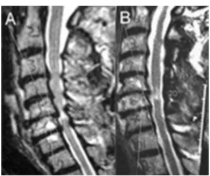
Figure 3. Sagittal IRM showing preoperative (A) and
Another complication relating to the laminoplasty tech
nique is the observance of C5 radiculopathy, which proba
bly results from traction on the nerve, as the cord migrates
dorsally after decompression. In published studies, this par
ticular complication rate varies from 5% to 14%22-25. Sasai et
al.25 discussed the role of prophylactic foraminotomies in de
creasing the incidence of motor palsy after laminoplasties.
Lee et al.1 performed concomitant foraminotomies in 11 of
105 patients undergoing open-door laminoplasties and re
ported improvements in 10 of 11 patients (91%). In our study, we had two cases of C5 radiculopathy (2%), but these cases exhibited spontaneous recovery. We attributed the low rate of complications observed in our series to the bilateral fo
raminotomy, which was made possible at this level by our technique of two-open-doors laminoplasty.
Another important advantage of our technique is that it exhibits the shortest surgery time compared with other techniques. his advantage allows the surgical procedure to
be performed in older patients with high-grade ASA (ante
rior spinal artery) conditions. Our series revealed a low rate of general clinical complications, even for the patient group with a median age of 63 years.
he literature reports an overall recovery rate after lami
noplasties ranging from 50% to 70%1,3,26,27. he positive out
come factors reported include the patient’s age, the time of
symptomatic myelopathy, and the degree of spinal compres
sion as determined by radiographic evaluation. Our series
demonstrates a clinical improvement of 88%.
Compared with the anterior approach, the incidence of axial neck pain and muscle atrophy is greater after
laminoplasty surgery. In a meta-analysis of 71 retrospective
papers, Sani et al.29 found an overall rate of axial neck pain
that varied from 6% to 60%. In our study, axial neck pain was observed in 8 patients (9%). Except for one patient who had persistent axial neck pain, all of these patients fully recov
ered after 3 months of physical therapy. We believe that this result is due to the surgical technique of dissection and de-in
sertion of the muscles that attach to the C2 spinous process.
Matsunaga et al.10 demonstrated that a post-surgery loss of
sagittal alignment was strongly associated with dissection and the subsequent nonhealing of the muscle insertion at C2. In our technique, all of the attachments of the C2 spinous
process are preserved.
Finally, laminoplasties cannot be used in kyphotic cervi
cal spines, owing to the risk of post-surgery complications
and instability1,3,5,26,27.
CONCLUSIONS
he two-open-doors laminoplasty technique used in this
study demonstrates a safe, easy and efective surgical proce
dure and provides an alternative for cases of multilevel cervi
cal spondylotic myelopathy without deformation. his meth
od is associated with a low rate of complications, a short operating time, and no incidence of post-surgery kyphosis or late instability. Furthermore, the two-open-doors lami
noplasty technique does not require a heterologous graft or instrumentation. herefore, this technique may represent a useful option for geriatric patients.
References
1. Lee TT, Green BA, Gromelski EB. Safety and stability of open-door cervical expansive laminoplasty. J Spinal Disord 1998;11:12-15.
2. Tomita K, Kawahara N, Toribatake Y, Heller JG. Expansive midline T-saw laminoplasty (modiied spinous process-splitting) for the management of cervical myelopathy. Spine 1998;23:32-37.
3. Wang MC, Kreuter W, Wolla CE, et al. Trends and variations in cervical spine surgery in the United States: Medicare beneiciaries, 1992 to 2005. Spine 2009;34:955-961.
4. Lam FC, Irwin BJ, Poskitt KJ, Steinbok P. Cervical spine instability following cervical laminectomies for Chiari II malformation: a retrospective cohort study. Childs Nerv Syst 2009;25:71-76.
5. White AA 3rd, Johnson RM, Panjabi MM, Southwick WO. Biomechanical analysis of clinical stability in the cervical spine. Clin Orthop 1975;109:85-96.
6. Nurick S. The pathogenesis of the spinal cord disorder associated with cervical spondylosis. Brain 1972;95:87-100.
7. Arantes A, Gusmão S, Rubinstein F, Oliveira R. Microsurgical anatomy of the recurrent laryngeal nerve: applications on the anterior approach to the cervical spine. Arq Neuropsiquiatr 2004;62:707-710.
8. Edwards, C, Heller, J, Morikami, H, et al. Corpectomy versus laminoplasty for multi-level cervical myelopathy: An independent matched cohort study. Spine 2002;27:1168-1175.
9. Vaccaro, AR, Falatyn, SP, Scuderi, GJ, et al. Early failure of long segment anterior cervical plate ixation. J Spinal Disord 1998;11:410-415.
10. Matsunaga, S, Sakou, T, Nakansisi, K, et al. Analysis of the cervical spine alignment following laminoplasty and laminectomy. Spinal Cord 1999;37:20-24.
11. Yonenobu, K, Okada, K, Fuji, T, Fujiwara, K, Yamashita, K, Ono, K. Causes of neurologic deterioration following surgical treatment of cervical myelopathy. Spine 1986;11:818-823.
12. Dai R, Ni B, Yuan W, Jia L. Radiculopathy after laminectomy for cervical compression myelopathy. J Bone Joint Surg Br 1998;80:846-84.
13. Hirabayashi K, Satomi K. Operative procedure and results of expansive open-door laminoplasty. Spine 1988;13:870-876.
14. Andrade GC, Silveira RL, Arantes A, Pinheiro N, Rocha EMM. Laminoplastia expansiva. Arq Neuropsiquiatr 2005;63:1005-1009.
15. Steinmetz MP, Resnick DK. Cervical laminoplasty. Spine J 2006;6(Suppl):S274-S281.
16. Wada E, Suzukim S, Kanazawa A. Subtotal corpectomy versus laminoplasty for multilevel cervical spondylotic myelopathy: a long-term follow-up study over 10 years. Spine 2001;26:1443-1447.
18. Yonenobu K, Hosono N, Iwasaki M, et al. Laminoplasty versus subtotal corpectomy: a comparative study of results in multisegmental cervical spondylotic myelopathy. Spine 1992;17:1281-1284.
19. Edwards C, Heller J, Morikami H, et al. Corpectomy versus laminoplasty for multi-level cervical myelopathy: An independent matched cohort study. Spine 2002;27:1168-1175.
20. Heller JG, Edwards CC 2nd, Murakami H, Rodts GE. Laminoplasty versus laminectomy and fusion for multilevel cervical myelopathy: an independent matched cohort analysis. Spine 2001;26:1330-1336.
21. Hirabayashi K, Watanabe K, Wakano K, et al. Expansive open-door laminoplasty for cervical spinal stenotic myelopathy. Spine 1988;8:693-699.
22. Fan D, Schwartz D, Vaccaro A, Hilibrand A, Albert T. Intraoperative neurophysiologic detection of iatrogenic C5 nerve root injury during laminectomy for cervical compression myelopathy. Spine 2002;27:2499-2502.
23. O’Brien MF, Peterson D, Casey ATH, Crockard HA. A novel technique for laminoplasty augmentation of spinal canal area using titanium miniplate stabilization: a computerized morphometric analysis. Spine 1996;21:474-483.
24. Satomi K, Nishu Y, Kohno T, Hirabayashi K. Long-term follow-up studies of open-door expansive laminoplasty for cervical stenotic myelopathy. Spine 1994;19:507-510.
25. Sasai K, Saito T, Akagi S, et al. Preventing C5 palsy after laminoplasty. Spine 2003;28:1972-1977.
26. Iwasaki M, Kawaguchi Y, Kimura T, et al. Long-term results of expansive laminoplasty for ossiication of the posterior longitudinal ligament of the cervical spine: more than 10 years follow-up. J Neurosurg 2002;96(Suppl):180-189.
27. Hale JJ, Gruson KI, Spivak JM. Laminoplasty: a review of its role in compressive cervical myelopathy. Spine J 2006;6(Suppl):S289-S298.
28. Martin-Benlloch JA, Maruenda-Paulino JI, Barra-Pla A, Laguia-Garzaram M. Expansive laminoplasty as a method for managing cervical multilevel spondylotic myelopathy. Spine 2003;28:680-684.
29. Sani S, Ratliff JK, Cooper PR. A critical review of cervical laminoplasty. Neurosurg Q 2004;14:5-16.