UNIVERSIDADE FEDERAL DE MINAS GERAIS Graduate Program in Molecular Medicine
Flávia Marques de Melo
WHOLE-EXOME SEQUENCING IDENTIFIES RXRG AND TH GERMLINE VARIANTS IN FAMILIAL ISOLATED PROLACTINOMA
Flávia Marques de Melo
WHOLE-EXOME SEQUENCING IDENTIFIES RXRG AND TH GERMLINE VARIANTS IN FAMILIAL ISOLATED PROLACTINOMA
Doctoral thesis submitted to the Graduate Program in Molecular Medicine of Universidade Federal de Minas Gerais as partial requirement for obtaining the title of PhD in Molecular Medicine.
Supervisor: Luiz Armando De Marco Co-supervisor: Luciana Bastos-Rodrigues Concentration area: Molecular Medicine
ACKNOWLEDGMENTS
I express my sincere gratitude and warmest affection to my supervisor Luiz Armando De Marco, PhD, a constant source of care, understanding, honesty, and knowledge. There are not enough words to describe how important he was to the many great turns my life had in the last couple years.
I thank my co-supervisor Luciana Bastos-Rodrigues, PhD for her friendship and caring advices. For being by my side at all times and always being open to new ideas and helping me to find ways to develop them.
I special thank Eitan Friedman, PhD, who in a few meetings has inspired me to be better and have more passion for each step I take on this journey.
Additionally, I would like to thank Eduardo Dias, PhD, who trusted our work and collaborated greatly with us.
I thank Patrícia Couto for the always pleasant company on the search for new knowledge and helping me to unveil parts of the present work, for being the first one to welcome me into the Lab, and always being so helpful.
I thank Allen Bale,PhD, Jessica Ng, PhD and Tom Curran, PhD whose involvement was essentialfor this research.
I thank Sergio D. Pena, PhD, Keith Mashiter, PhD and T.J. Peters, PhD for early insights and comments.
I thank the whole staff of Luiz’s Lab for the companionship, affection, and for at some point helping to develop this work.
I thank the Conselho Nacional de Pesquisa (CNPq), for granting the doctoral scholarship and financial support for this research.
“Start by doing what's necessary; then do what's possible; and suddenly you are doing the impossible.”
ABSTRACT
Pituitary adenomas are common intracranial tumors that occur sporadically. In some rare cases this condition is identified in familial clusters and has no involvement with other endocrine tumors, a disorder identified as Familial Isolated Pituitary Adenoma (FIPA). FIPA development has been associated with genetic abnormalities, especially in AIP gene, where germline mutations have been reported in approximately 20% of cases. Mutations in the MEN1 gene have been described in a subset of pituitary adenoma families, but with
bona fide multiple endocrine neoplasia type 1 feature. Mutations in prolactin receptor (PRLR) have also been associated to pituitary adenoma in animal models. Thus, in most FIPA cases the exact genetic defect that lead to disease development remains unknown. Therefore, the aim of this work is to determine the genetic basis of FIPA in a Brazilian family. The studied family is composed of three siblings presented with isolated prolactin-secreting pituitary adenoma diagnosed through clinical, biochemical and imaging testing. Sanger sequencing was used to genotype candidate genes AIP, MEN1 and PRLR. Further mutation screening was performed using whole-exome sequencing. In silico analysis and additional predictive algorithms were applied to prioritize likely pathogenic variants. No mutations in the coding and flanking intronic regions in the MEN1, AIP and PRLR genes were detected. Whole-exome sequencing revealed novel, predicted damaging, heterozygous variants in three different genes: RXRG, REXO4 and TH. The RXRG and TH
possibly pathogenic variants may be associated with isolated prolactinoma in the studied family and the possible contribution of these genes to additional FIPA families should be explored.
RESUMO
Adenomas hipofisários são tumores intracranianos comuns que ocorrem de forma esporádica. Em alguns casos raros, esta doença é identificada em grupos familiares e não tem envolvimento com outros tumores endócrinos, uma condição identificada como adenomas de hipófise familiais isolados (FIPA). O desenvolvimento de FIPA tem sido associado a anormalidades genéticas, especialmente no gene AIP, no qual mutações germinativas foram relatadas em aproximadamente 30% dos casos. Mutações no gene
MEN1 foram descritas em famílias com adenoma de hipófise, mas com fenótipo específico para neoplasia endócrina múltipla tipo 1. Mutações no receptor de prolactina (PRLR) também têm sido associadas ao desenvolvimento de adenoma hipofisário em modelos animais. Assim, na maioria dos casos de FIPA o defeito genético exato que leva ao desenvolvimento da doença permanece desconhecido. Portanto, o objetivo deste trabalho é determinar a base genética de FIPA em uma família brasileira. A família estudada é composta por três irmãos diagnosticados com adenoma hipofisário secretor de prolactina através de testes clínicos, bioquímicos e de imagem. O sequenciamento Sanger foi utilizado para genotipagem dos genes candidatos AIP, MEN1 e PRLR. Além disso, uma pesquisa por novas mutações foi realizada utilizando-se a técnica de sequenciamento de todo exoma. Uma análise in silico e algoritmos de previsão foram aplicados para priorizar variantes provavelmente patogênicas. Nenhuma mutação foi identificada na região codificadora e reguladora de MEN1, AIP e PRLR. O exoma revelou variantes novas e provavelmente patogênicas em três genes diferentes: RXRG, REXO4 e TH. As variantes em
RXRG e TH podem estar associadas ao prolactinoma isolado na família estudada e a possível contribuição desses genes para outras famílias FIPA devem ser exploradas.
LIST OF FIGURES
Figure 1. Creation of Adam (Michelangelo Buonarroti, 1508-1512) ... 14
Figure 2. Normal anatomy of the sellar and parasellar regions surrounding the pituitary gland in a coronal view. ... 15
Figure 3. Pituitary anatomy and cell types. ... 17
Figure 4. Control of the hypothalamic-pituitary-target-organ axes ... 18
Figure 5. Overview of PRL regulation. ... 20
Figure 6. Clinical manifestations of hyperprolactinemia ... 29
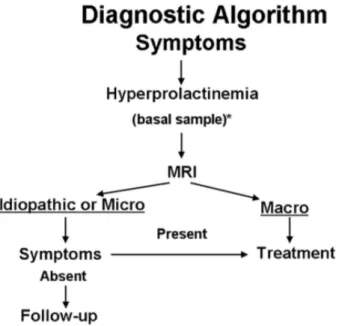
Figure 7. Recommended diagnostic algorism for prolactinomas (Casanueva et al., 2006). .... 30
Figure 8. Familial isolated prolactinoma pedigree ... 38
Figure 9. Flowchart of variant analyses method... 45
Figure 10. Diagram representing genes selected through different analyses. ... 50
Figure 11. Representative Electropherograms of validated genes for affected individuals through the family ... 53
Figure 12. RXRG protein structure.. ... 55
Figure 13. Pathway demonstrating TH probable interaction with hyperprolactinemia phenotype. ... 56
LIST OF TABLES
Table 1. Primer list and PCR conditions. ... 40
Table 2. Primer list for whole-exome sequencing validation. ... 47
Table 3. Description of sequence variants detected by whole exome sequencing. ... 49
Table 4. Genes selected through different analyses.. ... 51
Table 5. Combined exome results and analyses of single nucleotide variants (SNV) effect on protein structure and function by Polyphen, Sift, Provean and CADD. ... 52
Table 6. Small insertions and deletions analyses. ... 52
LIST OF ABREVIATIONS
a-MpT a-methyl-p-tyrosine
ACTH Adrenocorticotrophic Hormone
AHR Receptor aril hidrocarboneto
AIP Aryl-hydrocarbon receptor-interacting protein
AIP Aryl-hydrocarbon receptor-interacting protein gene
CDKN1B Cyclin-dependent kinase inhibitor 1B gene CRH Corticotrophic Releasing Hormone
D2R Dopaminergic receptor
FIPA Familial isolated pituitary adenoma
FSH Follicle-Stimulating Hormone
GH Growth Hormone
GHRH GH-Releasing Hormone
GnRH Hypothalamic-derived Gonadotropin-Releasing Hormone GPCRs G Protein Coupled Receptors
IGF1 Hepatic Insulin-like Growth Hormone
Kb Kilobase
kDa Kilodalton
LH Luteinizing Hormone
MEN1 Multiple endocrine neoplasia type 1
MEN1 Multiple endocrine neoplasia type 1 gene MEN4 Multiple Endocrine Neoplasia Type 4
NGS Next Generation Sequencing
PRF Prolactin releasing factors
PRKAR1A Protein kinase cAMP-dependent type I regulatory subunit alpha gene
PRL Prolactin
PRLR Prolactin receptor
PTTG Pituitary tumor-transforming gene
REXO4 REX4 Homolog, 3'-5' Exonuclease protein
REXO4 REX4 Homolog, 3'-5' Exonuclease gene RXRG Retinoid X Receptor, Gamma protein
RXRG Retinoid X Receptor, Gamma gene
T3 Tri-iodo-thyronine
TH Tyrosine hydroxylase protein
TH Tyrosine hydroxylase gene
TRH Thyrotropin-Releasing Hormone
TSH Thyrotrophin
TABLE OF CONTENTS
1. INTRODUCTION ... 13
1.1. Anatomy and physiology of the anterior pituitary gland ... 14
1.2. Prolactin regulation ... 19
1.3. Epidemiology of prolactinomas ... 21
1.4. Inherited prolactinomas ... 22
1.5. Genetics of prolactinomas ... 26
1.6. Clinical features and diagnosis of prolactinomas ... 28
1.7. Treatment of prolactinomas ... 30
1.8. Whole-exome sequencing ... 31
2. PURPOSE ... 34
3. MATERIALS AND METHODS ... 36
3.1. Clinical Case study (summary) ... 37
3.2. Sanger sequencing ... 39
3.3. Whole-exome sequencing (WES) ... 41
3.3.1. Variant Calling and annotation ... 42
3.3.2. Ingenuity® Variant AnalysisTM ... 43
3.3.3. Mendel, MD ... 44
3.3.4. Mutation Analysis ... 45
3.3.5. Indel analyses ... 46
3.3.6. Structural damage prediction and pathway analysis ... 46
4.1. AIP, MEN1 and PRLR mutation analyses ... 49
4.2. Whole exome sequencing analysis ... 49
4.3. Exome detected pathogenic variants validation via Sanger sequencing ... 53
4.4. Protein pathogenicity Prediction and pathway analysis ... 54
5. DISCUSSION ... 57
6. CONCLUSION ... 63
REFERENCES ... 65
1.1. Anatomy and physiology of the anterior pituitary gland
The pituitary is a small endocrine organ located within the sphenoid bone depression, called sella turcica. Together with the hypothalamus the pituitary gland orchestrates diverse body functions, including growth, reproduction and metabolic homeostasis. Scientists and artists have explored the anatomy and function of the hypothalamus and pituitary gland since the 2nd century AD. The importance of the hypothalamic-pituitary region has even influenced the work of the Renaissance artist Michelangelo Buonarroti in the Sistine Chapel ceiling at the Vatican, Italy (Figure 1). In this painting the creation of man sets in an arrangement that represents the brain outline, including the hypothalamic-pituitary region, suggesting the main role of this structure to life maintenance (Lechan and Toni, 2013).
Figure 1. Creation of Adam (Michelangelo Buonarroti, 1508-1512). (a) Photograph of the ceiling of
the Sistine Chapel at the Vatican, Italy; (b) The outline of the painting represents the midline sagittal
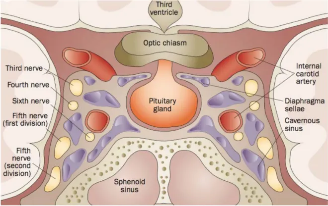
The adult pituitary weights about 600 mg and measures about 13 mm in the longest transverse diameter, 6 to 9 mm vertically, and around 9 mm anteroposteriorly (Melmed and Kleinberg, 2004). Dura mater surrounds the pituitary gland and form a roof over the sella turcica superiorly, such that the arachnoid membrane cannot enter the sella, thus pituitary gland maintains its anatomical and functional connections with the brain yet sits outside the blood-brain barrier (Nussey and Whitehead, 2001). The pituitary (Figure 2) is 5 mm beneath the optic chiasm, and positioned between the cavernous sinuses, that contain the internal carotid artery, oculomotor, trochlear and abducens cranial nerves, and also the first and second branches of the trigeminal nerve (Hong et al., 2016).
Figure 2. Normal anatomy of the sellar and parasellar regions surrounding the pituitary gland in a
The pituitary is attached directly to the median eminence of the hypothalamus and is composed of two morphologic and functional different components: the anterior lobe (adenohypophysis) and the posterior lobe (neurohypophysis) (Melmed and Kleinberg, 2004). The anterior pituitary gland derives from the invagination of the Rathke’s pouch, a primitive ectodermal tissue (Treier and Rosenfeld, 1996). The anterior pituitary is divided in three parts: pars intermedia, pars tuberalis and pars distalis (Figure 3a). The pars intermedia is composed of epithelial cells from the posterior limb of Rathke’s pouch, being rudimentary in humans. The pars tuberalis is a small rim of the adenohypophysis that involves the pituitary stalk. The bulk of the gland is the pars distalis,which represents 80% of the total pituitary volume (Asa and Ezzat, 2002; Drummond et al., 2003).
The adenohypophysis consists of five distinct types of differentially distributed hormone producing and secreting cells (Figure 3b). The functional development of these cell types involves complex spatiotemporal regulation of cell lineage-specific transcription factors expressed in pluripotential pituitary stem cells. The most frequent anterior pituitary cell line is the somatotroph, which comprises 45 to 50% of cells and produce growth hormone (GH). The lactotroph comprises between 9% of hormone-secreting anterior pituitary cells in males and nulliparous woman and up to 25% in multiparous females. These cells are specialized in prolactin (PRL) production. The corticotroph constitute 10 to 20% of anterior pituitary cells and produce adrenocorticotrophic hormone (ACTH). The gonadotrophic cells, 10 to 15% of cells, produce luteinizing hormone (LH) and follicle-stimulating hormone (FSH). The thyrotrophic cells account for 5% of hormone-secreting anterior pituitary cells and produce thyrotrophin (TSH) (Asa and Ezzat, 2002; Drummond
Figure 3. Pituitary anatomy and cell types. (a) Distinct parts of anterior pituitary; (b) Discrimination of pituitary cell types according to their hormone production (Asa and Ezzat, 2002).
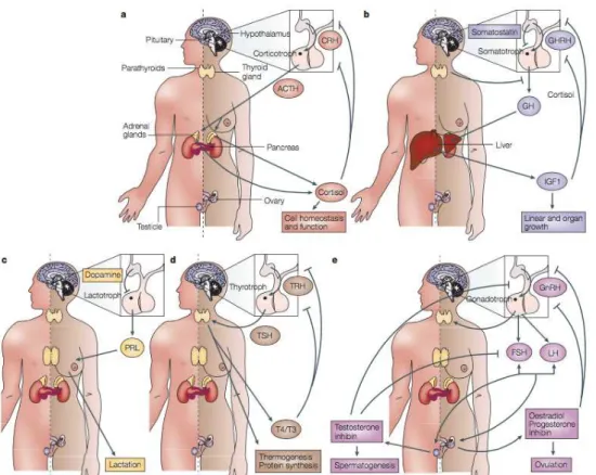
Different levels of control regulate the anterior pituitary hormone secretion. Hypothalamic control is mediated by adenohypophysiotropic hormones ,which are secreted into the portal system and bind directly to the anterior pituitary cell surface G-protein coupled receptors. A second control system is based on peripheral hormones, which act through negative feedback regulation of trophic hormones and their respective target hormones. The third regulation occurs inside the pituitary, where paracrine and autocrine soluble growth factors and cytokines locally regulate neighboring cell development and function. In consequence, a controlled pulsatile secretion of the six trophic pituitary hormones, ACTH, GH, PRL, TSH, FSH and LH, is achieved through these different regulatory mechanisms (Asa and Ezzat, 2002; Hong et al., 2016).
The regulation of ACTH secretion through corticotrophic cells is taken by hypothalamic-derived corticotrophic releasing hormone (CRH) and inhibited by cortisol. ACTH target organ is the adrenal gland where it regulates steroid secretion, leading to glucose, sodium and water homeostasis (Figure 4a). GH-releasing hormone (GHRH) and ghrelin, both hypothalamic hormones, induce GH secretion. Somatostatin (hypothalamus),
hepatic insulin-like growth hormone (IGF1), thyroid hormone and glucocorticoids inhibit GH secretion. GH regulates bone and muscle growth and maintains lean growth in adults (Figure 4b). Thyrotropin-releasing hormone (TRH), from the hypothalamus, and estrogen stimulate PLR secretion. PRL is negatively regulated by dopamine, released by cells in the median eminence. PRL-receptor signaling prepares and maintain the breast for postpartum and lactation (Figure 4c). TSH is positively regulated by TRH. TSH regulates thyroidal iodine metabolism, thyroid-hormone synthesis and thyroid growth, leading to thermogenesis and protein synthesis control. Tri-iodo-thyronine (T3), regulate TSH and TRH synthesis, providing control of TSH-directed thyroid hormone action (Figure 4d). Hypothalamic-derived gonadotropin-releasing hormone (GnRH) stimulates FSH and LH (Figure 4e). These hormones regulate sex-steroid synthesis and secretion, also participating in germ-cell development (Heaney and Melmed, 2004).
Figure 4. Control of the hypothalamic-pituitary-target-organ axes. (a) ACTH; (b)GH; (c)PRL;
1.2. Prolactin regulation
Prolactin is a 23 kDa polypeptide hormone that plays multiple homeostatic roles in the organism and is vital to mammogenesis, lactogenesis and galactopoiesis. The main cells that synthetize and secrete PRL are the lactotrophs, located at the anterior pituitary. Other organs and tissue are as well capable of PRL production and secretion, but little is known about the function of these PRL-secreting tissues (Freeman et al., 2000; Bernard et al., 2015b).
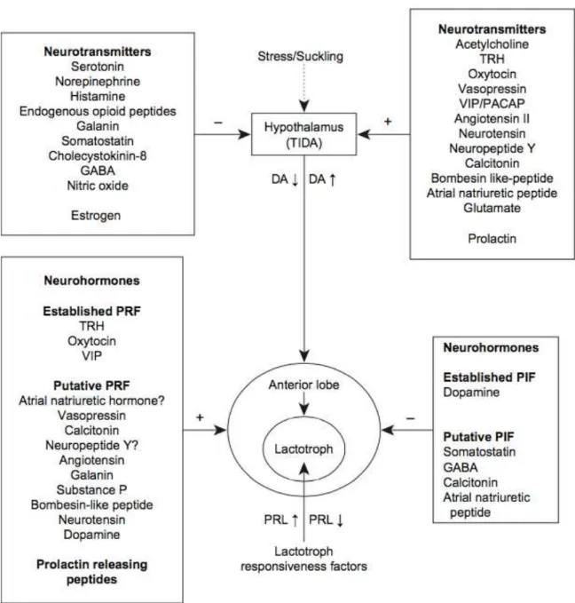
Synthesis and secretion of PRL by the lactotrophs are under the control of multiple prolactin releasing factors (PRF) and prolactin inhibitory factors (PIF). Dopamine secreted by tuberoinfundibular hypothalamic neurons (TIDA) is the primary inhibitory regulator of prolactin (Freeman et al., 2000; Mancini et al., 2008). Dopamine suppresses PRL though activation of D2 receptors. In fact, this is the physiologic basis for the therapeutic approach for hyperprolactinemia treatment, in which dopamine agonist is prescribed to in order to reduce PRL uncontrolled secretion (Neill, 1980).
Other PIF are somatostatin and gamma-aminobutyric acid. The major PRFs are TRH, oxytocin and neurotensin. These molecules are released into the long or short portal veins, as well across paracrine and autocrine mechanisms and target receptors in lactotrophic cells (Figure 5). Another regulating system is supported by PRL itself, which is capable of promoting feedback. PRL serum elevation activates PRL-receptors located at dopaminergic neurons and promote hypothalamic dopamine synthesis and increase the concentration of dopamine in the portal veins (Freeman et al., 2000; Mancini et al., 2008).
psychologic stress, treatment with dopamine receptor antagonist drugs and pituitary adenomas (Mancini et al., 2008; Vilar et al., 2008; Bernard et al., 2015b).
Figure 5. Overview of PRL regulation. Tuberoinfundibular dopaminergic system (TIDA) activity
controlling PRL in response to diverse stimulus. PIF and PRF are secreted by neuroendocrine neurons
and also regulate PRL secretion (Mancini et al., 2008).
1.3. Epidemiology of prolactinomas
Pituitary adenomas are monoclonal tumors, that is, the tumor arises from a single cell that has been transformed by genetic events that converted it into a neoplastic tissue. The transformations consist in acquisition of unique proliferative advantage and excessive proliferation of anterior pituitary hormone-producing cell lines (Asa and Ezzat, 2002; Melmed, 2011). Pituitary tumors rarely progress to become true metastatic carcinomas (Di Ieva et al., 2014). Despite exhibiting important growth, they present low mitotic activity when compared to other tumor types (Melmed, 2011).
Although not metastatic, these adenomas are associated to significant morbidity due to over-production of specific anterior pituitary hormones, leading to endocrine syndromes. Moreover, pituitary adenomas may promote a local space occupying effect. Prolactinomas arise from lactotrophic cells and secrete prolactin, causing symptoms such as hypogonadism, galactorrhea and bi-temporal hemianopsia (Asa and Ezzat, 2002; Heaney and Melmed, 2004; Hong et al., 2016).
European population-based studies report a pituitary adenoma prevalence of one per 1,277 individuals. These tumors account for 15% of all intracranial neoplasms, being the third most frequent tumor type after meningiomas and gliomas. The main pituitary adenomas are prolactinomas, which represent 50% of all cases on average (Aflorei and Korbonits, 2014).
(Ciccarelli, et al., 2005). Over 90% of prolactinomas are small, intrasellar tumors that rarely increase in size (Casanueva et al., 2006).
Prolactinomas have been reported in patients from two to 80 years, and its prevalence varies widely among different age groups, being the most prevalent pituitary adenoma type between the second and fourth decades of life (Mindermann and Wilson, 1994; Casanueva et al., 2006). There is also a difference of prolactinoma prevalence according to sex. In adults, prolactinomas arise more frequently in women than in men and become more diagnosed in men than in woman during the sixth decade of life (Ciccarelli et al., 2005; Aflorei and Korbonits, 2014).
1.4. Inherited prolactinomas
Although the vast majority of prolactinomas arise sporadically, some cases have been reported in family clusters, and can be defined as inherited prolactinomas. The classical familial syndromes that predispose patients to prolactinoma are Multiple Endocrine Neoplasia Type 1 (MEN1), Multiple Endocrine Neoplasia Type 4 (MEN4), Carney complex (CNC), and Familial Isolated Pituitary Adenomas (FIPA) (Ciccarelli et al., 2005; Lee and Pellegata, 2013).
anterior pituitary gland (Wermer, 1954). The most frequent tumors in MEN1 syndrome are those of the parathyroid glands (95% of cases), endocrine gastroenteropancreatic tract (30-80% of cases), and anterior pituitary (15-90% of cases) (Gribil et al., 2004). This disorder affects all age groups and has high penetration, with clinical manifestations developing in more than 80% of affected individuals by the fifth decade of life. Approximately 60% of pituitary adenomas occurring in MEN1 are PRL-secreting (Agarwal et al., 2009).
A variation of MEN1 syndrome, called MEN1-Burin, was described in four large kindred from the Burin peninsula, Canada. These patients have prominent features of prolactinomas in addition to carcinoids, and parathyroid tumors. These patients also show disruption in 11q13. A nonsense mutation in the MEN1 gene has been found to be responsible for the disease in all four MEN1-Burin families, suggesting that a common ancestral mutation in the MEN1-Burin phenotype is responsible for this prolactinoma variant of MEN1 (Olufemi et al., 1998).
suspected cases identified mutation in CDKN1B in only 11 patients (2.4%), of which four were asymptomatic (Georgitsi, 2010). Thus, this gene and disease related to it need further investigation.
Carney Complex is an autosomal dominant multiple endocrine neoplasia syndrome characterized by the complex of “myxomas, spotty pigmentation, endocrine overactivity, and schwannomas”. This disease was first described in by Carney in 1986 in a family in which symptoms occurred in three successive generations (Carney et al., 1986). Studies to unveil the genetic landscape of CNC identified mutations in protein kinase cAMP-dependent type I regulatory subunit alpha gene(PRKAR1A) in several families and also in patients with the sporadic form of the disease. Three unrelated families and one sporadic case shared the same 2bp deletion in exon 4B of PRKAR1A, suggesting it could be a hot spot for mutation (Kirschner et al., 2000). Actually, mutations in PRKAR1A have been reported in approximately 60% of patients with CNC. The hyperprolactinemia detected in such patients is for the most part asymptomatic and almost exclusively associated with clinical or subclinical acromegaly (Ciccarelli et al., 2005).
Familial Isolated Pituitary Adenoma classification was first mentioned in 2005 and has been a widely used concept since than (Ciccarelli et al., 2005; Daly and Beckers, 2014). Long before this classification, familial isolated pituitary adenomas were described. In 1967 Linquette and coworkers reported a family with isolated prolactinoma (Linquette
no further conclusions were taken (Berezin and Karasik, 1995).
FIPA is defined when two or more related individual reported with pituitary adenomas and no other syndromic features are diagnosed. In FIPA may occur pituitary tumors of the same type in all affected members of the same family (homogeneous presentation), or tumors of different cell types (heterogeneous presentation) (Daly and Beckers, 2014). Prolactinomas are the most commonly observed tumor (39.9%) in FIPA families, followed by GH-secreting or mixed GH-secreting and prolactin-secreting adenomas (30% and 7%, respectively) (Daly et al., 2006).
In about 20% of FIPA families, a mutation in Aryl hydrocarbon receptor-interacting protein (AIP) gene has been described and part of them have been associated to PRL-secreting pituitary adenomas (Daly and Beckers, 2014). Germline mutations in the
1.5. Genetics of prolactinomas
Although prolactinomas are common pituitary adenomas, the mechanisms that control the abnormal proliferation of this tumor type remains unclear for the most part. Studying the molecular profile of pituitary adenomas might be a challenging and technically difficult effort, since treatment might not include tissue resection and biopsy materials are rarely available; thus samples to provide information of when and how the lactotrophs acquire molecular modifications are scarce (Melmed, 2011). Based on the monoclonal nature of these neoplasms, evidence supports the hypothesis that pituitary tumors are caused by intrinsic pituitary-cell defects (Asa and Ezzat, 2002).
Common cancer-associated genes are rarely mutated in pituitary tumors (Melmed, 2011). This fact might explain the benign profile of these tumors. Besides, premature pituitary tumor senescence appears to bypass pro-proliferative signals and maintain cell viability. However, proto-oncogenes have been found to be mutated or overexpressed in prolactinoma, such as pituitary tumor-transforming gene (PTTG), which is expressed at high levels in most pituitary tissue. Experiments showed that besides overexpressed in pituitary tissue, this gene induces cellular transformation and is tumorigenic in nude mice (Melmed, 1997). However, PTTG role in pituitary tumorigenesis remains unclear (Asa and Ezzat, 2002)
As stated earlier, MEN1, MEN4, CNC and FIPA are familial syndromes that predispose patients to prolactinomas. Carney complex and MEN4 are rare causes of inherited forms of pituitary adenomas, especially prolactinomas. Thus mutations in MEN1
and AIP, although rare, are the most frequent type in such tumors (Agarwal et al., 2009; Daly et al., 2006). MEN1 and AIP are both located at the chromosome 11q13 locus (Lecoq
bona fide MEN1 features (Agarwal et al., 2009), AIP mutations have been reported in ~20% of FIPA cases (Daly and Beckers, 2014).
In addition, it has been shown that prolactin receptor (PRLR) knockout mice develop prolactinoma (Schuff et al., 2002) and that mutation in this same gene is associated to familial idiopathic hyperprolactinemia. The p.His188Arg variant of PRLR was found in three sisters with hyperprolactinemia, two of whom were presented with oligomenorrhea and the third with infertility (Newey et al., 2013). However, the role of
PRLR mutation in clinical manifestations has been discussed in the literature (Bernard et al., 2015a). Recently, Bernard et al. (2015a) investigated 88 patients with sporadic prolactinoma and found four PRLR mutations (p.Ile76Val, p.Ile146Leu, p.Glu108Lys and p.Glu554Gln) in 16 patients. However, the four variants were tested in vitro and had no effect on PRLR expression, localization and signaling after prolactin stimulation. Thus no phenotypically similar patients were reported to harbor inactivating germline mutations in this gene so far (Bernard et al., 2015b).
Experiments have been conducted in order to characterize other genes likely to be associated to prolactinoma tumorigenesis. The majority of studies is being held in the sporadic form of prolactinomas and has highlights new genetic targets. Large-scale expression profile analysis has pointed genes pertaining to prolactinoma formation, but further investigation is needed to access their role in lactotrophic proliferation (Evans et al., 2001; Evans et al., 2008; Jiang et al., 2010; Tong et al., 2012; Zhao et al., 2014; Seltzer et al., 2015; Zhou et al., 2015).
GADD45G, Gsp, MEG3a, MEN1, p53, Pdt-FGFR4, PKC, PRKAR1A, PTTG, RAS,
SSTR2/SSTR5, WIF and ZAC1 mutations. However PRDM2 emerged in this study as a drug-resistance and tumor recurrence driver (Gao et al., 2015).
Although current effort, it is still unclear how these new expression and genetic findings correlated to tumor development, and if any of these are also associated to familial settings.
1.6. Clinical features and diagnosis of prolactinomas
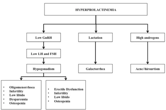
Prolactinomas are the most common causes of hyperprolactinemia (Ciccarelli, et al., 2005) and hyperprolactinemia is a well-established cause of hypogonadrotropic hypogonadism and anovulatory infertility. Scientific evidence suggests that PRL inhibits GnRH secretion and this leads to low circulating levels of LH and FSH and loss of ovarian stimulation, which can result in infertility (Bernard et al., 2015b).
In women, hyperprolactinemia is associated with oligo/amenorrhea in 90% of cases, 80% of patients also exhibit galactorrhea and may also manifest anovulatory infertility. Moreover, a chronic elevated PRL serum level is associated with reduced spinal bone mineral density. Hyperprolactinemia may also be identified in men and usually causes impotence, infertility and decreased libido (Figure 6). Due to the general aspects of symptoms and delayed recognition of them, men commonly present larger tumors than woman and are more susceptible to neurological symptoms (Casanueva et al., 2006).
palsies of cranial nerves 3, 4 or 6), or ipsilateral facial pain (from involvement of the V1 and V2 branches of the 5th nerve) (Melmed and Kleinberg, 2004; Hong, et al., 2016).
Figure 6. Clinical manifestations of hyperprolactinemia (modified from Vilar and Naves, 2012).
Figure 7. Recommended diagnostic algorism for prolactinomas (Casanueva et al., 2006).
1.7. Treatment of prolactinomas
Therapeutic goals for hyperprolactinemia include control of excessive hormone secretion and infertility, sexual dysfunctions, and osteoporosis, removal and relief of any disturbance in vision and cranial nerve function, and prevention of recurrence or progression (Auriemma et al., 2016).
Experimental therapies with somatostatin analogues, nerve growth factor, interferon-a and dopastatins are being developed to be used when first line therapy fails. These studies are in various phases of development, but none of these approaches has received approval or a demonstration to be advantageous. Thus, for medication non-responsive prolactinoma, surgical and radiation treatment are both available options, although are not frequently required (Capozzi et al., 2015).
1.8. Whole-exome sequencing
The genetic study of tumors uses the DNA sequencing, which is one of the main tools for medical research (Rabbani et al., 2014). The union of two techniques: the chain termination sequencing by Sanger et al. (1977), and the polymerase chain reaction (PCR) by Mullis and collaborators (1986), established many marked events such as the completion of the Human Genome Project (HGP). This approach provided a reference genome so that latter on genetic alterations could be associated to disease phenotypes (Sachidanandam et al., 2001; Venter, 2003; Rabbani et al., 2014).
data and the need for user-friendly software in the analysis of the raw sequence have to been addressed (Rabbani et al., 2014). The recent advances in these techniques are accelerating the pace of discovery in genetic disorders and cancer. As a result, they have entered the clinical practice and have been used to evaluate genes associated with phenotype for which no genetic abnormalities has been described (Bick and Dimmock, 2011).
Thus, next-generation sequencing technologies are useful tools to decipher the genetic events driving multiple diseases that lack known causal genetic mutation. Over the past few years, whole-exome sequencing has been used to detect causative mutations in endocrine related traits such as nonfunctioning pituitary adenomas. Genomic DNA from seven pituitary non-functioning pituitary adenoma were investigated and revealed 24 somatic variants identified and confirmed. However, DNA sequence analysis of these variants in a set of 24 pituitary non-functioning adenomas did not reveal any mutations, indicating that these genes are unlikely to contribute significantly in the etiology of sporadic pituitary (NEWEY et al., 2013).
prolactinomas Gao has used whole-exome sequencing to establish genetic difference between six dopamine-responsive and six dopamine-resistant prolactinomas. Multiple genes emerged at this study, however PRDM2 was pointed out as an important gene for prolactinoma tumorigenesis (Gao et al., 2015).
Thus, the present study applied whole exome sequencing in an attempt to identify the causative mutation in AIP, MEN1 and PRLR mutation-negative Brazilian family presenting with familial isolated prolactinoma.
This work aims to study a rare case o familial isolated prolactinoma and unveil the genetic characteristics of this family, and how they might be associated to tumor development. For that, the following specific purpose were considered:
2.1.Determine the presence of MEN1 mutation in this kindred. 2.2.Determine the presence of AIP mutation in this kindred. 2.3.Determine the presence of PRLR mutation in this kindred.
2.4.Perform whole exome in DNA samples from this kindred if genes referred to above had wild type alleles.
2.5.Analyze whole-exome sequencing results and select candidate single nucleotide variants.
3.1. Clinical Case study (summary)
3.2. Sanger sequencing
About 5ml of peripheral blood of all patients and controls were collected in vacuum tubes with EDTA after obtaining written informed consent of the patients. Genomic DNA was isolated from all study participants using saline concentration method of Lahiri and Nurnberger (Lahiri and Nurnberger, 1991). All participants gave informed consent which was approved by the Ethics Committee of the Universidade Federal de Minas Gerais.
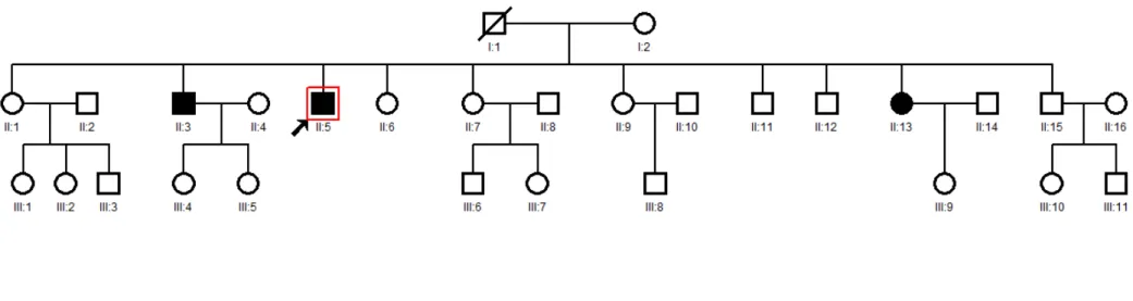
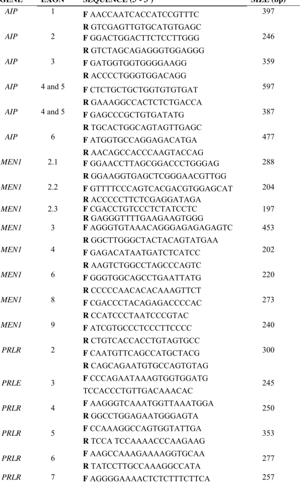
Genotyping for germline mutations in the MEN1, AIP, and PRLR genes was carried out in the three clearly affected family members (II.3, II.5, II.13 figure 8). Exon-specific flanking primers (Table 1) were either designed according to previously published studies (Vierimaa et al., 2006; Newey et al., 2013) or using PrimerBlast software, available at
http://www.ncbi.nlm.nih.gov/tools/primer-blast/. The DNA extracted from the blood patients was quantified and polymerase chain reactions (PCR) were performed using 50ng/ml DNA, 2.5ml of 10x Buffer IIB (40mM NaCl, 10mM TrisHCl, pH8.4, TritonX -100 0.1 %; 1.5MM MgCl2), dNTPs 2.5ml (0.2mm), 0.5ml of each primer pair (10 pmol/ml) and 0.25ml of Taq polymerase (0.625U). The products were amplified in a thermocycler Eppendorf Mastercycler Gradient, using the following steps: denaturation at 94° C for 3 minutes, and 35 cycles of denaturation at 94oC for 30 seconds. Annealing at 55-60oC for 30 seconds and extension at 72° C for 30 seconds. At the end of the cycles, the reactions undergone a final extension at 72° C for 5 minutes. PCR reaction products were gel-verified and purified using the PCRLinkTM Quick PCR Purification Kit (Life technologies, Carlsbad, California), then submitted to sequencing reaction with the ABI BigDye Terminator Cycle Sequencing Kit v3.1 on an ABI PRISM 3130 Genetic Analyzer (Applied Biosystems, Foster City, USA).
Table 1. Primer list and PCR conditions.
GENE EXON SEQUENCE (5´- 3´) SIZE (bp)
AIP 1 F AACCAATCACCATCCGTTTC 397
R GTCGAGTTGTGCATGTGAGC
AIP 2 F GGACTGGACTTCTCCTTGGG 246
R GTCTAGCAGAGGGTGGAGGG
AIP 3 F GATGGTGGTGGGGAAGG 359
R ACCCCTGGGTGGACAGG
AIP 4 and 5 F CTCTGCTGCTGGTGTGTGAT 597
R GAAAGGCCACTCTCTGACCA
AIP 4 and 5 F GAGCCCGCTGTGATATG 387
R TGCACTGGCAGTAGTTGAGC
AIP 6 F ATGGTGCCAGGAGACATGA 477
R AACAGCCACCCAAGTACCAG
MEN1 2.1 F GGAACCTTAGCGGACCCTGGGAG 288
R GGAAGGTGAGCTCGGGAACGTTGG
MEN1 2.2 F GTTTTCCCAGTCACGACGTGGAGCAT 204
R ACCCCCTTCTCGAGGATAGA
MEN1 2.3 F CGACCTGTCCCTCTATCCTC 197
R GAGGGTTTTGAAGAAGTGGG
MEN1 3 F AGGGTGTAAACAGGGAGAGAGAGTC 453
R GGCTTGGGCTACTACAGTATGAA
MEN1 4 F GAGACATAATGATCTCATCC 202
R AAGTCTGGCCTAGCCCAGTC
MEN1 6 F GGGTGGCAGCCTGAATTATG 220
R CCCCCAACACACAAAGTTCT
MEN1 8 F CGACCCTACAGAGACCCCAC 273
R CCATCCCTAATCCCGTAC
MEN1 9 F ATCGTGCCCTCCCTTCCCC 240
R CTGTCACCACCTGTAGTGCC
PRLR 2 F CAATGTTCAGCCATGCTACG 300
R CAGCAGAATGTGCCAGTGTAG
PRLE 3 F CCCAGAATAAAGTGGTGGATG 245 TCCACCCTGTTGACAAACAC
PRLR 4 F AAGGGTCAAATGGTTAAATGGA 250
R GGCCTGGAGAATGGGAGTA
PRLR 5 F CCAAAGGCCAGTGGTATTGA 353
R TCCA TCCAAAACCCAAGAAG
PRLR 6 F AAGCCAAAGAAAAGGTGCAA 277
R TATCCTTGCCAAAGGCCATA
GENE EXON SEQUENCE (5´- 3´) SIZE (bp)
R ACCATTTAAAACATATTTAGGGACA
PRLR 8 F GAA TGGAGGAAAACACTCTTGG 248
R TGACTATCATGATTGGGAGGAA
PRLR 9 F AGCTGCCAAACCAAGTCCTA 293
R AAGGCTGGCTGAAACTACCA
PRLR 10.1 F GGGA TGCTGA TTTGGAA TGT 500
R GGTAAGAGGATCTGGGGTTG
PRLR 10.2 F CCCTTTTGTCTGAAAAGTGTGA 400
R GGCGTATCCTGGTCAGTCTC
3.3. Whole-exome sequencing (WES)
There are different deep-sequencing platforms to choose from when performing whole exome sequencing. Hybridization is the most optimal and commonly used method for targeted exome. For this purpose, peripheral blood from the three clinically affected sibs (II.3, II.5, II.13 Figure 8) was collected and DNA extracted. Subsequently, DNA was subjected to whole-exome capturing and sequencing using the Roche NimbleGen V2 chip (Madison, Wisconsin) or Nextera (San Diego, California) with the Illumina HiSeq2000 sequencing platform (San Diego, California).
to the flow cell by hybridizing to oligos on its surface that are complementary to the ligated adaptors. The DNA-molecules are then amplified by a so called bridge amplification
which results in a hundred of millions of unique clusters. Finally, the reverse strands are cleaved and washed away and the sequencing primer is hybridized to the DNA-templates. During sequencing the huge amount of generated clusters are sequenced simultaneously. The DNA-templates are copied base by base using the four nucleotides (ACGT) which are fluorescently-labeled and reversibly terminated. After each synthesis step, the clusters are excited by a laser which causes fluorescence of the last incorporated base. After that, the fluorescence label and the blocking group are removed allowing the addition of the next base. The fluorescence signal after each incorporation step is captured by a built-in camera, producing images of the flow cell.
3.3.1. Variant Calling and annotation
Variant calling is the part of the process that perform the initial mapping of the reads, improvement of alignments and quality scores, variant identification, and recalibration of the variants quality scores. In general, a coverage of 20X to 50X at each nucleotide is considered acceptable when identifying variations.
All generated VCF files were analyzed as a familial group using three different tools. The first software used was Mendel, MD, developed by the Clinical Genomic Laboratory of Universidade Federal de Minas Gerais and available at
http://mendel.medicina.ufmg.br (Cardenas et al., 2015). VCF files were also clustered together and analyzed by Ingenuity® Variant AnalysisTM software, available at
www.ingenuity.com/variants. The third analysis was performed using the pipeline developed by Noam Shomron, Ofer Isakov and Marie Perrone at the Tel-Aviv University Medical School as previously detailed (Isakov, 2013a). For these analyses only variants with call quality of at least 40.0 and read depth of at least 20.0 were considered. Additionally, variants with allele frequency greater than or equal to 1.0% of the genomes reported in the 1000 genomes project (www.1000genomes.org), the public Complete Genomics (http://www.completegenomics.com/public-data/) or NHLBI ESP exomes (http://evs.gs.washington.edu/EVS/) were also excluded from further analyses. The selected genes carried identical homozygous or heterozygous sequence variants that co-occurred in all genotyped cases.
3.3.2. Ingenuity® Variant AnalysisTM
In addition to the above listed confidence and frequency criteria, variants associated with gain or loss of function, compound heterozygote, heterozygous ambiguous, haploinsufficiency, homozygous, or hemizygous that occurred in all WES genotyped samples at the variant level were chosen to be studied.
frameshift, in-frame indel, stop codon change, a missense unless predicted to be innocuous by SIFT or Polyphen-2, predicted to disrupt splice site up to 2.0 bases into intron, deleterious to a microRNA or a structural variant.
Considering the biological context, the following key words were selected from
Ingenuity® Variant AnalysisTM (Ingenuity biological analysis): hyperprolactinaemic disorder, prolactinoma, pituitary adenoma predisposition, prolactin excess, amount of prolactin-secreting pituitary gland adenoma (quantity of prolactinoma), amount of prolactin-producing pituitary adenoma (quantity of prolactinoma), autosomal dominant prolactin-producing pituitary adenoma (autosomal dominant prolactinoma), formation of secreting pituitary gland adenoma (formation of prolactinoma), prolactin-producing pituitary adenoma (prolactinoma), familial isolated pituitary adenoma or diseases consistent with phenotypes. Analysis that considered differentially expressed published prolactinoma genes was also applied. In this analysis, a list of differentially expressed genes in human prolactinomas reported from 1993 until 2015 was created. Gene list was extracted from six published papers (Evans et al., 2001; Evans et al., 2008; Jiang
et al., 2010; Tong et al., 2012; Zhao et al., 2014; Zhou et al., 2015). This list was used to filter if any of the mutations detected was noted within a gene that was previously found to be differentially expressed in prolactinomas.
3.3.3. Mendel, MD
Mendel, MD was also used to analyze indels. In this analysis, confidence, frequency and pathogenicity criteria were maintained and a filter for the pathogenicity impact of the alteration was added and only variants with high or moderate impact were selected.
3.3.4. Mutation Analysis
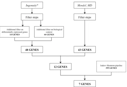
The results available from the Ingenuity® platform that considered a list of previously published differentially expressed proteins in prolactinomas was added to the list of genes selected through the Ingenuity® analysis of prolactinoma biological context and resulted in one mutated genes single list. This list was than combined to the analyses performed using Mendel, MD and only those genes that were present in both analyses were kept. Than, this single gene list was compared to the independent analysis performed using the Isakov-Shomron pipeline and the final single-nucleotide variant list that was detected by all three schemes was subsequently analyzed (Figure 9).
Figure 9. Flowchart of variant analyses method. Three methods of whole-exome variant analyses
3.3.5. Indel analyses
Using an annotation approach, genes that harbored indels were analyzed for their potential relevance to prolactinoma tumorigenesis. The following criteria were taken under consideration: (i) Pathway annotation, which includes all pathways in which a given gene product has reportedly been involved in. Pathway information was gathered from the Kyoto Encyclopedia of Genes and Genomes (KEGG) (http://www.genome.jp/kegg/); (ii) Interaction annotations from the STRING protein functional interactions database (http://string-db.org) (iii) Publications relating each gene to pituitary adenomas were accessed in the Medline database.
3.3.6. Structural damage prediction and pathway analysis
In order to obtain a homology model of mutated genes, a psiBLAST (ProteinDataBank, http://blast.ncbi.nlm.nih.gov/) was performed. The crystallographic coordinates were obtained from a PDB template with high homology to the translated protein sequence from the affected patients.
Pathway analysis was performed using Ingenuity® Variant AnalysisTM tool of pathway to phenotype, where a possible connection between protein and disease is established according to its protein to proteins interaction profile.
3.3.7. WES sequence data validation
family history of benign or malignant tumors (recruited under an Ethics Committee approved protocol from amongst individuals who currently attend the Geriatric Clinic of the Universidade Federal de Minas Gerais) were tested for selected sequence variants that were seemingly pathogenic and causative. After DNA isolation from the cases and controls, all relevant genes were amplified by PCR with primers specific for each region (Table 2). PCR products were purified using PCRLinkTM Quick PCR Purification Kit (Life technologies, Carlsbad, California) following manufacturer’s protocol and visualized on a silver-stained 6.5% polyacrylamide gel. Sequences were obtained on ABI 3130 Genetic Analyzer (Applied Biosystems, Foster City, CA). Bidirectional sequence data were analyzed by using Sequencer 4.9 software.
Table 2. Primer list for whole-exome sequencing validation.
GENE EXON SEQUENCE (5´-3´) SIZE (bp)
RXRG 1 F GGGGGGATGTGCAGAGCCATAAGTCAGG
R GCACTACCCAGAGGTTCATGCCCACGTG
464
REXO4 9 F CTCCACTCACCTGCACAGTC 223
R TGCTCTTTCACGAGGCTGAG
TH 11 F ACCAAGACCAGACGTACCAG 183
R TTCTCATCTGTGACCTGGGC
MAP2K3 17 F TGTGAAGCCCTCCAATGTCC 165
R ATCCTCTCCTGAGCCTGGG
PABPC1 18 F GTTATGATGGAGGGTGGTCG 287
4.1. AIP, MEN1 and PRLR mutation analyses
Mutations in the coding and flanking intronic regions of the AIP, MEN1 and PRLR
genes were not detected in any affected family members genotyped (data not shown).
4.2. Whole exome sequencing analysis
Variant calling from WES of the three genotyped patients resulted in 57,509 common variants in 8,498 genes, the mean base call quality was 1,547 and average read depth was X83. A average of 1,2337 missense single nucleotide variations were found in the three sequenced patients, and the mean number of nonsense variants were 125, mean silent SNV total of 12,702 and a mean total of 1,947 indels (Table 3).
Table 3. Description of sequence variants detected by whole exome sequencing.
Variants Patients Mean
II.3 II.5 II.13
Missense SNV 11231 11465 14317 12337
Nonsense SNV 89 97 189 125
Silent SNV 12116 12606 13384 12702
Total number of indels 2516 2578 747 1947
The Ingenuity® analysis, considering the biological context of prolactinoma, yielded a list of 44 mutated genes. The second analysis performed using the Ingenuity®
combined and only 12 genes emerged to be shared by both lists. The independent analysis performed using the Isakov-Shomron pipeline combined with the 12 genes list mentioned above resulted in seven selected genes (Figure 10, Table 4).
51 sel ected th rough different analyses. Shaded squa res indicate th e analy sis where the
. Bold genes
were those that em
erged in Ingenuity ® , Me n d el
and Isakov analy
Variants list were then filtered by PROVEAN, SIFT, Polyphen and CADD algorithms, resulted in six SNV in five genes predicted to be deleterious: RXRG, REXO4,
TH, PABPC1 and MAP2K3 (Table 5).
Table 5. Combined exome results and analyses of single nucleotide variants (SNV) effect on protein structure and function by Polyphen, Sift, Provean and CADD.
GENE CHR CODON
CHANGE
AA
CHANGE PROVEAN SIFT PPH2 CADD
RXRG 1 cGc/cAc p.R317H -4.58 0.022 1.000 34
TTN 2 cAa/cGa p.Q9198R -2.11 0.370 ND 7.214
AQP1 7 gTc/gGc p.V284G -1.99 0.072 ND 20.4
PABPC1 8 gAa/gGa p.E372G -6.14 0.000 0.999 27.6
PABPC1 8 Cgc/Tgc p.R374C -7.17 0.000 0.988 32
REXO4 9 tGg/tAg p.W195* ND ND ND 38
TH 11 Aag/Tag p.K474* ND ND ND 36
MAP2K3 17 tTg/tGg p.L215W -5.84 0.000 1.000 25.4
Shaded lines represent SNV that are highly expected to be deleterious according to the in silico analysis.
ND – No data; Cutoff: PROVEAN (-2.5), SIFT (0.05), PPH2 (0.95) CADD (15).
Indel analysis was also performed using data generated from all three samples from the affected patients. After filtration steps (see methods) a total of eight genes were selected (Table 6). Following the aforementioned steps of gene annotation, none of the indel listed genes were selected for further investigation.
Table 6. Small insertions and deletions analyses.
GENE CHR CODON CHANGE MUTATION FUNCTIONAL CLASS
HRNR 1 atg/ HOMO FRAME_SHIFT+START_LOST
ZNF717 3 ttt/ HETERO FRAME_SHIFT
MAP3K1 5 tcaaca/tca HOMO CODON_DELETION
PHPT1 9 tgtctg/ HETERO FRAME_SHIFT
ATRNL1 10 ccttct/cct HETERO CODON_DELETION
HYDIN 16 att/ HETERO FRAME_SHIFT
PKD1L2 16 aac/ HETERO FRAME_SHIFT
CNDP1 18 gtg/gTGCtg HETERO CODON_INSERTION
4.3. Exome detected pathogenic variants validation via Sanger sequencing
The missense variants found in PABPC1 and MAP2K3 genes were not validated or confirmed in the three affected siblings. Variants detected in the RXRG, REXO4 and TH
genes that were validated in all three affected family members and were subsequently sequenced in all available clinically unaffected family members (Figure 11, table 7) as well as in the 95 healthy ethnically matched controls. None of population controls carried any of the genotyped variants (data not shown). Family sequencing revealed that some clinically and serologically asymptomatic siblings (p.R317H RXRG: n=3, p.w195* REXO4: n=5; p.K474* TH: n=4) also harbored these variants.
Figure 11. Representative Electropherograms of validated genes for affected individuals
through the family. (a) TH sequencing results; (b) RXRG sequencing results; (c) REXO4
Table 7. Summary of validation sequencing results. Shaded squares indicate affected subjects.
SUBJECTS
RXRG REXO4 TH MAP2K3 PABPC1 PABPC1
p.R317H p.W195* p.K474* p.L215W p.E372G p.R374C
cGc/cAc tGg/tAg Aag/Tag tTg/tGg gAa/gGa Cgc/Tgc
I.2 G/G G/A A/A T/T A/A C/C
II.1 G/A G/A A/T T/T A/A C/C
II.3 G/A G/A A/T T/T A/A C/C
II.5 G/A G/A A/T T/T A/A C/C
II.6 G/G G/G A/A T/T A/A C/C
II.7 G/G G/A A/A T/T A/A C/C
II.9 G/G G/G A/A T/T A/A C/C
II.11 G/A G/G A/T T/T A/A C/C
II.12 G/G G/A A/T T/T A/A C/C
II.13 G/A G/A A/T T/T A/A C/C
II.15 G/A G/A A/T T/T A/A C/C
4.4. Protein pathogenicity Prediction and pathway analysis
The RXRG p.R317H mutation leads to major structural abnormality and predictably deleteriously affects protein function is located in RXRG-Retinoic acid binding site, a region evolutionarily highly conserved, as shown in Figure 12.
Figure 12. RXRG protein structure. (a) Overall structure of the tetrameric RXRG (PDB code
1G1U) shown in a cartoon diagram as A1 (light blue), B1 (green), B2 (yellow) and A2 (magenta). Side-view showing the location of the mutation p.R317H in orange spheres on each chain; (b) A close-up showing the side chain of amino acid 317 of RXR (Histidine, in green and Arginine in
pink) at an α helix.
a)
Arg317
His317
Figure 13. Pathway demonstrating TH probable interaction with hyperprolactinemia phenotype.
5.
DISCUSSION
The pituitary tumorigenesis is a complex process and the identification of genes that are critical for the characterization of the disease is a demanding task due to the infinitely possibilities. Thus familial cases, although rare, are an interesting starting point to seek for novel mutations that could occur throughout a family setting and promote pituitary adenoma tumorigenesis.
This work describes three affected siblings in a familial set. All affected individuals manifest typical hyperprolactinemia phenotypes and were responsive to dopamine-agonist treatment. In fact, dopaminergic agonists are the primary therapy for patients with hyperprolactinemia and prolactinoma (Casanueva et al., 2006), and as reported in the present study, these drugs normalize PRL levels and significantly reduce tumor volume (Heaney and Melmed, 2004). It is also interesting to notice that one of the reported patients presented spontaneous tumor remission after pregnancy. This event has actually been reported in two-thirds of pregnant patients (Almalki et al., 2015). This mechanism of remission could be related to the activity of lysosome enzymes in degradation of PRL granules pituitary cells, as previously shown in the lactotroph involution after cessation of lactation (De Marco et al., 1982).
Classically, PRL levels >250 ng/ml have been considered to be highly suggestive of the presence of a macroprolactinomas (Vilar et al., 2014), as shown in patient II.5. However, macroprolactinomas are associated to Circulating PRL levels of 100-200 ng/mL, but not infrequently they may be <100 ng/mL (Vilar et al., 2014), as observed in the two other patients described in this study, II.3 and II.13. Thus lower concentration of serum PRL might be present in prolactinoma patients since overlap in PRL values regardless of the etiology of hyperprolactinemia has been shown (Vilar et al., 2014).
affected family members were negative for mutations in these genes. Thus, a Whole-exome sequencing protocol was selected to continue the genetic studies of this family.
In this single Brazilian family with an isolated prolactinoma phenotype, three novel, seemingly pathogenic heterozygous variants in the RXRG, REXO4, and TH genes were identified. Of these, both REXO4 and TH variants are likely to be pathogenic as these are clearly inactivating (stop codon and premature termination of protein translation).
RNA exonuclease 4 (REXO4) is a nuclear expressed exonuclease, also known as
XPMC2H and REX4, located to the long arm of chromosome 9 (Kwiatkowska et al., 1997). Although the variation in this gene emerged as possibly pathogenic, its allele frequency in the ExAC Browser Exome Aggregation Consortium (http://exac.broadinstitute.orgI) is 6:1,000, which was considered high for a variant causing this disease. Also, based on the paucity of published data on the function of this gene product, no direct involvement of mutations in this gene can be inferred to contribute to prolactinoma formation and predisposition.
The second gene that emerged as a strong prolactinoma susceptibility gene was the Tyrosine Hydroxylase (TH) gene, located to the short arm of chromosome 11 (Craig et al., 1986) that carries the AIP and MEN1 genes (Lecoq et al., 2015). Not only the type of variant found in this gene and its location made it an attractive candidate prolactinoma formation gene, but also its function. TH enzyme converts L-tyrosine into L-3,4-dihydroxyphenylalanine (L-DOPA), the essential and rate-limiting step to formation of dopamine and other catecholamines. Since dopamine released from the hypothalamus negatively regulates the secretion of prolactin (PRL) from the anterior pituitary gland, TH is intimately involved in the prolactin signaling pathway (Bernard et al., 2015b).
since patients respond well to dopamine-agonist therapy, but might reside in genes that confer increased sensitivity to stimulatory neurohormones (Berezin and Karasik, 1995). It had been also speculated if the functional dopamine uncoupling from D2 receptor could contribute to the development of prolactin (PRL)-secreting pituitary tumors. However, mutations in the coding exons of the D2 could not be demonstrated (Friedman et al., 1994), Thus other elements of dopamine pathway might be disrupted in prolactinoma development and TH enters these criteria.
Experimental studies examined the role of TH expression in pituitary cell proliferation and have shown that hyper expression of tyrosine hydroxylase in human lactotroph adenomas enhances dopamine synthesis and diminishes prolactin secretion (Freese et al., 1996), as well as suppression of tumor growth (Williams et al., 2002). Studies using genetically modified organisms as model systems yielded inconclusive results in terms of teasing out the possible involvement of TH in prolactinoma development. Mice homozygotes for targeted null mutations are catecholamine deficient and usually die of cardiac failure (Kobayashi et al., 1995).
The missense variant found in the RXRG gene (R317H) was assigned a likely damaging score by several prediction algorithms. Although these are predictions and estimates, it has been shown that protein damage prediction algorithms have ~70% sensibility and ~15% specificity (Flanagan et al., 2010).
The RXRG gene is located to the long arm of chromosome 1 and encodes a protein member of the steroid/thyroid hormone superfamily of nuclear receptors, called retinoid X receptor gamma (Almasan et al., 1994). This gene is highly conserved across species and is expressed at low levels throughout the body, with higher levels in skeletal muscle, pituitary gland and certain areas of the brain. RXRG is involved in diverse cellular processes, from proliferation to metabolism. This receptor forms dimers with the retinoic acid, thyroid hormone, and vitamin D receptors, increasing both DNA binding and transcriptional function on their respective response elements. Noteworthy, the ligand-binding domain of the protein is where p.R317H variant found in this study locates to (Lefebvre et al., 2010). This missense heterozygous mutant was previously reported as a somatic mutation in endometrial cancer, but no association to pituitary adenomas was reported (COSMIC, http://cancer.sanger.ac.uk/cosmic).
It has been shown that RXRG is expressed in pituitary adenomas and may co-localize to Pit-1 (Sanno et al., 1999), a pituitary specific transcription factor that binds to and transactivates pituitary hormone genes such as PRL (INGRAHAM et al., 1997). Moreover, treatment with retinoic acid has been proved beneficial and well tolerated for Cushing’s disease patients, further supporting the possible importance of this pathway to pituitary tumor development (Giraldi et al., 2012).
pituitary tumor development but one could speculate that protein expression is needed to negatively regulate pituitary cells growth, and that an inactivating mutation could lead to accelerated proliferation and PRL protein overexpression.
REFERENCES
Aflorei ED, Korbonits M. Epidemiology and etiopathogenesis of pituitary adenomas. J Neurooncol. 2014; 117:379-394.
Agarwal SK, Ozawa A, Mateo CM, Marx SJ. The MEN1 gene and pituitary tumours. Horm Res. 2009; 2:131-138.
Almalk MH, Alzahrani S, Alsherbeni F, Alsherbeni S, Almoharb O, Aljohani N, Almagamsi A. Managing prolactinomas during pregnancy. FRontirs in Endocrinology. 2015; 6:85.
Almasan A, Mangelsdorf DJ, Ong ES, Wahl GM, Evans RM. Chromosomal localization of the human retinoid X receptors. Genomics. 1994; 20:397-403.
Asa SL and Ezzat S. The pathogenesis of pituitary tumours. Nat Rev Cancer. 2002; 2:836-49. Auriemma RS, Grasso LFS, Pivonello R and Colao Annamaria. The safety of treatments for
prolactinomas. Expert Opinion on Drug Safety. 2016; DOI: 10.1517/14740338.2016.1151493.
Berezin M, Karasik A. Familial prolactinoma. Clinical Endocrinology. 1995; 42:483-486. Bernard V, Bouilly J, Beau I, Broutin I, Chanson P, Young J, Binart N. Germline prolactin
receptor mutation is not a major cause of sporadic prolactinoma in humans. Neuroendocrinology 2015a; DOI:10.1159/000442981.
Bernard V, Young J, Chanson P, Binart N. New insights in prolactin: pathological implications. Nat Rev Endocrinol. 2015b; 11:265-275.
Bick D, Dimmock D. Whole exome and whole genome sequencing. Curr Opin Pediatr. 2011; 23:594-600.