Whole Genome Sequence of Multiple
Myeloma-Prone C57BL/KaLwRij Mouse Strain
Suggests the Origin of Disease Involves
Multiple Cell Types
Sarah R. Amend1, William C. Wilson1, Liang Chu1, Lan Lu1, Pengyuan Liu2, Daniel Serie5, Xinming Su1, Yalin Xu1, Dingyan Wang4, Anthony Gramolini4, Xiao-Yan Wen4,
Julie O’Neal1, Michelle Hurchla1, Celine M. Vachon5, Graham Colditz3, Ravi Vij1, Katherine N. Weilbaecher1, Michael H. Tomasson1*
1Division of Oncology, Washington University School of Medicine, St. Louis, MO, United States of America, 2Medical College of Wisconsin, Milwaukee, WI, United States of America,3Department of Surgery, Washington University School of Medicine, St. Louis, MO, United States of America,4Department of Physiology, University of Toronto, Toronto, Canada,5Department of Health Sciences Research, Division of Epidemiology, Mayo Clinic College of Medicine, Rochester, MN, United States of America
*tomasson@dom.wustl.edu
Abstract
Monoclonal gammopathy of undetermined significance (MGUS) is the requisite precursor to multiple myeloma (MM), a malignancy of antibody-producing plasma B-cells. The genetic basis of MGUS and its progression to MM remains poorly understood. C57BL/KaLwRij (KaLwRij) is a spontaneously-derived inbred mouse strain with a high frequency of benign idiopathic paraproteinemia (BIP), a phenotype with similarities to MGUS including progres-sion to MM. Using mouse haplotype analysis, human MM SNP array data, and whole exome and whole genome sequencing of KaLwRij mice, we identified novel KaLwRij gene variants, including deletion ofSamsn1and deleterious point mutations inTnfrsf22and
Tnfrsf23. These variants significantly affected multiple cell types implicated in MM patho-genesis including B-cells, macrophages, and bone marrow stromal cells. These data dem-onstrate that multiple cell types contribute to MM development prior to the acquisition of somatic driver mutations in KaLwRij mice, and suggest that MM may an inherently non-cell autonomous malignancy.
Introduction
Multiple myeloma (MM) is the second most common hematological malignancy and is charac-terized by accumulation of clonal plasma B-cells in the bone marrow, hypercalcemia, renal fail-ure, anemia, and lytic bone lesions. Despite impressive recent progress in treatments for MM, median survival is only 6 years [1]. The requisite precursor to MM is monoclonal gammopathy OPEN ACCESS
Citation:Amend SR, Wilson WC, Chu L, Lu L, Liu P, Serie D, et al. (2015) Whole Genome Sequence of Multiple Myeloma-Prone C57BL/KaLwRij Mouse Strain Suggests the Origin of Disease Involves Multiple Cell Types. PLoS ONE 10(5): e0127828. doi:10.1371/journal.pone.0127828
Academic Editor:Claire M. Edwards, University of Oxford, UNITED KINGDOM
Received:February 2, 2015
Accepted:March 10, 2015
Published:May 28, 2015
Copyright:© 2015 Amend et al. This is an open access article distributed under the terms of the
Creative Commons Attribution License, which permits unrestricted use, distribution, and reproduction in any medium, provided the original author and source are credited.
Data Availability Statement:All relevant data are within the paper and it's supporting information files. The KaLwRij whole genome sequence are deposited in GenBank under accession number SRP057008.
of undetermined significance (MGUS), a pre-neoplastic proliferation of clonally derived plas-ma cells without end organ daplas-mage.
Epidemiological studies suggest a genetic component to MM disease risk that is due to an increase MGUS development. African Americans have an increased risk of MM due to elevated MGUS risk, rather than an increased rate of conversion from MGUS to overt MM [2–4]. More-over, inherited risk variants for MM also confer a significant increase in risk for MGUS [5], providing further evidence that increased MM susceptibility is due to inherited predisposition to MGUS. Interestingly, the somatic mutations responsible for disease progression from MGUS to MM remain unknown. The majority of somatic mutations found in CD138-selected MM cells are present at similar frequencies in similar cells isolated from MGUS patients [6], suggesting that plasma-cell extrinsic factors may contribute to disease progression in MM.
Few experimental models exist to study the biology of MGUS and MM in a laboratory set-ting, confounding efforts to understand the biology of MM progression. Xenograft models to study human myeloma growth, e.g. SCID-hu, require the presence of a human bone marrow microenvironment [7], highlighting the importance of non-malignant cells within the myeloma microenvironment.
The C57BL/KaLwRij mouse (KaLwRij) is a spontaneously-derived inbred mouse strain identified nearly 40 years ago that is predisposed to myeloma [8]. KaLwRij mice develop be-nign idiopathic paraproteinemia (BIP), a condition analogous to human MGUS, at a high rate. Affected mice progress to myeloma at the same low rate as humans do, approximately 1% per year. The 5TGM1 cell line originally isolated from a sick KaLwRij mouse is often used as a model of myeloma because when transplanted back into KaLwRij mice, it causes disease with similar clinical features as human MM including lytic bone lesions [8]. Notably, while the 5TGM1/KaLwRij model recapitulates many features of human disease, the genetic basis of BIP susceptibility in KaLwRij mice remains unknown.
In this study, we investigated the genetic determinants underlying the BIP predisposition of the KaLwRij strain. Using an integrative genetics approach, including whole genome and exome sequencing, we identified novel KaLwRij gene variants, such as homozygous deletion of
Samsn1and deleterious point mutations in tumor necrosis factor receptor family members. KaLwRij genetic variants significantly affected multiple cell types implicated in MM pathogen-esis including B-cells, macrophages, and bone marrow stromal cells. These results illuminate pathways responsible for MM disease risk, and demonstrate for the first time that the develop-ment of myeloma involves multiple cell types prior to the acquisition of somatic mutations.
Results
We mapped genetic distances among myeloma-prone KaLwRij and eleven diverse inbred mouse strains using SNP arrays. KaLwRij was most closely related to its parent strain C57BL/6 (Fig 1A). Initially we hypothesized that KaLwRij predisposition to BIP would be reflected in a unique antibody response to immune challenge and that sustained serum immunoglobulin lev-els would provide a measurable quantitative phenotype to perform quantitative trait loci (QTL) mapping. Following immunization of these twelve strains (S1A Fig), analysis of serial serum samples by immunoglobulin ELISA demonstrated that the antibody response was highly heritable (IgG h2= 0.7247, IgM h2= 0.9551, IgA h2= 1.019), indicating influence by genetic background (S1B–S1D Fig). Serum protein electrophoresis (SPEP), a standard diagnostic test for human MGUS, was used to identify M-spikes indicative of BIP (S1E Fig). Most strains psented with an M-spike immediately following immunization, indicating a normal immune re-sponse (S1 Table). M-spike presentation may be due either to increased survival of plasma cells or increased activation of memory B-cells, but work beyond the scope of this manuscript is
Musculoskeletal Research Center (National Institutes of Health P30 AR057235) (to MH). The funders had no role in the study design, data collection and analysis, decision to publish, or preparation of the manuscript.
Fig 1. The KaLwRij strain was predisposed to BIP and intersecting mouse and human genetic analyses identified candidate genes that may influence murine BIP risk and human MM risk.(a) Phylogenetic tree demonstrating genetic distances of 12 inbred strains of mice. (b) Number of C57BL/6 and KaLwRij mice with positive M-spike by SPEP. (c) Haplotype analysis identified contiguous regions of non-shared polymorphic alleles between KaLwRij and C57BL/6 mice (red bars) in 419 genes. (D) GWAS between MM patients and healthy volunteers. SNPs in the 99th percentile (dashed line) fell in 178 genes. (e) Venn diagram representing combined analysis of (c) and (d), resulting in a candidate gene list of 5 genes.
necessary to dissect these possibilities. The highest frequency of an abnormal M-spike sus-tained to 18 months was found in KaLwRij (56%) while it had resolved in C57BL/6 mice (Fig 1B). The 18-month time frame and qualitative nature of the BIP phenotype prevented us from further pursuing QTL mapping.
We took advantage of the close genetic distance between resistant C57BL/6 and BIP-susceptible KaLwRij mouse strains to use haplotype mapping to identify BIP candidate genes. Of 562,061 single nucleotide polymorphisms (SNPs) queried, 21,133 SNPs varied between KaLwRij and C57BL/6 (3.76%). A ranked list, defined by blocks of five or greater physically consecutive divergent SNPs, identified 418 candidate genes different between C57BL/6 and KaLwRij (Fig 1C,S2 Table). To enrich for candidate genes relevant to human MM, we took an integrative cross-species approach. We performed genome-wide association analysis (GWAS) on genomic DNA isolated from normal tissue of 305 MM patients and 353 healthy controls to identify common genetic variants associated with MM. The relatively small patient population identified only one SNP (rs1029654 in an intergenic region) that reached genome-wide signifi-cance. To include additional genetic variants associated with MM risk, we queried SNPs in the 99th significance percentile (209 SNPs,Fig 1D) and generated a candidate gene list of 177 genes possibly influencing MM risk in humans (S3 Table). Importantly, this approach identi-fied SNPs in three of the seven previously published genetic loci associated with MGUS and MM risk (2p23.3, 3p22.1, and 7p15.3), validating our approach. The intersection of the KaL-wRij and C57BL/6 haplotype gene set (418 genes) and the human GWAS set (177 genes) con-tained five genes:Fstl4,Samsn1,Ccm2,Tenm3, andCsmd1(Fig 1E).
To characterize these loci at base-pair resolution and to identify additional genomic variants contributing to MM pathogenesis, we performed whole genome sequencing (WGS) and whole exome sequencing (WES). 926,326,580 reads were obtained by WGS and 75,950,592 by WES, with 96.0% and 98.9% mapping to the reference C57BL/6 genome respectively. These data were analyzed for large deletions, single nucleotide variants (SNVs), and small insertion or de-letion events (S4–S7Tables). 19,042 cross-validated SNVs were identified in the KaLwRij ge-nome (S5 Tableand data not shown). Of these SNVs, 1,128 (5.9%) resulted in
non-synonymous coding sequence changes (S5 Table), with 29 novel variants predicted to be dis-ruptive (Table 1).
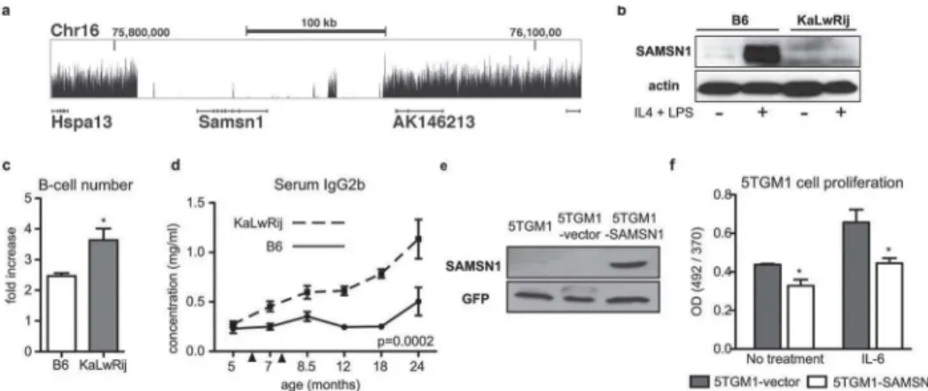
Analysis of structural variants identified two deletions. The first, confirmed by PCR and se-quence analysis, spanned 1.6 kb withinRfpl3s(data not shown). More compelling, however, was a large 180 kb deletion encompassing the entireSamsn1gene (Chr16: 75816190–
75996162;Fig 2A,S2A Fig), which was also identified in our cross-species approach.
To characterize the functional effect ofSamsn1deletion, we utilized a genetic mouse model with targeted deletion ofSamsn1on the C57BL/6 background.Samsn1-/-mice have increased B-cell proliferation in vitro and increased immunoglobulin response in the weeks following im-munization in vivo [9]. The 180 kb deletion resulted in an effectiveSamsn1knock-out in KaL-wRij mice, so we predicted KaLKaL-wRij mice would display similar B-cell phenotypes observed in
Samsn1-/-mice. Samsn1 was expressed in C57BL/6 but not in KaLwRij ex vivo stimulated splenic B-cells, confirming the loss of protein expression predicted by gene deletion (Fig 2B). B-cells isolated from young KaLwRij prior to onset of BIP and myeloma had significantly in-creased proliferation following stimulation compared to C57BL/6 (Fig 2C). Additionally, KaL-wRij mice had a significant and progressive elevation in immunoglobulin IgG2b levels following immunization (Fig 2D).
We next tested whetherSamsn1expression played a role in fully transformed myeloma cells. The 5TGM1 myeloma cell line, originally isolated from a myeloma-bearing KaLwRij mouse [10], was confirmed asSamsn1-null (Fig 2E,S2A Fig). Stable re-expression ofSamsn1
stimulated conditions (Fig 2F). Taken together, these data demonstrate thatSamsn1regulated B-cell proliferation in non-tumor bearing mice and restrained MM tumor cell growth in part through a plasma cell intrinsic mechanism.
SAMSN1(HACS1) was first cloned on the basis of its differential expression in multiple my-eloma with low expression in human mymy-eloma cell lines [11]. To further investigate whether
SAMSN1participates in human myeloma, we queried plasma-cellSAMSN1gene expression in patient samples.SAMSN1was expressed at lower levels in both human MGUS and MM cells compared to normal human plasma cells (S3 Fig), suggesting that plasma-cell intrinsic
SAMSN1may also play a role in human MM progression.
Samsn1expression was not restricted to the B-cell lineage (S2B Fig), so we hypothesized thatSamsn1might influence MM development via effects on additional cell types in the tumor stroma. We examined two cell types known to participate in MM pathogenesis: macrophages (Samsn1expressors) and bone marrow stromal cells (BMSCs,Samsn1non-expressors;S2B Fig). Microarray analysis of BIP-resistant C57BL/6 and BIP-susceptible KaLwRij primary bone marrow macrophages identified 281 differentially expressed genes (Fig 3A,S8 Table). KaLwRij macrophages had increased proliferation (Fig 3B) and increasedChi3l3(1.80 fold),Chi3l1
Table 1. KaLwRij novel germline missense, stoploss, and stopgain mutations.
Chr Position (bp) Gene ID Gene Name Variant
2 20861102 Arhgap21 Rho GTPase activating protein 21 c.C3215T:p.T1072I
3 95130195 Scnm1 Sodium channel modifier 1 c.T559C:p.stop187R
4 88403392 Focad Focadhesin c.A4979T:p.N1660I
7 140274118 5830411N06Rik Riken cDNA 5830411N06 gene c.C1033T:p.Q345stop
7 140466879 Olfr533 Olfactory receptor protein 533 c.G677A:p.R226H
7 140466884 Olfr533 Olfactory receptor protein 533 c.C682T:p.R228C
7 140503539 Olfr536 Olfactory receptor protein 536 c.C919T:p.R307C
7 140691588 Olfr45 Olfactory receptor protein 45 c.C682T:p.R228C
7 143643365 Tnfrsf22 Tumor necrosis factor receptor superfamily, member 22 c.A236C:p.Q79P 7 143680034 Tnfrsf23 Tumor necrosis factor receptor superfamily, member 23 c.A206C:p.Q69P
8 53513574 Aga Aspartylglucosaminidase c.C160A:p.L54M
11 3179430 Sfi1 Sfi1 homolog, spindle assembly associated (yeast) c.G344A:p.W115stop
11 3923487 Tcn2 Transcobalamin 2 c.C858G:p.F286L
11 4075493 Sec14l3 SEC14-like 3 (S. cerevisiae) c.C1016A:p.T339N
11 5144386 Emid1 EMI domain containing 1 c.A124G:p.T42A
11 43490831 C1qtnf2 C1q and tumor necrosis factor related protein 2 c.G379A:p.G127R
11 49294204 Olfr1392 Olfactory receptor protein 1392 c.A882T:p.K294N
11 50833599 Zfp879 Zincfinger protein 879 c.G410T:p.G137V
11 51007734 Olfr51 Olfactory receptor protein 51 c.C761T:p.A254V
11 51027442 Olfr54 Olfactory receptor protein 54 c.G439T:p.A147S
11 52144841 Olfr1373 Olfactory receptor protein 1373 c.G688A:p.V230M
11 107179035 Nol11 Nucleolar protein 11 c.C988T:p.P330S
13 21423407 Pgbd1 PiggyBac transposable element derived 1 c.C505T:p.Q169stop
16 32753901 Muc4 Mucin 4, cell surface associated c.C3776A:p.T1259N
17 46555608 Srf Serum response factor c.G221T:p.G74V
17 73535535 Galnt14 UDP-N-acetyl-alpha-D-galactosamine:polypeptide N-acetylgalactosaminyltransferase 14 c.C519A:p.N173K
18 67402164 Tubb6 Tubulin, beta 6 class c.T1132G:p.F378V
X 20853392 Araf v-raf murine sarcoma 3611 viral oncogene homolog c.G632A:p.R211H
X 51130161 Mbnl3 Muscleblind-like 3 (Drosophila) c.C715A:p.Q239K
(2.48 fold), andCxcl2(3.45 fold) expression, transcriptional markers for pro-tumor M2 macro-phage polarization. Notably, there were no variants identified in these genes in the KaLwRij ge-nome.Chi3l3expression was also significantly increased in macrophages isolated from
Samsn1-/-mice, confirming that these macrophage expression changes were due specifically to
Samsn1deficiency (Fig 3C).
To test the function ofSamsn1in tumor-associated macrophages in vivo, we injected either wild-type orSamsn1-/-ex vivo polarized M2 macrophages into established 5TGM1 tumors. Compared to wild-type,Samsn1-/-M2 macrophages significantly increased the growth of mye-loma tumors (Fig 3D). These findings indicate thatSamsn1regulates a pro-tumor function of macrophages in the myeloma microenvironment.
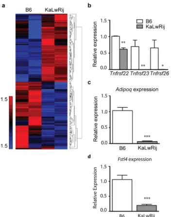
Interestingly, microarray analysis ofSamsn1-nonexpressing BMSCs from C57Bl/6 and KaL-wRij also showed many gene expression differences (Fig 4A).Tnfrsf22,Tnfrsf23, andTnfrsf26, identified in the WGS (S5 Table), showed lower expression in KaLwRij BMSCs compared to C57BL/6 (Fig 4B,S8 Table). Consistent with other reports, expression ofAdipoq, previously implicated in MM biology in mice and patients [12], was also significantly reduced in BMSCs from KaLwRij (Fig 4C).Fstl4, initially identified by our integrative genetics approach, was also depleted at steady-state levels in BMSCs (Fig 4D).
Discussion
High throughput WGS analyses of human MM cell genomes have revealed striking genetic di-versity but have failed to clarify the etiology of MM [13,14]. The spontaneously-derived KaL-wRij inbred mouse strain is commonly used as a model for MM but the KaLKaL-wRij genome has not previously been sequenced. Here, we present the WGS of the KaLwRij mouse as well as provide gene expression profiles for MM-supportive macrophages and BMSCs as evidence for
Fig 2.Samsn1is deleted in KaLwRij and was a negative regulator in B-cells and transformed myeloma cells.(a) Genome sequence coverage is shown for theSamsn1locus. Physical distance shown along the x-axis and the size and location of the region are indicated. Height of the curve represents accumulated sequencing reads. There are virtually no reads acrossSamsn1indicating a total deletion of this gene.Hspa13gene 5’ofSamsn1(shown and labeled) is not affected, and neither are 3’genes AK146213 (shown and labeled) or 4930578N18Rik (first two exons shown but not labeled).Samsn1is encoded in reverse orientation. (b) Western blot analysis of SAMSN1 in CD43- splenic B-cells from C57BL/ 6 or KaLwRij mice stimulated with IL-4 and LPS for 72 hours. (c) CD43- splenic B-cells were stimulated with IL-4 and LPS for 72 hours and cell number counted pre- and post-stimulation to determine proliferation. (d) Mice were immunized (arrowheads indicate primary and secondary immunization) and serial serum samples collected. IgG2b levels were determined by ELISA. (e) Western blot for Samsn1 in parental 5TGM1 cells, control vector cells (5TGM1-vector), and cells overexpressing Samsn1 (5TGM1-SAMSN1). (f) 5TGM1-vector and 5TGM1-SAMSN1 cells were stimulated with IL-6 for 24 hours. Cell proliferation was measured by BrdU incorporation.
multiple cell lineages contributing to MM prior to overt disease manifestation. Rather than being confined to the B-lineage, the presumed cell of origin of MM, our results suggest that ge-netic susceptibility alleles are expressed in both the pre-malignant B-cell and in supportive host microenvironment cells.
Combined analysis of loci that contribute to inherited disease risk to both MM in humans and BIP in mice identifiedSamsn1as a likely candidate to influence disease susceptibility. Sur-prisingly, we discovered thatSamsn1is homozygously deleted in the KaLwRij genome [15]. KaLwRij mice have a similar phenotype toSamsn1-/-mice including increased proliferation of B-cells in ex vivo culture and elevated IgG levels with age [9], supporting the conclusion that absence ofSamsn1drives KaLwRij BIP.Samsn1add-back inhibited proliferation in trans-formed MM cells suggesting that the gene can play a plasma-cell intrinsic tumor suppressor role in KaLwRij mice, but we also demonstrated that the absence ofSamsn1in macrophages also contributes to MM progression, making the role of this pathway more complex.
Most compelling were the non-plasma cell contributions ofSamsn1to MM. The majority of genetic alterations in MM cells are already present in MGUS plasma cells [6], suggesting that plasma cell extrinsic factors contribute to the conversion of MGUS to MM. Rather than being
Fig 3. Loss of Samsn1 enhanced pro-tumor macrophage function.(a) Microarray analysis of gene expression in C57BL/6 and KaLwRij primary bone marrow macrophages. (b) Proliferation of C57BL/6 and KaLwRij macrophages was measured by MTT assay. (c) Macrophage M2 polarization marker Chi3l3 in bone marrow macrophages. (d) Wild-type orSamsn1-/-M2 macrophages polarized ex vivo were injected directly
into established 5TGM1 tumors and tumor burden was monitored by bidirectional caliper measurement.
confined to the affected plasma cell, the genetic susceptibility alleles in KaLwRij also are ex-pressed in supportive host microenvironment cells.Samsn1-null macrophages had increased M2 macrophage markers and potently increased myeloma tumor growth in vivo. These data placeSamsn1as an inhibitor of pro-tumorigenic macrophage polarization and as a plasma-cell extrinsic regulator of MM growth.
Our findings also strongly suggest that gene loci in addition toSamsn1are likely to be im-portant to MM pathogenesis in the KaLwRij model. We found significant gene expression dif-ferences between C57BL/6 and KaLwRij BMSCs, a myeloma-supportive cell type in the tumor microenvironment that does not expressSamsn1(Fig 4). Through WGS, we identified addi-tional KaLwRij genes that may explain differences between KaLwRij and C57BL/6 BMSCs. No-tably, we identified deleterious variation and decreased expression in multiple
BMSC-expressed genes, includingTnfrsf22,Tnfrsf23, andTnfrsf26. These genes encode decoy recep-tors for the TRAIL cytokine [16]. TRAIL-OPG signaling has been implicated in MM progres-sion in humans [17], and these KaLwRij variants may support MM via paracrine signaling.
WGS also identified a novel variant in a paralog of adiponectin (Adipoq),C1qtnf2, predicted to be disruptive.C1qtnf2is a ubiquitously expressed lipokine with similar function to adipo-nectin.Adipoqhas been shown to have anti-myeloma effects in KaLwRij mice and in humans [12], and we confirmed significantly lowerAdipoqexpression in KaLwRij BMSCs. Together, these results provide additional lines of evidence that multiple pathways and cell types are
Fig 4. KaLwRij bone marrow stromal cells (BMSCs) have altered gene expression profiles independent ofSamsn1.(a) Microarray analysis of B6 and KaLwRij primary BMSCs. (b-d) RT-qPCR analysis ofTnfrsf22,Tnfrsf23,Tnfrsf26,Adipoq, andFstl4mRNA levels in B6 and KaLwRij BMSCs. *P>0.05,**P>0.005,***P<0.0005.
involved in KaLwRij MM predisposition, and validate our combined genetics approach as a method for identifying pathways involved in MM pathogenesis.
SAMSN1expression is decreased in MGUS and MM patient samples (S3 Fig) and has re-duced expression in human MM cell lines [11], butSAMSN1is not the target of somatic muta-tion in human MM [14]. Further work to map the SAMSN1 pathway in humans is needed before we can determine the significance ofSAMSN1to human MM. WhileSAMSN1is con-served in humans, one of its murine binding partners, PIRB is not [18]. SAMSN1 is also re-ported to bind cortactin, a component of integrin signaling and cell migration that is conserved in humans [19], but it is not known whether this interaction is necessary forSAMSN1's role in MM. We pursued investigation of the role ofSamsn1in KaLwRij mouse BIP susceptibility on the basis of a SNP in the humanSAMSN1locus identified in a MM patient GWAS (Fig 2). WhileSAMSN1is not deleted in human MM, components of a SAMSN1 pathway appear likely to involved in myeloma susceptibility and/or disease progression.
Our observation that the susceptible background of KaLwRij mice involves multiple cell types helps explain two persistent dilemmas in the myeloma field: i) the marked clonal hetero-geneity observed in human MM samples and ii) the inability of human MM cells to engraft in immunocompromised mice without co-transplantation of myeloid and stromal cells [7]. Drug resistance and MM cell survival is well-known to involve several stroma cell types, but the as-sumption in the field has been that it is the malignant MM cells that subvert normal bone mar-row cells to create a pro-tumor milieu. Our findings suggest for the first time that multiple cell lineages are involved in MM pathogenesis prior to disease manifestation and independently of tumor cell somatic mutations. Further evaluation of the interaction between somatic and germ-line genetic events in the KaLwRij model system may provide additional insights into human MM.
Materials and Methods
Ethics statement
The study cohort was approved by the Human Research Protection Office at Washington Uni-versity School of Medicine and at the Mayo Clinic. Informed written consent from the patients was obtained in accordance with the Declaration of Helsinki. Mice were housed in shared path-ogen-free conditions according the guidelines of the Division of Comparative Medicine, Wash-ington University School of Medicine. The WashWash-ington University Animal Studies Committee approved all experiments.
Mice
129S1/SvImJ, A/J, AKR/J, BALB/cByJ, CBA/J, C3H/HeJ, C57BL/6, DBA/2J, FVB/NJ, NOD/ ShiLtJ, SJL/J, and NOD-scid–IL2Rγmice were purchased from The Jackson Laboratory. C57Bl/KaLwRij mice were originally obtained from Gregory Mundy at Vanderbilt University. Mice were housed in shared pathogen-free conditions according the guidelines of the Division of Comparative Medicine, Washington University School of Medicine. Mice were euthanized by asphyxiation using CO2chambers. The animal ethics committee approved all experiments.
Samsn1-/-mice were originally obtained from Dingyan Wang [9].Samsn1-/-andSamsn1+/+
Primary cell culture
Splenic B-cells were negatively selected via MACS with anti-CD43 beads (Miltenyi Biotec). Cells were cultured in RPMI 1650 media, 10% FBS, 0.00035% BME, 1% penicillin-streptomy-cin and stimulated with 10 ng/ml LPS and 20 ng/ml IL-4 for 72 hours. Cells were character-ized by FACS (CD43- / B220+ / IgD+,S5 Fig). To generate bone marrow macrophages (BMMs), whole bone marrow was cultured inαMEM, 10% FBS, 1% penicillin-streptomycin, 50 ng/ml MCSF for 3 days. Cells were characterized by FACS (GR1- / F4/80+,S7 Fig). Prolif-eration was measured by standard MTT assay (Sigma-Aldrich). M2 polarized macrophages were generated by stimulating BMMs with 5 ng/ml IL-4 for 24 hours. Bone marrow stromal cells (BMSCs) were generated by plating whole bone marrow cells in ascorbic acid-free αMEM, 10% FBS, 1% penicillin-streptomycin for 7 days in 5% oxygen followed by negative selection via MACS with anti-CD45 beads (Miltenyi Biotec). Cells were characterized by FACS (CD45-,S6 Fig). Flow Cytometric Analysis was performed using FACSCALIBUR (BD Biosciences) and analyzed with FlowJo software (Tree Star). Antibodies used: APC-CD45 (BD Pharmingen), FITC-Gr1 (eBioscience), APC-F4/80 (BioLegend), FITC-CD43 (BD Pharmin-gen), APC-B220 (eBioscience), and PE-IgD (eBioscience).
5TGM1-GFP cell culture
The 5TGM1-GFP (5TGM1) cell line was originally obtained from Gregory Mundy at Vander-bilt University.Samsn1cDNA was subconed into an MSCV-PGK-Puro plasmid.
MSCV-PGK-Puro plasmid and lentiviral vectors pCMVΔ8.9 and pM2G were transfected into HEK293T cells. Cell supernatant containing lentivirus was then plated on 5TGM1 cells and se-lected with puromycin for 72 hours. Cells were maintained in DMEM, 10% FBS, 1% penicillin-streptomycin. Proliferation was measured by BrdU ELISA (Roche Diagnostics).
Immunoblotting
Antibodies: SAMSN1 (Sigma-Aldrich), actin (Sigma-Aldrich), and GFP (Santa Cruz Biotech-nology). Blots were incubated with horseradish conjugated secondary antibodies (GE Health-care) and visualized by chemiluminescence (Pierce Biotechnology).
5TGM1
in vivo
tumors
1x1065TGM1 cells were injected subcutaneously into the right flank of NOD-scid-IL2Rγ fe-male mice. 14 days following tumor inoculation, 0.8x10⁶ex vivo M2 polarized macrophages were injected directly into the tumor. Tumor volume was monitored by bidirectional precision caliper measurements (1/2 x length x width2). Maximum tumor volume was approximately 2cm3.
Gene expression microarrays
RNA was extracted from C57BL/6 and KaLwRij BMMs (3 mice/strain) and BMSCs (2 mice/ strain) using RNeasy Mini kit (Qiagen). RNA samples were submitted to the Genome Technol-ogy Access Center at Washington University School of Medicine for hybridization using the GeneChip Mouse Gene 1.0 ST array (Affymetrix). Differentially expressed genes were defined as1.5 fold changed between C57BL/6 and KaLwRij.
We re-analyzed the microarray data of CD138+ plasma cells from healthy donors, MGUS patients, and MM patients published by Fonseca, R. et al. (GSE6477) [20,21].
Quantitative reverse transcription PCR
RNA was extracted using RNeasy Mini kit (Qiagen) and cDNA generated using iScript (Bio-Rad). Quantitative PCR was completed using SsoFast EVA Green Supermix or iQ Supermix (Bio-Rad). All samples run with biological replicates of2. Primer sequences are inS1 Methods.
Statistics
Data are shown as mean +/- SEM. Unless otherwise indicated, experiments were analyzed using Student’s t-test to compare 2 groups or ANOVA to compare multiple groups.p<0.05; p<0.01;p<0.001.
Supporting Information
S1 Fig. Immune responses are significantly different among mouse strains.Schema for im-munization and serial serum sample protocol. Serum was collected at baseline (T0–5 months), post-primary immunization (T1–7 months), post-boosting immunization (T2–8.5 months), 12 months (T3), and 18 months (T4). Analysis of serial serum samples by ELISA for (b) immuno-globulin isotype G, (c) immunoimmuno-globulin isotype M, and (d) immunoimmuno-globulin isoype A. (e) Rep-resentative SPEP of mouse serum samples negative (-) and positive (+) for M-spike.
(PDF)
S2 Fig.Samsn1is deleted in KaLwRij and expression varies by cell type in C57BL/6.(a)
PCR amplification of the regions surrounding the 180kb deletion includingSamsn1identified via WGS of the KaLwRij strain. Primers flanking the breakpoint amplified a product in KaL-wRij genomic DNA and KaLKaL-wRij-derived 5TGM1 myeloma cell line DNA, but not C57BL/6 genomic DNA. (b)Samsn1mRNA expression was measured by RT-qPCR in multiple cell types. CD43- B cells were analyzed pre- and post-stimulation for 72hrs with IL4 and LPS. Mac-rophages were analyzed pre- and post-polarization to a M2 phenotype using IL4. CD45-BMSCs were also analyzed forSamsn1expression.
P<0.005,P<0.0005. (PDF)
S3 Fig.SAMSN1is expressed at a lower level in human MGUS and MM plasma cells.
Micro-array data from CD138+ plasma cells from human healthy donors, MGUS patients, and MM patients, first published by R. Fonsecaet al. in 2006 (GEO accession: GSE6477) was analyzed forSAMSN1expression levels.P<0.05,P>0.0001.
(PDF)
S4 Fig. FACS analysis of splenic B-cells.Isolated mouse splenocytes were negatively selected by magnetic immunodepletion, using anti-CD43 beads (Miltenyi Biotec). The negative fraction was then analysed by FACS. The CD43−(middle panel) and naive B-cell (B220+, IgD+, right panel) populations are shown.
(PDF)
S5 Fig. FACS analysis of bone marrow stromal cells.Whole bone marrow cells were cultured ascorbic acid-freeαMEM, 10% FBS, 1% penicillin-streptomycin for 7 days in 5% oxygen (left panels,“pre-sorted”). On day 7, cells were negatively selected by MACS with anti-CD45 beads (right panels).
(PDF)
population is enriched for GR1- / F4/80+ macrophages. (PDF)
S7 Fig. FACS analysis of bone marrow macrophages.Whole bone marrow was cultured in αMEM, 10% FBS, 1% penicillin-streptomycin, 50 ng/ml MCSF for 3 days. After 3 days, the cell population is enriched for GR1- / F4/80+ macrophages.
(PDF)
S1 Methods. Supplemental section with expanded details regarding the experimental meth-ods used in this manuscript.
(DOCX)
S1 Table. Mice with positive M-spike on serum protein electrophoresis. (XLSX)
S2 Table. Candidate genes underlying genetic susceptibility to BIP in KaLwRij mice. (XLSX)
S3 Table. Candidate genes underlying genetic susceptibility to multiple myeloma in hu-mans.
(XLSX)
S4 Table. KaLwRij germline structural variants. (XLSX)
S5 Table. KaLwRij germline SNVs. (XLSX)
S6 Table. KaLwRij germline Indels. (XLSX)
S7 Table. Frameshift and splice site mutations in the KaLwRij germline. (DOCX)
S8 Table. Microarray analysis of BMM and BMSC gene expression. (XLSX)
Author Contributions
Conceived and designed the experiments: SRA WCW LC XS KNW MHT. Performed the ex-periments: SRA WCW LC LL XS YX MH JO. Analyzed the data: SRA WCW PL DS CMV MH. Contributed reagents/materials/analysis tools: DW AG CMV GC RV XYW. Wrote the paper: SRA WCW KNW MHT JO.
References
1. Kumar SK, Dispenzieri A, Lacy MQ, Gertz MA, Buadi FK, Pandey S, et al. Continued improvement in survival in multiple myeloma: changes in early mortality and outcomes in older patients. Leukemia. 2013.
2. Landgren O, Gridley G, Turesson I, Caporaso NE, Goldin LR, Baris D, et al. Risk of monoclonal gam-mopathy of undetermined significance (MGUS) and subsequent multiple myeloma among African American and white veterans in the United States. Blood. 2006; 107(3):904–6. PMID:16210333 3. Kyle RA, Durie BG, Rajkumar SV, Landgren O, Blade J, Merlini G, et al. Monoclonal gammopathy of
4. Kyle RA, Therneau TM, Rajkumar SV, Larson DR, Plevak MF, Offord JR, et al. Prevalence of monoclo-nal gammopathy of undetermined significance. The New England jourmonoclo-nal of medicine. 2006; 354 (13):1362–9. PMID:16571879
5. Weinhold N, Johnson DC, Rawstron AC, Forsti A, Doughty C, Vijayakrishnan J, et al. Inherited genetic susceptibility to monoclonal gammopathy of unknown significance. Blood. 2014; 123(16):2513–7. doi: 10.1182/blood-2013-10-532283PMID:24449210
6. Davies FE, Dring AM, Li C, Rawstron AC, Shammas MA, O'Connor SM, et al. Insights into the multistep transformation of MGUS to myeloma using microarray expression analysis. Blood. 2003; 102
(13):4504–11. PMID:12947006
7. Epstein J, Yaccoby S. The SCID-hu myeloma model. (1543–1894 (Print)).
8. Radl J, Hollander CF, van den Berg P, de Glopper E. Idiopathic paraproteinaemia. I. Studies in an ani-mal model—the ageing C57BL/KaLwRij mouse. Clinical and experimental immunology. 1978; 33 (3):395–402. PMID:367647
9. Wang D, Stewart AK, Zhuang L, Zhu Y, Wang Y, Shi C, et al. Enhanced adaptive immunity in mice lack-ing the immunoinhibitory adaptor Hacs1. FASEB journal: official publication of the Federation of Ameri-can Societies for Experimental Biology. 2010; 24(3):947–56.
10. Garrett IR, Dallas S, Radl J, Mundy GR. A murine model of human myeloma bone disease. Bone. 1997; 20(6):515–20. PMID:9177864
11. Claudio JO, Zhu YX, Benn SJ, Shukla AH, McGlade CJ, Falcioni N, et al. HACS1 encodes a novel SH3-SAM adaptor protein differentially expressed in normal and malignant hematopoietic cells. Onco-gene. 2001; 20(38):5373–7. PMID:11536050
12. Fowler JA, Lwin ST, Drake MT, Edwards JR, Kyle RA, Mundy GR, et al. Host-derived adiponectin is tumor-suppressive and a novel therapeutic target for multiple myeloma and the associated bone dis-ease. Blood. 2011; 118(22):5872–82. doi:10.1182/blood-2011-01-330407PMID:21908434 13. Lohr JG, Stojanov P, Carter SL, Cruz-Gordillo P, Lawrence MS, Auclair D, et al. Widespread genetic
heterogeneity in multiple myeloma: implications for targeted therapy. (1878–3686 (Electronic)). 14. Chapman MA, Lawrence MS, Keats JJ, Cibulskis K, Sougnez C, Schinzel AC, et al. Initial genome
se-quencing and analysis of multiple myeloma. Nature. 2011; 471(7339):467–72. doi:10.1038/ nature09837PMID:21430775
15. Sarah Amend LC, Daniel Serie, Celine M Vachon, Lan Lu, Ravi Vij, Graham A Colditz, Katherine N Weilbaecher, Michael H. Tomasson, editor Deletion of samsn1 underlies genetic susceptibility to MGUS in mice. 55th American Society of Hematology Annual Meeting and Exposition; Dec. 9th, 2013; New Orleans, LA: 55th American Society of Hematology Annual Meeting and Exposition.
16. Schneider P, Olson D Fau—Tardivel A, Tardivel A Fau—Browning B, Browning B Fau—Lugovskoy A, Lugovskoy A Fau—Gong D, Gong D Fau—Dobles M, et al. Identification of a new murine tumor necro-sis factor receptor locus that contains two novel murine receptors for tumor necronecro-sis factor-related apo-ptosis-inducing ligand (TRAIL). (0021–9258 (Print)).
17. Colucci S, Brunetti G, Rizzi R, Zonno A, Mori G, Colaianni G, et al. T cells support osteoclastogenesis in an in vitro model derived from human multiple myeloma bone disease: the role of the OPG/TRAIL in-teraction. Blood. 2004; 104(12):3722–30. PMID:15308561
18. Zhu YX, Benn S, Li ZH, Wei E, Masih-Khan E, Trieu Y, et al. The SH3-SAM adaptor HACS1 is up-regu-lated in B cell activation signaling cascades. The Journal of experimental medicine. 2004; 200(6):737–
47. PMID:15381729
19. von Holleben M, Gohla A, Janssen KP, Iritani BM, Beer-Hammer S. Immunoinhibitory adapter protein Src homology domain 3 lymphocyte protein 2 (SLy2) regulates actin dynamics and B cell spreading. The Journal of biological chemistry. 2011; 286(15):13489–501. doi:10.1074/jbc.M110.155184PMID: 21296879
20. Chng WJ, Kumar S, Vanwier S, Ahmann G, Price-Troska T, Henderson K, et al. Molecular dissection of hyperdiploid multiple myeloma by gene expression profiling. Cancer research. 2007; 67(7):2982–9. PMID:17409404