Heart Rate Recovery after Treadmill Electrocardiographic Exercise
Stress Test and 24-Hour Heart Rate Variability in Healthy Individuals
Ivana Antelmi, Eliseu Yung Chuang, Cesar José Grupi, Maria do Rosário Dias de Oliveira Latorre, Alfredo José Mansur Instituto do Coração - Faculdade de Medicina da Universidade de São Paulo, São Paulo, SP - Brazil
Mailing address: Ivana Antelmi •
Rua Belgrado, 199 - Vila Moinho Velho - 04285-040, São Paulo, SP - Brazil E-mail: [email protected]
Manuscript received July 28, 2007; revised manuscript received September 21, 2007; accepted November 21, 2007.
Summary
Background: Heart rate recovery after treadmill electrocardiographic exercise stress test is modulated by the autonomic nervous system. Analysis of heart rate variability can provide useful information about autonomic control of the cardiovascular system.
Objective: The aim of the study was to test the hypothesis of association between heart recovery after treadmill electrocardiographic exercise test and heart rate variability.
Methods: We studied 485 healthy individuals aged 42±12.1 (range 15-82) years, 281(57.9%) women, submitted to treadmill electrocardiographic exercise stress tests and heart rate variability evaluations over time (SDNN, SDANN, SDNNi, rMSSD, pNN50) and frequency (LF, HF, VLF, LF/HF ratio) domains in 24-hour ambulatory electrocardiographic monitoring.
Results: Heart rate recovery was 30±12 beats in the 1st minute and 52±13 beats in the 2nd minute after exercise. Younger
individuals recovered faster from the 2nd to the 5th minute after exercise (r= 0.19-0.35, P< 0.05). Recovery was faster
in women than in men (4±1.1 beats lower in the 1st minute, p<0.001; 5.7±1.2 beats lower in the 2nd minute, p<0.01;
4.1±1.1 beats lower in the 3rd minute, p<0.001). There was no significant correlation between heart rate recovery
and heart rate variability in 1st and 2nd minutes after exercise. SDNN, SDANN, SDNNi, rMSSD, and pNN50 indices
demonstrated a significant correlation with heart rate recovery only at the 3rd and 4th minutes.
Conclusion: The hypothesis of association between heart rate recovery and 24-hour heart rate variability in the first two minutes after exercise was not substantiated in this study. Heart rate recovery after exercise was associated with age and gender. (Arq Bras Cardiol 2008; 90(6): 380-385)
Key words: Heart rate, variability; exercise; exercise test.
follow-up (5% vs 1%; hazard ratio 4.26; 95% confidence interval 2.65-6.68; P<0.001)2. In an additional study of 2,193
patients referred for treadmill electrocardiographic exercise stress test for evaluation of chest pain, heart rate recovery lower than 22 beats per minute in the 2nd minute after exercise was
significantly associated with higher mortality (hazard ratio 2.6; 95% confidence interval 2.4-2.8; p < 0.05)3.
Study samples in these reports consisted of patients referred for treadmill electrocardiographic exercise stress test due to clinical indications1-3, including patients referred for coronary
angiography1,3. There are few recent reports of patterns of
heart rate recovery after exercise in patients without any evidence of heart disease after careful clinical examination.
Heart rate recovery immediately after exercise is considered to be a function of reactivation of the parasympathetic drive and a decrease in the sympathetic drive that usually occurs during the first 30 seconds of recovery after exercise4.
Abnormalities in parasympathetic activation and drive were suggested as a potential pathophysiological link to the observed association between reduced or impaired heart rate in early recovery after treadmill electrocardiographic exercise
Introduction
Recent studies have demonstrated that impaired heart rate recovery after exercise was associated with less favorable prognosis in patient follow-up. In a study of 2,428 consecutive adults referred for a first symptom-limited electrocardiographic exercise stress test and single-photon emission computed tomography (SPECT) with thallium between September 1990 and December 1993, the decrease of 12 beats or less in the 1st minute of recovery relative to heart rate at peak exercise
was associated with higher mortality rate during follow-up (adjusted relative risk 2; 95% confidence interval 1.5 – 2.7; p < 0.001)1. Another study examined 9,454 consecutive patients
referred for treadmill electrocardiographic exercise stress test: heart rate recovery <12 beats per minute in the 1st minute
stress test and increased mortality during the follow-up period2.
Hence, the study of autonomic nervous system physiology in this setting may be warranted.
Heart rate variability in time and frequency domains is a noninvasive tool useful for evaluation of autonomic nervous system physiology5. Time domain analysis of heart rate
variability estimates the variation of differences between successive RR intervals through indices developed by statistical methods. Frequency domain analysis of heart rate variability estimates respiratory-dependent high frequency and low frequency power through spectral analysis. High frequency power is considered to be mediated mainly by vagal activity, while low frequency power has been suggested to represent predominantly sympathetic modulation6,7.
We had the opportunity of evaluating a sample of asymptomatic healthy individuals without any evidence of heart disease after careful clinical and laboratory examination.
We hypothesized that different rates of heart rate recovery after treadmill electrocardiographic exercise stress test would be associated with different indices of heart variability in healthy men and women of different ages as an expression of the balance between parasympathetic and sympathetic drives. Specifically, we tested the hypothesis that individuals with higher indices of parasympathetic activity would have higher rate of recovery of heart rate after treadmill electrocardiographic exercise stress test.
The aim of this study was to evaluate the association between heart rate variability indices obtained during 24-hour ambulatory electrocardiographic monitoring and heart rate recovery after treadmill electrocardiographic exercise stress test in a large sample of subjects without any evidence of heart disease after careful clinical and laboratory examination.
Methods
Study protocol - A cohort of asymptomatic individuals with no evidence of heart disease after careful clinical examination was established to evaluate clinical and cardiovascular variables. Patients were from a General Outpatient Clinic Unit of a tertiary care university hospital dedicated to cardiology that provides also primary and secondary care.
Clinical evaluation included a detailed medical examination, 12-lead electrocardiogram and chest X-ray. The asymptomatic individuals with normal clinical examination, as well as normal electrocardiogram and chest X-ray, were considered eligible for the study, and were invited to participate. After informed and written consent, participants were submitted to further laboratory work-up, including treadmill electrocardiographic exercise stress test, two-dimensional transthoracic Doppler echocardiography, and 24-hour ambulatory electrocardiographic monitoring. In addition, laboratory work-up included hemoglobin, hematocrit, leukocyte count, serum glucose, serum cholesterol, triglycerides, uric acid, thyroid-stimulating hormone, and creatinine.
Inclusion criteria - We included in the study men and women older than 15 years of age, asymptomatic and with normal clinical examination, as well as normal electrocardiogram, chest X-ray, echocardiogram and treadmill electrocardiographic exercise stress test.
Exclusion criteria - We excluded symptomatic patients, as well as patients with past medical history of cardiovascular disease, systemic hypertension, diabetes mellitus, thyroid-stimulating hormone lower than 0.05 or higher than 8 mg/dl, chronic obstructive pulmonary disease, asthma, renal failure, chronic inflammatory diseases, osteoarticular diseases, chronic anemia or neoplasia, and abnormal 12-lead resting electrocardiogram, echocardiogram or treadmill electrocardiographic exercise stress test.
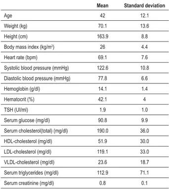
Study sample - 485 individuals were enrolled in the study; 204 (42.1%) men and 281 (57.9%) women. Ages ranged between 15 and 82 (mean 42, standard deviation 12.1) years. Demographic, clinical and laboratory characteristics of the study sample are presented in Table 1.
Treadmill exercise stress test - Tests were performed on the Fukuda Denshi MI – 8000 Star model according to the Bruce/ Ellestad protocol. Predicted peak heart rate was calculated as 220 - age. Individuals were encouraged to exercise until they experienced limiting symptoms, even if 85% of maximum predicted heart rate was achieved. Criteria for exercise termination were physical exhaustion or maximum heart rate greater than the age-predicted maximum. During each exercise stage and recovery stage, symptoms, blood pressure, heart rate and exercise workload in metabolic equivalents (METS) were recorded. Following peak exercise, individuals walked for a two-minute cool-down period at 1.5 mph and 2.5% grade. Heart rate was measured during each minute of exercise, at maximum exercise, and during recovery at 1, 2, 3, 4 and 5 minutes in the standing position. Heart rate recovery was defined as maximum heart rate minus heart rate at specified time period after exercise and represented
Table 1 - Baseline clinical characteristics of the 485 participants*
Mean Standard deviation
Age 42 12.1
Weight (kg) 70.1 13.6
Height (cm) 163.9 8.8
Body mass index (kg/m2) 26 4.4
Heart rate (bpm) 69.1 7.6
Systolic blood pressure (mmHg) 122.6 10.8
Diastolic blood pressure (mmHg) 77.8 6.6
Hemoglobin (g/dl) 14.1 1.4
Hematocrit (%) 42.1 4
TSH (UI/ml) 1.9 1.0
Serum glucose (mg/dl) 90.8 9.9
Serum cholesterol(total) (mg/dl) 190.0 36.0
HDL-cholesterol (mg/dl) 51.9 30.0
LDL-cholesterol (mg/dl) 119.1 33.0
VLDL-cholesterol (mg/dl) 23.6 18.7
Serum triglycerides (mg/dl) 112.9 71.1
Serum creatinine (mg/dl) 0.8 0.1
the drop in heart rate during that time interval. The exercise tests were performed, analyzed and reported with a standard protocol utilizing a computerized database. In this study, all tests were terminated due to exhaustion.
Heart rate variability - All subjects underwent 24-hour electrocardiographic recording. Mean recording time of Holter tapes was 22.6 ± 1 hour. All measurements were obtained with a Marquette 8000 portable recorder and processed by Marquette MARS 8000 equipment with 125-Hz sampling using MARS software version 4.0 (Milwaukee, Wisconsin). Each beat was classified and labeled with respect to the site of origin using template-matching techniques. The program eliminates 1 RR interval before and 2 after each non-sinus beat. An experienced observer manually reviewed and corrected all tracing. Recording with non-sinus beats that comprised > 2% of the total number of beats were excluded. Corresponding algorithms supplied by the manufacturer were used to analyze HRV. The following time-domain indices were studied: Standard deviation (SD) of all normal sinus RR intervals during 24 hours (SDNN), SD of averaged normal sinus RR intervals for all 5-minute segments (SDANN), mean of the SDs of all normal sinus RR intervals for all 5-minute segments (SDNNi), and root-mean-square of the successive normal sinus RR interval difference (rMSSD), and percentage of successive normal sinus RR intervals >50 ms (pNN50). In the frequency domain (fast-Fourier transform), the following indices were compared: very low frequency (VLF) of 0.0003 to 0.04 Hz, low frequency (LF) of 0.04 to 0.15 Hz, high frequency (HF) of 0.15 to 0.40 Hz, and the LF/HF ratio (LF/HF).
Statistical analysis - After descriptive statistics, the association between heart rate recovery, age, gender and heart rate variability indices was assessed with Pearson correlation and multiple linear regression models. A P value <0.05 was considered significant.
Ethical aspects - The study protocol was approved by the Committee of Ethics on Human Research of the Hospital. Subjects who agreed to participate in the study signed an informed consent.
Results
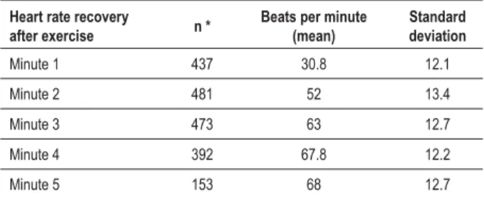
Heart rate recovery - Heart rate recovery after exercise was more pronounced in the first 2 minutes after exercise. Mean decrease in heart rate was 30.8 beats (standard deviation 12.1) in the first minute after exercise and 52 beats (standard deviation 13.4) in the second minute after exercise. Further data are presented in table 2. Heart rate usually did not return to pre-exercise levels within 5 minutes.
Subjects with higher increase in heart rate during exercise demonstrated a faster recovery rate from the second to the fifth minute after exercise (r=0.16, 0.36, 0.48 P<0.05, respectively 2nd, 3rd and 4th minutes); this relationship was
not demonstrated in the first minute after exercise, even after controlling for age. Resting heart rate correlated significantly with heart rate recovery at the 4th and 5th minutes after exercise
(r= 0.2, P<0.05).
Heart rate recovery relative to age and gender - Heart rate recovery after exercise demonstrated a statistically significant correlation with age: younger individuals recovered faster than
Table 2 - Heart rate recovery after exercise relative to peak heart rate during exercise in standing position
Heart rate recovery
after exercise n *
Beats per minute (mean)
Standard deviation
Minute 1 437 30.8 12.1
Minute 2 481 52 13.4
Minute 3 473 63 12.7
Minute 4 392 67.8 12.2
Minute 5 153 68 12.7
1XPEHURILQGLYLGXDOVZLWKUHFRYHU\UDWHUHFRUGHG
older ones from the second to the fifth minute after exercise (r= 0.19-0.35 P<0.05). Heart rate recovery in women was more rapid than in men: the rate was 4±1.1 (<0.05) beats more rapid at the first minute, 5.7±1.2 (p<0.05) beats more rapid at the second minute, and 4.1±1.1 (p<0.05) beats more rapid than in men at the third minute. The difference in heart rate recovery between men and women from the 4th minute
after exercise was not statistically significant (figure 1).
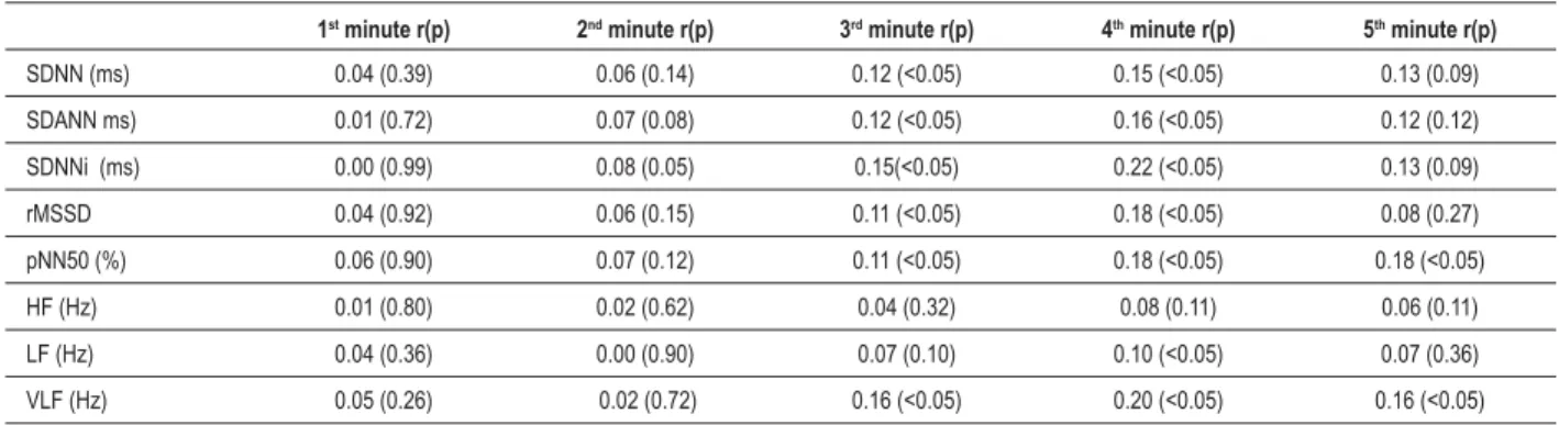
Heart rate recovery relative to heart rate variability - We found no association between heart rate recovery and heart rate variability for the 1st and 2nd minutes of recovery after
exercise, neither in time nor in frequency domains. For heart rate variability indices in the time domain, there was an association between heart rate recovery and SDNN, SDANN, SDNNi, rMSSD, and pNN50 indices in the time domain of heart rate variability only at the 3rd and 4th minutes
of recovery after exercise (table 3). Correlations were not more statistically significant for the 5th minute of recovery
after exercise.
For heart rate variability indices in the frequency domain, there was an association between heart rate recovery and very low frequency (VLF) in the frequency domain only at the 3rd and 4th minutes of recovery after exercise. We found
no significant correlation between high frequency power (HF) and low frequency to high frequency power ratio (LF/HF) at any minute of recovery (table 3). The strongest correlation between heart rate recovery and heart rate variability was identified at 4th minute of recovery for LF indices.
Correlation between SDNN and heart rate recovery after exercise at 3rd and 4th minutes remained statistically
significant after controlling for age and gender in multiple regression analysis.
Discussion
We studied heart rate recovery after exercise in a sample of men and women without any evidence of heart disease after careful clinical and laboratory evaluation. This is an interesting characteristic of this study sample, since previous studies included subjects with and without heart disease8,9 in a
range from normal to several degrees of pathologic conditions. Other important studies included only men10-12. In addition,
our sample was composed of non-athlete, sedentary subjects with maximal oxygen consumption below 44.9ml/kg/min13
Figure 1 -%R[SORWGLVWULEXWLRQLQWHUTXDUWLOHUDQJHEHWZHHQ¿UVWDQGWKLUGTXDUWLOHVPHGLDQRIKHDUWUDWHUHFRYHU\LQWKH¿UVWXSWRWKHthPLQXWHRIUHFRYHU\LQZRPHQ
Q DQGPHQQ
Table 3 -$VVRFLDWLRQVEHWZHHQKHDUWUDWHUHFRYHU\DIWHUH[HUFLVHDQGLQGLFHVRIKHDUWUDWHYDULDELOLW\GHULYHGIURPKRXUDPEXODWRU\ electrocardiographic monitoring
1st minute r(p) 2nd minute r(p) 3rd minute r(p) 4th minute r(p) 5th minute r(p)
SDNN (ms) 0.04 (0.39) 0.06 (0.14) 0.12 (<0.05) 0.15 (<0.05) 0.13 (0.09)
SDANN ms) 0.01 (0.72) 0.07 (0.08) 0.12 (<0.05) 0.16 (<0.05) 0.12 (0.12)
SDNNi (ms) 0.00 (0.99) 0.08 (0.05) 0.15(<0.05) 0.22 (<0.05) 0.13 (0.09)
rMSSD 0.04 (0.92) 0.06 (0.15) 0.11 (<0.05) 0.18 (<0.05) 0.08 (0.27)
pNN50 (%) 0.06 (0.90) 0.07 (0.12) 0.11 (<0.05) 0.18 (<0.05) 0.18 (<0.05)
HF (Hz) 0.01 (0.80) 0.02 (0.62) 0.04 (0.32) 0.08 (0.11) 0.06 (0.11)
LF (Hz) 0.04 (0.36) 0.00 (0.90) 0.07 (0.10) 0.10 (<0.05) 0.07 (0.36)
VLF (Hz) 0.05 (0.26) 0.02 (0.72) 0.16 (<0.05) 0.20 (<0.05) 0.16 (<0.05)
In our sample, mean heart rate recovery was much higher than that associated with poor prognosis in published studies1-3.
It is also noteworthy that, in this study, heart rate decrease after exercise was different in men and women; decrease being faster in women. This difference in the rate of decline relative to sex has not been addressed in previous studies that enrolled men and women1,2. This finding may be related to
the previous suggestion of higher parasympathetic drive in women in comparison to men as found in studies of heart rate variability13-16. Additionally, during exercise women are
usually considered to have lower cardiorespiratory fitness, higher cardiac outputs for comparable workloads and lower systolic volume, providing a balance between O2 demand and supply from increased heart rate17-19. These physiological bases
may have been operative in our findings. Sedentary women in menopause displayed lower aerobic capacity than men in the same age range20. To compensate for lower capacity,
aerobic power in women during dynamic exercise increased more than in men.
Recovery rate after exercise was also modulated by age as found by others. Maximum heart rate and cardiac output were lower in older individuals, partly because of decreased beta-adrenergic responsivity21 and partly because of lower
workload. Fifty percent of the increase in heart rate may be attributed to sympathetic stimulation, mainly beta-adrenergic stimulation and circulating catecholamines.
References
1. Cole CR, Blackstone EH, Pashkow FJ, Snader CE, Lauer MS. Heart rate recovery immediately after exercise as a predictor of mortality. N Engl J Med. 1999; 341: 1351-7.
2. Nishime EO, Cole CR, Blackstone EH, Pashkow F, Lauer MS. Heart rate recovery and treadmill exercise score as predictors of mortality in patients referred for exercise ECG. JAMA. 2000; 284: 1392-8.
3. Shelter K, Marcus R, Frolicker VF, Vera S, Chalkiest D, Parakeet M, et al. Heart rate recovery: validation and methodology issues. J Am Coll Cardiol. 2001; 38: 1980-7.
4. Imai K, Sato H, Hori M, Kusuoka H, Ozaki H, Yokoyama H, et al. Vaguely mediated heart rate recovery after exercise is accelerated in athletes but blunted in-patients with chronic heart failure. J Am Coll Cardiol. 1994; 24: 1529-35.
5. Heart rate variability: standards of measurement, physiological interpretation and clinical use. Task Force of the European Society of Cardiology and the North American Society of Pacing and Electrophysiology. Circulation. 1996; 93 (5):1043-65.
6. Akselrod S, Gordon D, Ubel FA, Shannon DC, Berger AC, Cohen RJ. Power spectrum analysis of heart rate fluctuation: a quantitative probe of beat-to-beat cardiovascular control. Science. 1981; 213 (4504): 220-2.
7. Pomeranz B, Macaulay RJ, Caudill MA, Kutz I, Adam D, Gordon D, et al. Assessment of autonomic function in humans by heart rate spectral analysis.
Am J Physiol. 1985; 248 (1 Pt 2): H151-3.
8. Cole CR, Food JM, Blackstone EH, Laver MS. Heart rate recovery after suboptimal exercise testing as a predictor of mortality in a cardiovascular healthy cohort. Ann Intern Med. 2000; 132: 552-5.
9. Watanabe J, Thamilarasan M, Blackstone EH, Thomas JD, Laver MS. Heart rate recovery immediately after treadmill exercise and left ventricular systolic dysfunction as predictors of mortality: the case of stress echocardiography. Circulation. 2001; 104: 1911-6.
10. Lipinski MJ, Vetrovec GW, Froelicher VF. Importance of the first two minutes of heart rate recovery after exercise treadmill testing in predicting mortality and the presence of coronary artery disease in men. Am J Cardiol. 2004; 93: 445-9.
11. Linder L, Andren B. Heart rate recovery after exercise is related to the insulin resistance syndrome and heart rate variability in elderly men. Am Heart J. 2002; 144: 666-72.
12. Javorka M, Zila I, Balharek T, Javorka K. Heart rate recovery after exercise: relations to heart rate variability and complexity. Braz J Med Biol Res. 2002; 35: 991-1000.
13. Antelmi I, DePaula RS, Shinzato AR, Peres CA, Mansur AJ, Grupi CJ. Influence of age, gender, body mass index and functional capacity on heart rate variability in a cohort of subjects without heart disease. Am J Cardiol. 2004; 93: 445-50.
modulated mainly by parasympathetic stimuli such as rMMSD and pNN50 in the time domain did reveal an association with heart rate recovery after exercise in the 3rd and 4th minutes.
Surprisingly, SDNN, SDANN and SDNNi indices, which are an expression of both sympathetic and parasympathetic influences, were also associated with heart rate recovery in the 3rd and 4th minutes. This was an unexpected finding
because high-frequency component, rMMSD and pNN50 are more influenced by the vagal tone, as demonstrated in controlled experiments with atropine in 5-minute recording16.
The 24-hour recording conditions may also have modulated this finding. In a recent study of 70 men over 70 years of age12, no significant relationship between heart rate recovery
and rMSSD, pNN50, high frequency component and low-frequency to high-low-frequency component ratio was detected, except in the low frequency component during the first minute of recovery after exercise.
Our study has limitations. Heart rate recovery after exercise is modulated by a procedure of cool-down as was performed in this study (first 2 min of 1.5 mph at 2.5%). The impact of exercise protocol and cool-down period on heart rate recovery after exercise is uncertain, and recovery protocols have not yet been standardized in clinical practice. The mechanism of this modulated recovery is less well known. Differences might well vary with immediate cessation of exercise and assumption of the supine position without a cool-down protocol20.
Conclusion
In conclusion, heart rate recovery after exercise was associated with age and gender. Younger individuals recovered
faster than older ones from the second to the fifth minute after exercise and heart rate recovery in women was more rapid than in men. The hypothesis of association between heart rate recovery and indices of 24-hour heart rate variability in the first two minutes after exercise was not substantiated in this study. There was an association between heart rate recovery and Standard deviation (SD) of all normal sinus RR intervals during 24 hours (SDNN), SD of averaged normal sinus RR intervals for all 5-minute segments (SDANN), mean SDs of all normal sinus RR intervals for all 5-minute segments (SDNNi), root-mean-square of the successive normal sinus RR interval difference (rMSSD), percentage of successive normal sinus RR intervals >50 ms (pNN50) in the time domain only at the 3rd
and 4th minutes of recovery after exercise. Our data may add
to the evaluation of heart rate recovery in the first 5 minutes after exercise in subjects without any evidence heart disease after careful clinical and laboratory evaluation. Such a finding may be useful for further studies in the field.
Potential Conflict of Interest
No potential conflict of interest relevant to this article was reported.
Sources of Funding
There were no external funding sources for this study.
Study Association
14. Arai Y, Saul JP, Albrecht P, Hartley LH, Lilly LS, Cohen RJ, et al. Modulation of cardiac autonomic activity during and immediately after exercise. Am J Physiol. 1989; 256: 132-41.
15. Bigger JT, Fleiss JL, Steinman RC, Rolnitzky LM, Kleiger RE, Rottman JN. Frequency domain measures of heart period variability and mortality after myocardial infarction. Circulation. 1992; 85: 164-71.
16. Kuo TBJ, Lin T, Yang CCH, Li CL, Chen CC, Chou P. Effects of aging on gender differences in neural control of heart rate. Am J Physiol. 1999; 277: 2233-9.
17. Ryan SM, Goldberg AL, Pincus SM, Mietsus J, Lipsitz LA. Gender and age related differences in heart rate dynamics: are women more complex than men? J Am Coll Cardiol. 1998; 5: 141-9.
18. Yamasaki Y, Kodama M, Matsuhisa M. Diurnal heart rate variability in healthy subjects: effects of aging an sex differences. Am J Physiol. 1996; 271: 303-10.
19. Froelicher V, Myers JN. Exercise and heart. Philadelphia: WB Saunders Company; 2000.
20. Gallo L Jr, Maciel BC, Marin Neto JA, Martins LEB, Lima Filho EC, Golfetti R, et al. Control of heart rate during exercise in health and disease. Braz J Med Biol Res. 1995; 28: 1790-184.