http://dx.doi.org/10.1590/bjpt-rbf.2014.0159 298 Braz J Phys Ther. 2016 July-Aug; 20(4):298-305
Analysis of agreement between cardiac risk stratification
protocols applied to participants of a center for
cardiac rehabilitation
Ana A. S. Santos1, Anne K. F. Silva1, Franciele M. Vanderlei1,
Diego G. D. Christofaro2, Aline F. L. Gonçalves1, Luiz C. M. Vanderlei1
ABSTRACT | Background: Cardiac risk stratiication is related to the risk of the occurrence of events induced by exercise. Despite the existence of several protocols to calculate risk stratiication, studies indicating that there is similarity between these protocols are still unknown. Objective: To evaluate the agreement between the existing protocols on cardiac risk rating in cardiac patients. Method: The records of 50 patients from a cardiac rehabilitation program were analyzed, from which the following information was extracted: age, sex, weight, height, clinical diagnosis, medical history, risk factors, associated diseases, and the results from the most recent laboratory and complementary tests performed. This information was used for risk stratiication of the patients in the protocols of the American College of Sports Medicine, the Brazilian Society of Cardiology, the American Heart Association, the protocol designed by Frederic J. Pashkow, the American Association of Cardiovascular and Pulmonary Rehabilitation, the Société Française de Cardiologie, and the Sociedad Española de Cardiología. Descriptive statistics were used to characterize the sample and the analysis of agreement between the protocols was calculated using the Kappa coeficient. Differences were considered with a signiicance level of 5%. Results: Of the 21 analyses of agreement, 12 were considered signiicant between the protocols used for risk classiication, with nine classiied as moderate and three as low. No agreements were classiied as excellent. Different proportions were observed in each risk category, with signiicant differences between the protocols for all risk categories. Conclusion: The agreements between the protocols were considered low and moderate and the risk proportions differed between protocols.
Keywords: cardiology; exercise; protocols; rehabilitation.
BULLET POINTS
• Studies indicating similarity between cardiac risk stratiication protocols are important in clinical practice. • Most protocols for cardiac risk stratiication present low or moderate agreements.
• The protocols have shown good applicability to most patients.
HOW TO CITE THIS ARTICLE
Santos AAS, Silva AKF, Vanderlei FM, Christofaro DGD, Gonçalves AFL, Vanderlei LCM. Analysis of agreement between cardiac risk stratiication protocols applied to participants of a center for cardiac rehabilitation. Braz J Phys Ther. 2016 July-Aug; 20(4):298-305. http://dx.doi.org/10.1590/bjpt-rbf.2014.0159
1 Departamento de Fisioterapia, Faculdade de Ciências e Tecnologia, Universidade Estadual Paulista (UNESP), Presidente Prudente, SP, Brazil 2 Departamento de Educação Física, Faculdade de Ciências e Tecnologia, UNESP, Presidente Prudente, SP, Brazil
Received: Apr. 09, 2015 Revised: Sept. 15, 2015 Accepted: Nov. 26, 2015
Introduction
Cardiovascular diseases (CVD) are the leading cause of death in most countries, including Brazil, accounting for about 20% of all deaths in individuals over 30 years of age1,2. In addition to the high mortality rate, these diseases may be responsible for physical disability and contribute signiicantly to increased spending on health1. This scenario demonstrates the need for effective interventions, of which cardiac rehabilitation (CR) seems to be one example. According to the World Health Organization3, CR is the range of proposed
activities to ensure better living conditions for an individual with heart disease, as well as contributing to the improvement in functional capacity4, having an important role in preventing cardiovascular events and reducing mortality from these conditions5.
functional status of the patient which, among other things, provides a patient risk stratiication, related to the possible risk of adverse events induced by exercise during performance of the CR program8, guiding the
form and intensity of the work to be performed with the cardiac individual.
In a literature review conducted by our group7, eight risk stratiication protocols were found, developed, and validated by various national and international entities, devised for the participation of individuals in exercise programs and CR.
The criteria for stratiication consider factors associated with an increased risk of morbidity and mortality during physical exercise, and based on these criteria, the individual is usually classiied as low, moderate, or high risk7,9. In addition to the knowledge of the risk level, stratiication provides information for the proper direction of the patient throughout the CR process and planning of the program10, aiding the professional to determine the appropriate level of monitoring in accordance with the risk level of the patient8.
However, the existence of multiple risk stratiication protocols may hamper the selection of the most suitable to be used during the CR process. A search in the literature found no studies evaluating the similarity between the risk stratiication protocols, demonstrating gaps in the literature that raise the following questions: Would an individual be classiied in the same risk level in different protocols? Are there agreements between the risk ratings used in the protocols? If so, which ones can be considered similar and which differ? This information may contribute to researchers and clinicians who act in CR programs, giving safer direction for adopted behaviors and exercise prescription with cardiac patients and even identifying differences between speciic protocols.
One of the few studies that compared protocols was carried out by Paul-Labrador et al.11 that evaluated American patients and found that the ability of the guidelines (American Association of Cardiovascular and Pulmonary Rehabilitation [AACVPR]9, American
Heart Association [AHA]12, American College of Cardiology [ACC]13, and American College of Physicians [ACP]14) to predict complications in patients at high risk was low.
In this context, the aim of this study was to evaluate the level of agreement between existing protocols on the cardiac risk score in heart disease. It was hypothesized that despite the differences between the protocols, there would be agreement between them
regarding the prediction of the risk of developing complications during exercise.
Method
This is a descriptive/analytical cohort study, transversal, with a restropective characteristic, developed from data drawn from 50 medical records of individuals seen in an outpatient exercise program for patients with cardiovascular disorders, between April and May 2014, with no restrictions concerning age or gender.
These patients received information about the objectives and procedures of the study and signed a consent form authorizing the use of their data. All procedures were approved by the Research Ethics Committee of Universidade Estadual Paulista Júlio de Mesquita Filho (UNESP), Presidente Prudente, SP, Brazil, under protocol number 792.373 of 05/09/14.
Data collection
The patient records were analyzed and the following information extracted: age, gender, weight, height, clinical diagnosis for which the patient was referred to the heart disease unit, medical history, risk factors (RF) for the development of CVD, associated diseases, and the results of recent laboratory tests and complementary tests related to the evaluation of the cardiovascular system (exercise testing, echocardiography, Holter monitoring, cardiac catheterization, echo-stress, myocardial scintigraphy, electrocardiogram, and coronary angiogram). This information was tabulated and subsequently used for risk stratiication of patients using the different protocols evaluated in this study. A single evaluator performed all data collection.
Characterization of participants
The characterization was based on information regarding age, weight, height, and clinical diagnosis. Body mass index (BMI) was calculated from weight and height using the formula: body weight (kg)/height2(m) to determine the obesity risk factor
according to the criteria of the Brazilian Obesity Guidelines – 3rd edition15.
Clinical diagnosis
300 Braz J Phys Ther. 2016 July-Aug; 20(4):298-305
with stent placement, CABG and stent, or conservative treatment. Patients with heart failure (HF) were also subdivided according to the New York Heart Association (NYHA) Functional Classiication16. Patients without the presence of diagnosed heart disease were allocated to the preventive group.
Medical history
The following information was extracted from the medical records: the presence and number of cardiopulmonary arrests, number of days of hospitalization, complications during hospital stay or after performing an invasive procedure, and current complications.
Risk factors
The information contained in the medical records was analyzed for the presence of the following risk factors (RF): sex/age (men over the age of 45 years and women over 55), family history (considering the presence of CVD in irst-degree relatives), hypertension, dyslipidemia, and smoking. For the obesity risk factor, the value obtained in the calculation of BMI was used, considering patients obese with a BMI≥30 kg/m2 15.
Associated diseases
Associated diseases were considered as any musculoskeletal, neurological, pulmonary, or metabolic dysfunction, with the data obtained from the medical records.
Laboratory tests and complementary tests
The most recent exams recorded in the medical records were analyzed, regardless of the results contained in these exams. Blood glucose, triglycerides, total cholesterol, HDL-cholesterol, and LDL-cholesterol were obtained from the laboratory records. Only complementary tests that assessed the cardiovascular system were used. The results for patients who had undergone Holter monitoring were classiied according to Lown and Wolf17, which takes into account ventricular premature beats for determining the appropriate risk class, based on the frequency and severity with which they appear.
Stratification of cardiac risk
For risk stratiication, we used the following protocols described in the study of Silva et al.7: American College of Sports Medicine (ACSM)18, Brazilian Society of Cardiology (BSC)19, AHA12, the protocol designed by
Pashkow10, AACVPR9, French Society of Cardiology
(FSC)20, and Spanish Society of Cardiology (SSC)21. Risk stratiication was performed on each protocol using the same complementary tests for each patient. In all protocols, the patients were classiied as low, moderate, or high risk, and the presence of any characteristics in a higher cardiac risk band ranked the individual in that category.
The guidelines of the ACSM18 were used as a basis for classifying information on age, health status, symptoms, and RF. The BSC protocol19 is based mainly on maximal exercise test results to identify myocardial ischemia, ventricular dysfunction, cardiac arrhythmias, and atrioventricular conduction disturbances.
Unlike previous guidelines, AHA12 classiies patients into risk classes (A, B, C, and D) and takes into account the presence of symptoms or heart disease, RF, and indings of the exercise test. For this protocol, patients classiied as class A were considered low-risk patients, class B as moderate risk, and class C as high risk. According to this classiication, patients in class D should not participate in a CR program12, therefore this class was not included in the study. In 1993, Pashkow10 developed a model of risk stratiication based on important guidelines at the time as well as a new means of risk identiication that bases stratiication on the results of tests such as the progressive stress test, electrocardiogram, and echocardiogram.
The guidelines of the AACVPR9 stratiies patients
based mainly on the indings of the ergometric test. According to these guidelines, patients who do not undergo this test before entering the program or those with undiagnosed exercise tests could be categorized inappropriately and therefore risk stratiication should be approached with caution.
The FSC protocol20 is adapted from the recommendations of the European Society of Cardiology, and the AACVPR and is based mainly on the indings of the stress test and echocardiogram for the classiication of the patient. The SSC protocol21, published in the Practice Guidelines on Cardiovascular Prevention and Rehabilitation, stratiies patients using clinical data and indings of examinations, especially echocardiography and the ergometric test.
Statistical analysis
Data normality was tested by the Shapiro-Wilk test. To characterize the sample, descriptive statistics were used, with the results presented as mean, standard deviation, absolute and relative values. Comparison of risk ratios (low, moderate, or high risk) between the protocols was performed by applying the chi-square tests. To analyze the agreement among the protocols, the Kappa index was used. The agreement was considered weak for k values below 0.40, moderate for values between 0.40 and 0.75, and excellent for k values greater than 0.75. Differences were considered signiicant if p<0.05. The software used was SPSS version 15.0.
Results
Table 1 presents the characteristics of the study population. Of the patients who had a main diagnosis of CAD, 37.0% (n=10) underwent angioplasty with stent placement, 33.3% (n=9) CABG, 7.4% (n=2) both stent and CABG, and 22.2% (n=6) performed conservative treatment. Among the patients diagnosed with IC it was observed that 22.2% (n=2) were classiied in functional class I and 77.8% (n=7) in functional class II, according to the NYHA Functional Classiication16.
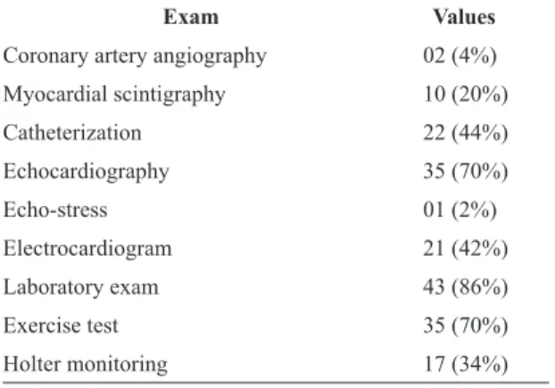
Table 2 shows the complementary tests present in the records. The most commonly used were laboratory examinations, echocardiography, and an ergometric test.
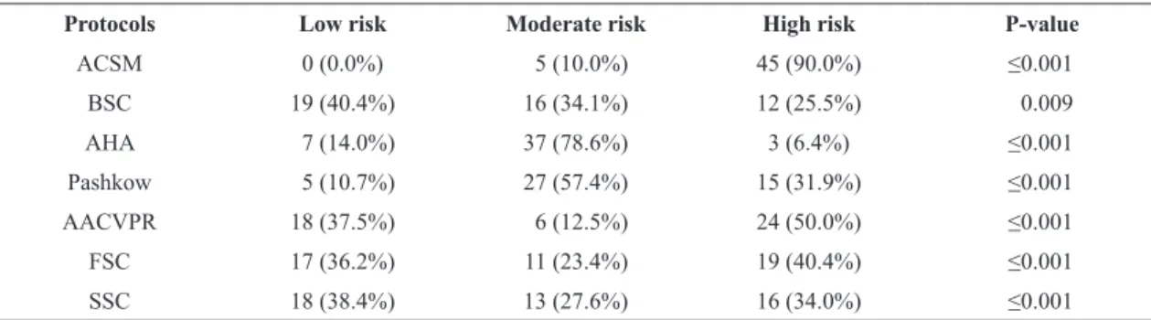
Table 3 shows the percentage distribution of risk classiication for each protocol. Different proportions were observed in each category of risk stratiication among the evaluated protocols, with less accurate proportions between the protocols of the AHA and ACSM. It should be noted that there were statistically signiicant differences between the protocols for all risk categories.
The agreement between risk classifications obtained from the protocols can be seen in Table 4. Of the 21 agreements obtained, 12 were considered signiicant, nine classiied as moderate, and three as low, according to the Kappa coeficient. A signiicant p value was observed in all moderate agreements.
The agreement table also shows that the ACSM protocol had no agreement with any of the analyzed protocols and that the AHA protocol presented agreement only with the FSC and SSC, though the agreement was low. Low agreement was also observed between the BSC and SSC protocols, and the highest agreement was found between the BSC protocol and the Pashkow protocol.
Discussion
This study aimed to evaluate the level of agreement between existing risk stratiication protocols in individuals who regularly attended a CR program. According to the results observed in this study, there were only 12 agreements between the evaluated protocols, which were classiied as low or moderate, without any agreement considered excellent. Different proportions in each category of risk stratiication were
Table 1. Characteristics of the study population (n=50).
Characteristics Values
Sex (M/F) 33 (66%) / 17 (34%)
Age (years) 65.50±9.56 [39 – 81]
Weight (Kg) 79.30±13.09 [54.00 – 105.50]
Height (m) 1.65±0.08 [1.46 – 1.78]
BMI (Kg/m2) 29.13±4.22 [20.68 – 38.25] Principal Diagnosis
Coronary Insuficiency 27 (54%)
Heart Failure 09 (18%)
Constrictive Pericarditis 01 (2%)
Atrial Fibrillation 01 (2%)
Valvulopathy 01 (2%)
Coronary Artery Dissection 01 (2%)
AMI 03 (6%)
Cardiovascular risk factors 07 (14%)
Values expressed as mean±standard deviation [Minimum value – Maximum value] or in absolute values and percentages. M: male; F: female; Kg: kilogram; m: meters; BMI: body mass index; m2: meters
squared; AMI: acute myocardial infarction.
Table 2. Absolute values and percentage of complementary tests
found in the patient’s medical records.
Exam Values
Coronary artery angiography 02 (4%)
Myocardial scintigraphy 10 (20%)
Catheterization 22 (44%)
Echocardiography 35 (70%)
Echo-stress 01 (2%)
Electrocardiogram 21 (42%)
Laboratory exam 43 (86%)
Exercise test 35 (70%)
302 Braz J Phys Ther. 2016 July-Aug; 20(4):298-305
observed among the evaluated protocols, this ratio being less accurate between the AHA and ACSM.
This is the irst study to evaluate the existence of agreements between protocols developed to stratify the risk of individuals in exercise programs and CR. Different proportions in each category of risk stratiication were observed in the majority of the evaluated protocols. In the ACSM protocol, 90% of
patients were classiied as high risk in contrast to the AHA, which ranked only 6.4% of these patients in the same risk category. One of the reasons for this discrepancy is the fact that the ACSM protocol is the only one that uses the presence of cardiovascular, metabolic or pulmonary disease as criteria for classifying the individual as high risk7,18 and the majority of the sample (45 individuals) comprised individuals
Table 3. Absolute values and percentage of the risk classiication of patients according to the protocols analyzed.
Protocols Low risk Moderate risk High risk P-value
ACSM 0 (0.0%) 5 (10.0%) 45 (90.0%) ≤0.001
BSC 19 (40.4%) 16 (34.1%) 12 (25.5%) 0.009
AHA 7 (14.0%) 37 (78.6%) 3 (6.4%) ≤0.001
Pashkow 5 (10.7%) 27 (57.4%) 15 (31.9%) ≤0.001
AACVPR 18 (37.5%) 6 (12.5%) 24 (50.0%) ≤0.001
FSC 17 (36.2%) 11 (23.4%) 19 (40.4%) ≤0.001
SSC 18 (38.4%) 13 (27.6%) 16 (34.0%) ≤0.001
Values expressed in absolute values and percentages. ACSM: American College of Sports Medicine; BSC: Brazilian Society of Cardiology; AHA: American Heart Association; AACVPR: American Association of Cardiovascular and Pulmonary Rehabilitation; FSC: French Society of Cardiology; SSC: Spanish Society of Cardiology.
Table 4. Agreement of risk classiication between the protocols used.
Protocols K P-value
ACSM vs BSC 0.06 0.241
ACSM vs AHA 0.01 0.537
ACSM vs Pashkow 0.04 0.417
ACSM vs AACVPR 0.00 0.999
ACSM vs FSC 0.02 0.683
ACSM vs SSC 0.02 0.690
BSC vs AHA 0.04 0.711
BSC vs Pashkow 0.74 ≤0.001
BSC vs AACVPR 0.52 ≤0.001
BSC vs FSC 0.57 ≤0.001
BSC vs SSC 0.39 0.006
AHA vs Pashkow 0.00 0.931
AHA vs AACVPR 0.04 0.550
AHA vs FSC 0.19 0.028
AHA vs SSC 0.22 0.016
Pashkow vs AACVPR 0.57 ≤0.001
Pashkow vs FSC 0.72 ≤0.001
Pashkow vs SSC 0.56 ≤0.001
AACVPR vs FSC 0.65 ≤0.001
AACVPR vs SSC 0.44 0.001
FSC vs SSC 0.68 ≤0.001
with these disorders, which contributed to the high prevalence of high-risk individuals in this protocol.
Depending on the stratiication criteria adopted by the protocols, not all were able to stratify all individuals in the sample. In the BSC, FSC, SSC, and Pashkow protocols, it was not possible to stratify three patients who did not have complementary tests or information that allowed allocation into one of the stratiication criteria. In the AACVPR protocol, only one of these three patients was stratiied, because although the patient had no complementary test, their clinical condition satisied one of the stratiication criteria of this protocol.
In the AHA protocol, three patients could not be stratiied. Two patients had complementary tests, but basic conditions (atrial ibrillation and constrictive pericarditis) did not meet any of the criteria for determining the risk class, and one patient had no complementary tests to determine the risk class, although the underlying disease was among the criteria.
The above data highlight the importance of complementary tests in the risk stratiication process of these patients. Complementary tests related to the evaluation of the cardiovascular system were present in 94% of the analyzed records. Among the patients with complementary tests, only one had not undergone the ergometric test or echocardiography, which are examinations used as stratiication criteria for the majority of protocols9,10,12,19-21, however the patient had an echo-stress, which enables not only the evaluation of contractile function but also the veriication of the presence of ischemia with stress. The ergometric test and echocardiogram are very important in the stratiication process, since the protocols that use them consider their indings in more than one level and in many cases are the deining tests for the risk presented by the patient.
Regarding the agreement between the protocols, it was observed that the risk classiication of the ACSM protocol did not agree with any of the other protocols used, which is related to the fact that this protocol uses the clinical history of the patient as the criterion for stratifying, without the use of complementary tests.
Due to the low number of similarities in the risk ranges between the AHA protocol and other protocols, this protocol did not agree with four of the six guidelines and the two agreements that were found, with the protocols of the FSC and the SSC, were classiied as low according to the Kappa coeficient (K<0.40).
The FSC and Pashkow were the protocols that obtained the highest number of moderate agreements
(each with four protocols) with other protocols. The highest agreement was observed between the BSC and Pashkow protocols (K=0.74) and it was close to the value considered as excellent (K>0.75). These protocols have similarities regarding the criteria for risk classiication that may explain, at least in part, the agreement found. In addition, the criteria use the most prevalent examinations in the study sample.
The majority of protocols examined in this study are based on the indings of complementary tests for cardiac risk classiication9,10,12,19-21. Among the tests used for risk stratiication of heart patients, the ergometric test stands out due to its well-established methodology22.
This test is used to identify myocardial ischemia and arrhythmia induced by exercise and it provides the value of the metabolic equivalent (MET), which is utilized in all protocols that use complementary tests as one of the main references to determine the risk level7. Despite its importance, 30% of the patients had no ergometric test in their medical records.
Echocardiographic data are also used by six of the seven protocols, and ejection fraction is a reference in ive protocols9,12,19-21. The value of the ejection fraction obtained by echocardiography is considered a risk predictor23. As well as an ergometric test, 30% of the patients included in the study did not have an echocardiogram in their records.
Some factors that contributed to the disagreements between the protocols were: i) a number of the protocols9,10,19-21 are directed mainly at patients who suffered acute myocardial infarction; ii) many of the tests used have discrepancies between the protocols, i.e. not all information concerning the ergometric test and echocardiogram was used to characterize the individual in relation to the risk classiication.
This study has some limitations that should be pointed out. The fact that some records contained incomplete medical history without some details necessary to some protocols, such as the presence of complications during hospitalization, reinfarction, and the occurrence of cardiac arrest, may have produced errors in the stratiication of patients in some protocols (AACVPR9, AHA12, FSC20, and SSC21). It is suggested
304 Braz J Phys Ther. 2016 July-Aug; 20(4):298-305
The results of this study do not allow us to determine the best protocol to be used, however it can be stated that the ACSM protocol is the most suitable for patients who have no complementary tests. All of the other protocols can be used in patients who have complementary tests, however according to the tests they have and the patient’s clinical history, some protocols may be more suitable for assessing the risk of cardiac events during exercise.
The SSC protocol21 is the only one that takes into account the occurrence of reinfarction among its criteria for classiication and a history of cardiac arrest is a criterion used by the AACVPR9, AHA12, and FSC
protocols20. The FSC protocol20 also differs from the others in the use of the Lown classiication in their criteria, a protocol indicated for patients who have the Holter monitoring among their complementary tests. Myocardial scintigraphy information is used by the SSC21, BSC19, FSC20, and Pashkow10 protocols, with the last two also addressing echo-stress test results.
The AHA protocol12, despite being introduced as an extensive method for the stratiication of patients, does not possess all existing cardiac diseases in its criteria and thus is not feasible to determine the risk for all patients included in a CR program. In contrast, this is the only protocol that takes into account the NYHA Functional Classiication16 as one of the stratiication criteria.
The majority of the evaluated protocols presented low or moderate agreement. However, despite their limitations, all protocols showed good applicability for the cardiac risk assessment of the majority of the patients, which is important for professionals acting in the CR area.
Conclusion
Based on the indings of the present, it can be concluded that the agreements between cardiac risk classiication protocols in cardiac patients undergoing a CR program were considered low to moderate. Furthermore, the risk ratios differed between protocols according to each category of risk stratiication.
References
1. HerdyAH, López-JiménezF, TerzicCP, MilaniM, SteinR, Carvalho T, et al. Consenso sul-americano de prevenção e reabilitação cardiovascular.Arq Bras Cardiol. 2014;103(2 Suppl 1):1-31. PMid:25387466.
2. Mansur AP, Favarato D. Mortalidade por doenças cardiovasculares no Brasil e na região metropolitana de São
Paulo: atualização 2011.Arq Bras Cardiol. 2012;99(2 ):755-61. http://dx.doi.org/10.1590/S0066-782X20120050000):755-61. PMid:22735870.
3. BrownRA. Rehabilitation of patients with cardiovascular diseases. Report of a WHO expert committee.World Health Organ Tech Rep Ser. 1964;270:3-46. PMid:14128604. 4. MuelaHCS, BassanR, SerraSM. Avaliação dos benefícios
funcionais de um programa de reabilitação cardíaca.Rev Bras Cardiol.2011;24(4):241-50.
5. GhisiGLM, SantosRZ, SchveitzerV, BarrosAL, Recchia TL, OhP, et al. Desenvolvimento e validação da versão em português da escala de barreiras para reabilitação cardíaca.Arq Bras Cardiol. 2012;98(4):344-52. http://dx.doi. org/10.1590/S0066-782X2012005000025. PMid:22426990. 6. AraújoCGS, Carvalho T, CastroCLB, CostaRV, Moraes RS, OliveiraJAFo, et al. Normatização dos equipamentos e técnicas da reabilitação cardiovascular supervisionada.Arq Bras Cardiol. 2004;83(5):448-52. http://dx.doi.org/10.1590/ S0066-782X2004001700012. PMid:15543365.
7. SilvaAKF, BarbosaMPCR, BernardoAFB, VanderleiFM, PacagnelliFL, VanderleiLCM. Cardiac risk stratification in cardiac rehabilitation programs: a review of protocols.Rev Bras Cir Cardiovasc. 2014;29(2):255-65. PMid:25140477. 8. GobleAJ, WorcesterMUC. Best practice guidelines for
cardiac rehabilitation and secondary prevention. Melbourne: The Heart Research Centre; 1999.
9. American Association of Cardiovascular and Pulmonary Rehabilitation. Diretrizes para reabilitação cardíaca e programas de prevenção secundáriaMarx AG, translator. 4th ed. São Paulo: Roca; 2007. 244 p.
10. PashkowFJ. Issues in contemporary cardiac rehabilitation: a historical perspective.J Am Coll Cardiol. 1993;21(3):822-34. http://dx.doi.org/10.1016/0735-1097(93)90116-I. PMid:8436764. 11. Paul-LabradorM, VongvanichP, MerzCNB. Risk stratification
for exercise training in cardiac patients: do the proposed guidelines work?J Cardiopulm Rehabil. 1999;19(2 ):118-25. http://dx.doi.org/10.1097/00008483-199903000-00006. PMid:10200918.
12. FletcherGF, BaladyGJ, AmsterdamEA, ChaitmanB, Eckel R, FlegJ, et al. Exercise standards for testing and training: a statement for healthcare professionals from the American Heart Association.Circulation. 2001;104(14):1694-740. http://dx.doi.org/10.1161/hc3901.095960. PMid:11581152. 13. ParmleyWW. Position report on cardiac rehabilitation.
Recommendations of the American College of Cardiology. J Am Coll Cardiol. 1986;7(2):451-3. http://dx.doi.org/10.1016/ S0735-1097(86)80526-5. PMid:3711468.
14. Health and Public Policy Comittee, American College of Physicians. Cardiac rehabilitation services.Ann Intern Med. 1988;109(8):671-3. http://dx.doi.org/10.7326/0003-4819-109-8-671. PMid:3421577.
15. Associação Brasileira para o Estudo da Obesidade e da Síndrome Metabólica – ABESO. Diretrizes Brasileiras de Obesidade 2009/2010 / ABESO – Associação Brasileira para o Estudo da Obesidade e da Síndrome Metabólica.3rd ed. Itapevi: AC Farmacêutica; 2009.
2002;31(4):262-70. http://dx.doi.org/10.1067/mhl.2002.124554. PMid:12122390.
17. LownB, WolfB. Approaches to sudden death form coronary heart disease.Circulation. 1971;44(1):130-42. http://dx.doi. org/10.1161/01.CIR.44.1.130. PMid:4104697.
18. American College of Sports Medicine. Diretrizes do ACSM para o teste de esforço e sua prescrição. Taranto G, translator. 7th ed. Rio de Janeiro: Guanabara Koogan; 2007. 239 p. 19. MoraesRS. Diretriz de Reabilitação Cardíaca.Arq Bras
Cardiol. 2005;84(5):431-40.
20. Monpère C, SellierP, MeurinP, AeberhardPB, D’Agrosa BoiteuxM, IliouMet al. Recommandations de la Société française de cardiologie concernant la pratique de la réadaptation cardiovasculaire chez l’adulte. Version 2. Arch Mal Coeur.2002;95(10):962-97.
21. VelascoJA, CosínJ, MarotoJM, MuñizJ, CasasnovasJA, PlazaI, et al. Guías de práctica clínica de la Sociedad Española de Cardiología en prevencíon cardiovascular y rehabilitación cardíaca.Rev Esp Cardiol. 2000;53(8 ):1095-120. http://dx.doi.org/10.1016/S0300-8932(00)75211-0. PMid:10956605.
22. SecundoPFC, SantosBFO, SecundoJAJr, SilvaJB, Souza AR, FaroGBA, et al. Parâmetros clínicos e ecocardiográficos associados a baixo índice cronotrópico em pacientes não idosos.Arq Bras Cardiol. 2012;98(5):413-20. http://dx.doi. org/10.1590/S0066-782X2012005000033. PMid:22481642. 23. Dagres N, Hindricks G. Risk stratification after myocardial infarction: is left ventricular ejection fraction enough to prevent sudden cardiac death?Eur Heart J. 2013;34(26):1964-71. http://dx.doi.org/10.1093/eurheartj/ eht109. PMid:23644180.
Correspondence
Ana Alice Soares dos Santos
Universidade Estadual Paulista Faculdade de Ciências e Tecnologia Departamento de Fisioterapia