©Revista Brasileira de Fisioterapia
MECHANIC CRITERIA FOR PROGRESSION IN INTERNAL AND
EXTERNAL ROTATION EXERCISES OF THE SHOULDER IN THE
SAGITTAL PLANE
T
OLEDOJM, R
IBEIRODC & L
OSSJF
School of Physical Education, Federal University of Rio Grande do Sul, Porto Alegre, RS - Brazil
Correspondence to: Joelly Mahnic de Toledo, Av. Baltazar de Oliveira Garcia, 3221, apto 407 bloco 4, Bairro Jardim Leopoldina, CEP 91180-001, Porto Alegre, RS – Brazil, e-mail: [email protected]
Received: 06/03/2006 - Revised: 30/06/2006 - Accepted: 07/11/2006
ABSTRACT
Introduction: Knowledge of torque and force production capacity and moment arm patterns throughout the movement, and their influence on the torque produced, are essential for understanding human movement and may be of great use for controlling the overload imposed on the muscle-tendon structure. Objective: To present mechanical criteria for progression in internal rotation (IR) and external rotation (ER) exercises of the shoulder in the sagittal plane. Method: Six individuals were assessed using an isokinetic dynamometer and an electrogoniometer. From the data collected, the mean torque, mean resultant force and weighted mean moment arm were calculated using the SAD32 and Matlab® software. Results: The angles at which the peak ER and IR
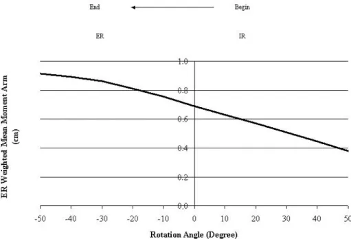
torque occurred were -34° and 6º with values of 43 Nm and 69 Nm, respectively. The peaks for ER and IR muscle force were at 35º and -14º, and the values at these angles were 10227 N and 8464 N, respectively. The weighted mean moment arm for ER presented an increasing pattern over the whole range of motion (ROM) and the peak was at the end of the ROM, i.e. at -50º (0.91 cm). The weighted mean moment arm for IR was almost constant with its peak at 50º (0.96 cm). Conclusion: The mechanical criteria for progression in internal and external rotation exercises of the shoulder are torque, force and weighted mean moment arm because different overloads on the muscle-tendon structure can be caused according to their patterns over the ROM.
Key words: shoulder, exercise, rotation, rehabilitation.
INTRODUCTION
Rehabilitation of the shoulder joint may be difficult not only because of its complex function, which involves anatomical and functional integrity, but also due to the physiological and biomechanical contributions of structures such as the scapula1,2. Generally speaking, shoulder rehabilitation programs make use, in most cases, of exercises with progressive loads and intensities according to the type of injury and surgical procedure that was performed1,2,3. Even though these characteristics are decisive for exercise progression, knowledge of the joint mechanics is fundamental for appropriately choosing the exercises3.
Joint movements are consequences of the rotation of one segment in relation to another. This rotational effect of an applied force is called torque or moment. The torque that a muscle generates on the joint is influenced by the moment arm range or the force production capacity of the joint4,5,6. The moment arm (perpendicular distance) is the smallest distance between the line of muscle action and the center of joint rotation4,7,8,9. The magnitude of the moment arm represents the mechanical advantage of a muscle in a joint,
and its measurement may assist in understanding how the muscle functions5.
for understanding human movement and may be of great use for controlling the overload imposed on the muscle-tendon structure, as well as for better planning for exercise progression in a rehabilitation program16,17,18.
The objective of this study was to present mechanical criteria for the progression of internal rotation (IR) and external rotation (ER) exercises of the shoulder, when performed in the sagittal plane.
MATERIALS AND METHODS
This study was approved by the Ethics Committee of the IPA Methodist University Center (registration no. 1211) and all the participants signed a free and informed consent statement.
The sample consisted of six male individuals, with ages between 22 and 32 years (mean: 25.1 ± 4.0) and height between 167 and 192 cm (mean: 182.6 ± 9.8), who were regularly doing physical activities (at least twice a week). All individuals in the sample participated in all the stages of the study. The shoulder evaluated was the right shoulder (dominant limb) and none of the individuals presented histories of injuries or dysfunctions in the evaluated shoulder.
The data collection consisted of measuring the maximum ER and IR torque produced at 60º/sec in the sagittal plane. For this, an isokinetic dynamometer was used (Cybex Norm model, Dataq Instruments, Inc., Ohio, United States). With the aim of recording joint positions with greater precision, an electrogoniometer was used (XM 180 model, Biometrics Ltd (Cwmfelinfach, Gwent, United Kingdom), adapted together with the isokinetic dynamometer. The isokinetic dynamometer and the electrogoniometer were connected to a Pentium III 650 MHz microcomputer by means of a 16-channel analog-digital converter. For data processing, the SAD32 software (a data acquisition system developed by the Mechanical Measurements Laboratory of the Federal University of Rio Grande do Sul) and the MATLAB 7.0® software (MathWorks Inc, Massachusetts, United States) were used.
The collection procedures were divided into five phases: preparation, positioning, calibration, test familiarization and testing.
Preparation: warm-up and stretching of the right arm. Positioning of the individuals: dorsal decubitus with the right arm positioned at 90º abduction and the elbow flexed at 90º.
Calibration: the ER and IR ranges of motion (ROM) were determined according to the maximum ROM at which the individual was capable of producing maximum torque. The zero angle of rotation on the electrogoniometer was
submaximal concentric contractions were performed.
Test: five repetitions of ER and IR maximal concentric contractions were performed at an angular velocity of 60°/sec19.
The data of the torque generated and the angle were filtered using a low-pass third-order Butterworth digital filter with a frequency cutoff of 3 Hz for the angle data and 10 Hz for the torque data. After the signal filtering, the mean of the five repetitions was calculated. The convention used for the angular positions was that the ER would have negative values and the IR would have positive values8.
From the ER and IR torque values, it was possible to estimate the magnitude of the resultant force exerted by the external and internal rotations, through the ratio between the torque and the moment arm of force application. Since many muscles are able to perform ER or IR, a simplification was made, to make it possible to determine equation (1):
T = dp x Fm
(1)
In which: T = torque; Fm = muscle force;
dp = moment arm (between the muscle force action line and the rotation center of the shoulder)8.
For this, the mean moment arms of all the internal rotator and external rotator muscles were calculated. This mean was weighted by the physiological cross-sectional area of each muscle, thus resulting in the weighted mean moment arm (WMMA). The muscles used for the calculation were the supraspinatus, infraspinatus, teres minor, posterior deltoid, middle deltoid and anterior deltoid for ER; and the pectoralis major, latissimus dorsi, teres major, posterior deltoid, middle deltoid and anterior deltoid for IR. The physiological cross-sectional muscle area and the moment arms of the muscles were obtained from the literature 8.
RESULTS
The ER torque behavior is presented in Figure 1. At the beginning of the movement, there was an increase and then the curve tended to remain constant over the intermediate section of the ROM. After maintaining this plateau, there was a slight increase representing the torque peak. At the end of the movement, the torque curve presented a descending phase. The ER torque peak occurred at an angle of -34°, at which the shoulder is rotated externally with a mean torque of 43 Nm (100%).
ER: external rotation; IR: internal rotation
Figure 2. Weighted mean moment arm of the external rotation.
ER: external rotation; IR: internal rotation.
Figure 1. Percentage mean torque of the external rotation, with standard error.
.
.
.
.
.
Figure 3. Percentage resultant force of the external rotation, with standard error.
ER: external rotation; IR: internal rotation.
Figure 4. Percentage mean torque of the internal rotation, with standard error.
Figure 5. Weighted mean moment arm of the internal rotation.
ER: external rotation; IR: internal rotation.
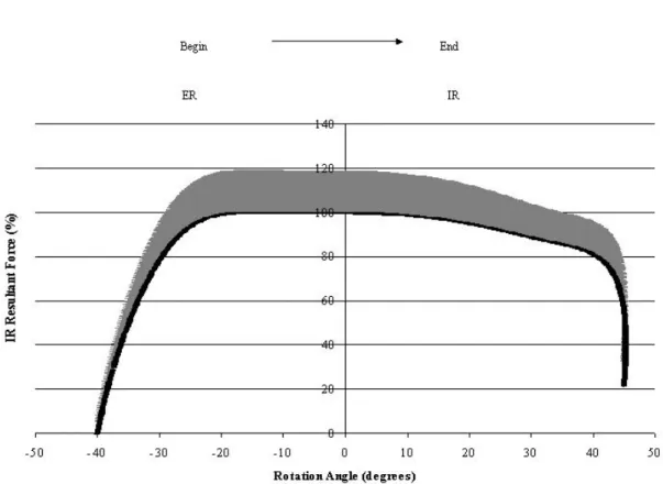
Figure 6. Percentage resultant force of the internal rotation, with standard error.
ER: external rotation; IR: internal rotation. .
.
.
.
.
.
of the movement. Unlike the torque behavior, the peak force occurred when the shoulder was internally rotated, when it was stretched, before reaching the neutral position and without presenting any plateau throughout the ROM. The ER peak force occurred at an angle of 35°, with a mean value of 10227N (100%).
The IR torque behavior (Figure 4) was very similar to the ER pattern (Figure 1). At the beginning of the movement, this curve also presented an increase and then tended to remain constant over the intermediate section of the ROM. However, differing from the ER, the IR peak torque occurred at an approximate angle of 6° during this plateau, with a mean of 69 Nm (100%), when the shoulder was internally rotated. At the end of the movement, this torque curve also presented a descending phase.
The behavior of the WMMA of the IR was practically constant over the course of the ROM (Figure 5). At the beginning of the movement, there was an ascending phase, which finished approximately at the intermediate section of the ROM, when the WMMA was practically constant. There was then a new ascending phase, culminating at the WMMA peak at the end of the movement. The largest WMMA of the IR occurred at a rotation of 50°, with a moment arm of 0.96 cm.
The behavior of the resultant IR force curve (Figure 6) was similar to the IR torque curve (Figure 4), but with different magnitudes. At the beginning of the movement, this curve also presented an increase and then tended to remain constant during the intermediate section of the ROM. At the plateau, the peak force of the IR occurred at an approximate angle of -14º, with a mean of 8464N (100%), when the shoulder was externally rotated. At the end of the movement, this curve also presented a descending phase.
DISCUSSION
During ER, it was observed that the torque plateau that occurred in the middle of the ROM was maintained because of the antagonistic behavior of the WMMA and the resultant ER force. Since the ER peak torque occurred when the shoulder was externally rotated, it can be inferred that the WMMA was more important for torque generation in this ROM and for plateau maintenance than was the length versus tension relationship represented by the curve of resultant force. It can also be noted that the peak torque and peak force did not occur at the same angles, since they depended on the length-tension relationship of the muscle and its respective moment arm11.
The behavior of the ER force curve was very similar to the behavior of the curve of the length-tension relationship of the sarcomere that was presented by Gordon et al.20. The
for the formation of cross-bridges. Since the muscles are slightly stretched, there is a contribution from the elastic elements of the muscles, to force production21,22,23. After this point, the force decreases because of the muscle shortening and because of the reduced possibility of forming new cross-bridges11,23,24.
Regarding the IR, the torque and resultant force curves can be analyzed simultaneously because of their similar behavior. This is because of the behavior of the WMMA, which remains more or less constant, with a very low rate of increase. These findings are similar to those of Rassier et al.11, who reported that the torque-angle relationship of a muscle is determined by the length-tension relationship and the moment arm. When the moment arm remains constant throughout the movement, the behavior of the torque curve reflects the resultant force curve. It can thus be presumed that the length versus tension relationship is the main factor responsible for the IR torque behavior.
The objective of rehabilitation is to recover ROM and strengthen muscles, especially the rotators, which are important for stabilizing and protecting the joint structures from injuries. The exercises must have progressive loads and respect the mechanics of joint functioning, and a rehabilitation program must be efficient in order to achieve the objectives and respect the particular features of the shoulder25. In the specific case of the shoulder, in which the rotation movement is performed by means of synergistic action of different muscles, it is important to evaluate the weighted mean moment arm and the resultant force production capacity as criteria for progression in the intensity and loads of the exercises.
length-tension relationship may promote less overload in the muscle-tendon structure. On the other hand, if the peak resistance is applied at amplitudes at which the moment arm and the length-tension relationship are unfavorable, there will be greater overload.
In injuries of the teres minor and infraspinatus, the imposed load may vary, thus modifying the angular section at which the peak torque resistance occurs. During the initial phase of a rehabilitation program, muscular reinforcement is recommended, with small loads in order to optimize the healing process16,17. This work can be done with the peak resistance torque situated between the neutral and final positions of the ER, at a shoulder abduction of 90º, since there is a mechanical advantage in this section (larger moment arm) during torque production and lower force production levels are needed. Consequently, a lower number of motor units will be activated, thus generating less overload on the muscle. In the intermediate phase of rehabilitation, the peak resistance torque could occur between the neutral position and the maximum IR since, during this section, the moment arm is smaller and the force production capacity is the main factor responsible for torque production. With the same torque resistance as cited in the earlier example, the teres minor and infraspinatus will be subjected to greater overload, since a larger number of motor units must be recruited to compensate for the decrease in the moment arm, with the purpose of generating the same torque.
CONCLUSION
The mechanical criteria for progression in internal and external rotation exercises are the torque, force and weighted mean moment arm because, based on their behavior, it is possible to promote different overloads on the muscle-tendon structure. Although these are theoretical elaborations, these criteria are based on principles of muscle-tendon healing. Thus, this study represents a first step towards structuring mechanical criteria for progression in the overloads imposed on the muscle-tendon structure.
REFERENCES
1. Kibler WB, McMullen J, Uhl T. Shoulder rehabilitation strategies, guidelines and practice. Orthopedic Clinics of North America. 2001;32(3):527-38.
2. Rubin BD, Kibler WB. Fundamental Principles of Shoulder Rehabilitation: Conservative to Postoperative Management. Arthroscopy. 2002;15(9):29-39.
3. Hayes K, Ginn KA, Walton JR, Szomor ZL, Murrell GAC. A randomized clinical trial evaluating the efficacy of physiotherapy after rotator cuff repair. Australian Journal of Physiotherapy. 2004;50:77-83.
4. Otis JC, Jiang CC, Wickiewicz TL, Peterson MGE, Warren RF, Santner TJ. Changes in the moment arms of the rotator cuff and deltoid muscles with abduction and rotation. The Journal of Bone and Joint Surgery. 1994;76(5):667-76.
5. Liu J, Hughes RE, Smutz WP, Niebur G, An KN. Roles of deltoid and rotator cuff muscles in shoulder elevation. Clinical Biomechanics. 1997;12(1):32-8.
6. Wilde LD, Audenaert E, Barbaix E, Audenaert A, Soudan K. Consequences of deltoid muscle elongation on deltoid muscle performance: the computerized study. Clinical Biomechanics. 2002;17:499-505.
7. Kuechle DK, Newman SR, Itoi E, Morrey BF, An KN. Shoulder muscle moment arms during horizontal flexion and elevation. Journal of Shoulder and Elbow Surgery. 1997;6:429-39.
8. Kuechle DK, Newman SR, Itoi E, Niebur GL, Morrey BF, An KN. The relevance of the moment arm of shoulder muscles with respect to axial rotation of the glenohumeral joint in four positions. Clinical Biomechanics. 2000;15:322-9.
9. Graichen H, Englmeier KH, Reiser M, Eckstein F. An in vivo technique for determining 3D muscular moment arms in different joint positions and during muscular activation – application to the supraspinatus. Clinical Biomechanics. 2001;16:389-94.
10. Proske U, Morgan L. Do cross-bridges contribute to the tension during stretch of passive muscle? Journal of Muscle Research and Cell Motility. 1999;20:433-42.
11. Rassier DE, MacIntosh BR, Herzog W. Length dependence of active force production in skeletal muscle. Journal Applied Physiology. 1999;86(5):1445-57.
12. Huxley AF, Niedergerke R. Structural changes in muscle during contraction. Interference microscopy of living muscle fibres. Nature. 1954;173:971-3.
13. Huxley H, Hanson J. Changes in cross-striations of muscle during contraction and stretch and their structural interpretation. Nature. 1954;173:973-6.
14. Huxley AF. Muscle structure and theories of contraction. Prog Biophys Biophys Chem. 1957;7:255-318.
15. Huxley AF, Simmons RM. Proposed mechanism of force generation in striated muscle. Nature. 1971;233:533-8.
16. Wilk KE, Harrelson GL, Arrigo C. Reabilitação do Ombro. In: Andrews JR, Harrelson GL, Wilk KE. Reabilitação Física das Lesões Desportivas. 3ª ed. Rio de Janeiro: Guanabara Koogan; 2005. p. 545-622.
17. Magee DJ, Reid DC. Shoulder Injuries. In: Zachazewski JE, Magee DJ, And Quillen WS. Athletic Injuries and Rehabilitation. Philadelphia: Saunders; 1996. p. 509-39.
18. Walmsley RP, Szibbo C. A Comparative Study of the Torque Generated by the Shoulder Internal and External Rotator Muscles in Different Positions and at Varying Speeds. The Journal of Orthopaedic and Sports Physical Therapy. 1987;9(6):217-22.
19. Divir Z. Isokinetics of the shoulder muscles. In: Divir Z. Isokinetic: Muscles testing, interpretation and clinical applications. Edinburg: Churchill Livingstone; 1995. p. 171-91.
20. Gordon AM, Huxley AF, Julian FJ. The variation in isometric tension with sarcomere length in vertebrate muscle fibres. Journal of Physiology. 1966;184:170-92.
Biomechanics. 2003;36:1309-16.
23. Schachar R, Herzog W, Leonard TR. The effects of muscle stret-ching and shortening on isometric force on the descending limb of the force-length relationship. Journal of Biomechanics. 2004;37:917-26.