O
RIGINALA
RTICLE Revista Brasileira de FisioterapiaEffects of low-level laser therapy on epidermal
oxidative response induced by wound healing
Efeitos da laserterapia de baixa potência na resposta oxidativa epidérmica
induzida pela cicatrização de feridas
Silveira PCL, Silva LA, Tuon T, Freitas TP, Streck EL, Pinho RA
Abstract
Background: Therapeutic use of low-level laser in physical therapy has increased significantly. Objective: To assess the effects of low-level laser therapy on the oxidative parameters of wound healing in rats. Methods: Eighteen Wistar rats were randomly divided into three groups (control, 5 days, n=6; 2 J/cm2, 5 days, n=6; 4 J/cm2, 5 days, n=6). A single circular wound measuring 8 x 8 mm was surgically created on the rats’ backs. Thirty minutes after the last irradiation, the rats were euthanized and the irradiated tissue was surgically removed and stored at -70°C. We determined the activity of the respiratory chain enzymes DCIP oxidoreductase (complex II) and soluble succinate dehydrogenase (SDH); the activity of cytochrome c oxidase (complex IV); the production of superoxide anion; and the activity of superoxide dismutase (SOD) and catalase (CAT). Lipid peroxidation was assessed by means of the TBARS assay.
Results: There was a decrease in the complex II activity in the groups irradiated for 5 days with 2 and 4 J/cm2, while superoxide anion production decreased significantly in the group irradiated for 5 days with 4 J/cm2 when compared with the control group. There was also a significant increase in CAT activity in the group irradiated for 5 days with 2 J/cm2as well as a decrease in lipid peroxidation activity in the two irradiated groups. Conclusions: The results of the present study indicate that laser stimulates antioxidant activity and protects cells against oxidative damage during the wound healing process in rats.
Keywords: wound healing; low-level laser therapy; oxidative stress; respiratory chain; free radical.
Resumo
Introdução:O uso terapêutico do laser de baixa potência na fisioterapia tem aumentado significativamente. Objetivo: Avaliar os efeitos da laserterapia de baixa potência nos parâmetros oxidativos na cicatrização de feridas em ratos. Métodos: Dezoito ratos Wistar foram divididos randomicamente em 3 grupos (controle 5 dias,n=6; 5 dias/2 J/cm2, n=6; 5 dias/4 J/cm2, n=6). Uma única ferida circular medindo 8 X 8 mm foi cirurgicamente realizada no dorso do rato. Trinta minutos após a última irradiação, os ratos foram submetidos à eutanásia, e o tecido irradiado foi removido cirurgicamente e armazenado a -70oC. Foi determinada a atividade das enzimas da cadeia respiratória: DCIP oxirredutase (complexo II) e succinato desidrogenase solúvel (SDH), atividade do citocromo c oxidase (complexo IV), produção de ânion superóxido, atividade da superóxido dismutase (SOD) e catalase (CAT). A lipoperoxidação foi avaliada pela técnica de TBARS. Resultados: Os resultados mostram uma diminuição na atividade do complexo II nos grupos irradiados por 5 dias com 2 e 4 J/cm2, enquanto a produção de ânion superóxido mostrou uma diminuição significativa no grupo irradiado por 5 dias com 4 J/cm2 em relação ao grupo controle. Além disso, um aumento significativo na atividade da catalase foi observado no grupo irradiado por 5 dias com 2 J/cm2, como também uma diminuição da peroxidação lipídica nos dois grupos irradiados. Conclusões: Os resultados do presente estudo indicam que o laser estimula a atividade antioxidante e protege a célula contra danos oxidativos durante o processo de cicatrização de feridas cutâneas em ratos.
Palavras-chave: cicatrização; terapia a laser de baixa intensidade; estresse oxidativo; cadeia respiratória; radicais livres.
Received: 14/02/2008 – Revised: 26/08/2008 – Accepted: 12/12/2008
Graduate Program in Health Sciences, Universidade do Extremo Sul Catarinense (UNESC), Criciúma (SC), Brazil
Introduction
he application of low-level laser therapy as therapeutic technology in the area of physical therapy has grown sig-niicantly. he healing properties of laser radiation, associated with the treatment’s safety, seem to be the most responsible for that growth and for the increased interest by biomedical researchers in investigating the action mechanisms and the therapeutic efects of the low-level lasers1. Laser therapy has
a signiicant efect on the ulcerative process and reduces heal-ing time. his response allows the individual to resume normal activities more quickly2. However, some mechanisms involved
in this response are still obscure, especially with regard to the efects of laser on the mitochondrial respiratory chain and on oxidative stress biomarkers3.
According to Karu4, laser exposure causes an increase
in mitochondrial electrochemical activity and a concomi-tant increase in ATP synthesis. Eells et al.5 suggest that
cytochrome c oxidase is the main photoreceptor of laser light. Additionally, the low-level laser has a cascade effect on the cell signaling, which promotes cellular proliferation and cytoprotection6. Some authors also postulate that
la-ser therapy influences oxidative stress parameters such as changes in antioxidant enzyme activity and the production of reactive oxygen species (ROS)7-10.
he absorption of laser light accelerates the transfer of electrons (respiratory chain) and induces an initial ROS pro-duction, speciically increasing the production of superoxide anion7. he excessive production of these species can damage
cell components such as lipids, proteins and nucleic acids11.
he cell membrane seems to be the irst target. he efects of la-ser irradiation on the cellular mechanism and its inluence on oxidative parameters are still unclear, with conlicting results in the literature12. However, it is possible that, depending on the
dosage, exposure time and intensity, laser therapy can change the defense mechanisms that counter excessive ROS produc-tion7. herefore, the aim of the present study was to evaluate
the efects of low-level laser therapy on mitochondrial respira-tory chain activity and oxidative stress parameters in response to wound healing in rats.
Methods
Animals
Eighteen adult male Wistar rats (250-300g) of the vivar-ium of Universidade do Extremo Sul Catarinense (UNESC) were used in this study. he rats were kept at a constant
temperature of 22°C with a 12-hour light/dark cycle and free access to water and standard diet. he procedures have been approved by the Research Ethics Committee of UNESC, pro-tocol number 167/2005.
Ulceration and low-level laser therapy
After anesthesia with ketamine (80 mg/Kg, i.p.), the ani-mals’ dorsal region was shaved and disinfected with alcohol 70%. In the mid-dorsal region, between the infrascapular line and the tail, a circular area of the skin of approximately 8 mm in diameter was removed with a punch13. he animals were
randomly divided into 3 groups (n:6): injury without treatment (control); injury with treatment (2 J/cm2); injury with
treat-ment (4 J/cm2). After the injury, the wounds were immediately
treated for 5 consecutive days with low-level laser. All of the animals were anesthetized before each application, including the control group (ketamine - 80 mg/Kg, i.p.).
he low-level laser used in this study was gallium ar-senide (GaAs), pulse waveform, invisible beam, wavelength of 904 nm, peak power of 15 mW, frequency of 2000 Hz, pulse time of 180 ns and beam cross-section of 0.07 cm2 (Laserpulse
- Ibramed). he application time was 40 seconds (2 J/cm2)
and 80 seconds (4 J/cm2). Non-contact application
(ap-proximate distance of 1 mm) was used with the applicator perpendicular to the injury on ive spots around the wound 1cm apart14. hirty minutes after the last irradiation, all of
the animals were euthanized (guillotine). he tissue around the wound was removed, processed, fractioned and stored at -70°C for subsequent biochemical analyses.
Biochemical analyses
Respiratory chain enzyme activity
Tissue preparation: the tissue around the wound was ho-mogenized (1:10 w/v) in SETH bufer, pH 7.4 (250 mM sucrose, 2 mM EDTA, 10 mM Trizma base, 50 IU/mL heparine). he homogenate was centrifuged at 800 x g for 10 minutes and the supernatant stored at -70°C for determination of enzymatic ac-tivity. he maximum period between the homogenization and the enzymatic analysis was ive days.
Complex II + succinate dehydrogenase (SDH) activity: the enzymatic activities were measured according to the method described by Fischer et al.15, in which the decrease in the
absor-bance of the 2.6-DCIP in 600 nm was used to calculate complex II activity. For the SDH calculation, the same system was used in the presence of phenazine methosulfate.
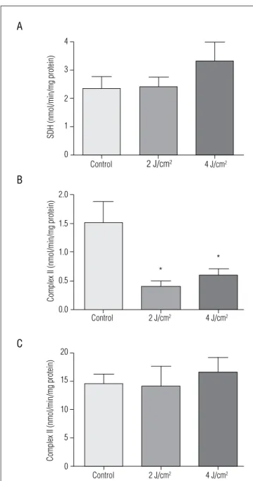
Figure 1. Effect of low-level laser therapy on (A) succinate dehydrogenase activity; (B) mitochondrial respiratory chain complex II; (C) mitochondrial respiratory chain complex IV. The values are presented as mean±SEM, and the results are expressed as nmol/min/mg protein.
Control 4
3
2
1
0
SDH (nmol/min/mg protein)
A
B
C
Complex II (nmol/min/mg protein)
Complex II (nmol/min/mg protein)
2 J/cm2 4 J/cm2
Control 2 J/cm2 4 J/cm2
Control 2 J/cm2 4 J/cm2
2.0
1.5
1.0
0.5
0.0
20
15
10
5
0
*
* decrease in absorbance caused by the oxidation of reduced
cytochrome c, measured in 550 nm.
Superoxide anion: determined by the adrenaline oxidation rate showed through the spectrophotometer at 480 nm, as de-scribed by McCord and Fridovich17.
Superoxide dismutase (SOD) and catalase (CAT) activity:
the enzymatic SOD activity was determined by the inhibition of adrenaline auto-oxidation measured through the spectro-photometer (480 nm)18. CAT activity was determined by the
fall in absorbance (240 nm) corresponding to the consumption of hydrogen peroxide19.
Lipid peroxidation: as an index of lipid peroxidation, we veriied the formation thiobarbituric reactive substances (TBARS) measured with the spectrophotometer (532 nm)20.
Protein determination: the amount of protein in biochem-ical trials was measured using the Lowry et al.21 method.
Statistical analysis
he data were expressed as means and standard deviation and statistically analyzed using one-way analysis of variance (ANOVA), followed by the Tukey post-hoc test. he α level con-sidered for the analysis was set at 0.05. he software SPSS (Statis-tical Package for the Social Sciences) version 12.0 was used.
Results
Mitochondrial enzyme activity
According to Figure 1B, there was a signiicant decrease in complex II activity in the groups irradiated with 2 J/cm2
(0.39±0.25) and 4 J/cm2 (0.59±0.27) compared to the group
without treatment (1.51±0.92), however no signiicant difer-ence was observed in the SDH and complex IV activity.
Superoxide anion production
According to Figure 2, only the group irradiated with 4 J/cm2
(25.04±2.23) showed a signiicant decrease in superoxide anion production compared to the untreated group after laser treat-ment (51.33±3.95).
Superoxide dismutase and catalase activity
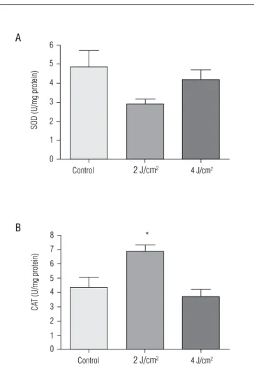
he results show that there was no signiicant change in SOD activity in the groups irradiated for ive days with 2 and 4 J/cm2 compared to the control group (Figure 3A). However, the
CAT activity had a signiicant increase in the group irradiated
with 2 J/cm2 (6.85±0.45) compared to the group without
treat-ment (4.31±0.71; Figure3B).
Lipid peroxidation
he TBARS results seen in Figure 4 show a signiicant de-crease in lipid peroxidation in the groups irradiated for ive days with 2 J/cm2 (0.62±0.10) and 4 J/cm2 (0.42±0.09) compared
to the group without treatment (1.19±0.23).
Figure 4. Effect of low-level laser therapy on levels of lipid peroxidation. The values are presented as mean±SEM, and the results are expressed as nmol MDA/mg protein.
Figure 2. Effect of low-level laser therapy on superoxide anion production. The values are presented as mean±SEM, and the results are expressed as nmol/min/mg protein.
O2
(nmol/min/mg protein)
Control 2 J/cm2 4 J/cm2
60
50
30 40
20
0 10
*
The significant difference used was p<0.05(*).
Figure 3. Effect of low-level laser therapy on (A) superoxide dismutase activity and (B) catalase activity. The values are presented as mean±SEM, and the results are expressed as U/mg protein.
Control 4
5 6
3
2
1
0
SOD (U/mg protein)
A
2 J/cm2 4 J/cm2
CA
T (U/mg protein)
B
Control 4
5 7
6 8
3
2
1 0
2 J/cm2
4 J/cm2 *
The significant difference used was p<0.05(*)
TBARS
(nmol/mg proten)
Control 2 J/cm2 4 J/cm2
1.50
1.25
0.75 1.00
0.50
0.00 0.25
*
*
The significant difference used was p<0.05(*).
Discussion
he healing process has an essential role in the protective response of the epidermal injury through tissue repair22. his
process stimulates inlammatory mediators, such as cytokines and ROS, which have harmful efects on the tissue23. Recent
studies have reported evidence of the important role of ROS in microvascular dysfunction, tissue damage and inlammatory processes which precede the tissue healing24-27.
he present study evaluated the efects of low-level laser therapy (904 nm) with varied irradiation intensity on mito-chondrial respiratory chain activity and some oxidative stress markers. With regard to the respiratory chain activity, Figure 1, the results show a signiicant decrease in complex II activity in the groups irradiated with 2 J/cm2 and 4 J/cm2, respectively
(Figure 1B). It is possible that the inhibitory efect of complex II is directly linked to the excessive irradiation, which can produce complete oxidation and consequently a reduction in the complexes28. his inhibition is not directly associated with
the electron transfer, but with a partial or total structural rear-rangement, which leads to a bioinhibitory efect29.
Previous studies indicate that irradiation with energy density above 4 J/cm2 has a high energy luence with
inhibi-tory characteristics30. However, it was observed that this
in-hibitory characteristic also occurred with the energy density of 2 J/cm2 and 4 J/cm2 on the complex II activity. It is possible
that this diferentiated response results from the laser type and wavelength used in the studies, responsible for the high energy luence.
According to Kreisler et al.31, the stimulation of the
chain and the change in the ATP levels are not well estab-lished, and they are the subject of several discussions. It is commonly accepted that both the stimulatory and inhibi-tory effects of laser on cells are dependent on dosage and wavelength. Low-level irradiation performs biomodulative functions in cellular activity32. The initial molecular
absorp-tion of laser light is still unknown and, depending on the wavelength, the effects are changed due to the different chromophores which can be available as photoreceptors. It is likely that the process of photomodulation is only one among many photosignaling phenomena33.
Figures 1A and 1C show that the complex IV and SDH activity did not change after laser treatment. he reasons for these results are still unknown and merit further investiga-tion. However, previous studies show that irradiation with 2.4 and 3 J/cm2 increases complex II and IV activity after 10 days
of irradiation27,28. It is believed that this fact occurs due to the
longer exposure time, which activates the chromophores of the respiratory chain, especially cytochrome c oxidase. he results of this study also show a signiicant reduction in su-peroxide anion production (Figure 2) ive days after injury in the group irradiated with 4 J/cm2.
The inflammatory response induced by the epidermal injury provokes the migration of neutrophils and mac-rophages, leading to rapid oxygen consumption. This mech-anism activates the NADPH-oxidase, catalyzing the electron transfer from the NADPH to the oxygen to form superoxide34.
Laser therapy reduces this migration of neutrophils and macrophages and stimulates leukocyte phagocytosis and shortening the inflammatory phase thus reducing super-oxide anion production35. It is believed that the reduction
observed in the present study can also be associated with a mitochondrial mechanism of reassertion, suggesting that the superoxide anion can be a source of electrons for the oxidative phosphorylation of ADP7.
It must also be pointed out that there was no significant difference between the group irradiated with 2 J/cm2 and
the control group. Therefore, there may be a dosage and time-dependent ratio of laser therapy on superoxide anion production. The SOD enzyme represents the first line of enzymatic defense against the intracellular production of free radicals, catalyzing the dismutation of the superoxide anion. The resulting product of the reaction catalyzed by SOD is hydrogen peroxide (H2O2), which is catalyzed by the catalase and other peroxidases11.
It has been postulated that the use of low-level laser induces an increase in SOD activity in diferent models, contributing to a decrease in tissue damages and to the maximization of the healing process36,37. As demonstrated
in Figure 3A, there was no signiicant diference in the SOD activity, however there was an important increase in the CAT activity in the group irradiated with 2 J/cm2 (Figure 3B).
Re-garding the CAT activity, the results are in agreement with Fulton and Shitabata38 in that the activity increased in the
rats’ skin after irradiation with low-level laser.
Clinically, this change in the CAT activity in the epithelial tissue after laser exposure can be produced by the generation of free radicals caused by rotational changes in the macro-molecules due to the photostimulation. his stimulation can be important because antioxidants enzymes are controlled by dosage or exposure duration39. Low-level laser irradiation has
been eicient in the reduction of oxidative damages in difer-ent models and situations40.
The results, according to Figure 4, show a significant decrease in lipid peroxidation in the groups treated with 2 J/cm2 and 4 J/cm2. These results suggest that low-level
laser therapy stimulates the defense mechanisms against the oxidative damages to membrane lipids. Although SOD activity did not increase, and CAT activity only increased in the group irradiated with 2 J/cm2, it is possible that
other enzymatic and non-enzymatic antioxidants are involved in the protection against lipid oxidation. Addi-tionally, photostimulation can increase tissue resistance against lipid peroxidation. These variables can justify the observed results.
Fillipin et al.36 observed significantly reduced TBARS
values in rats tendons irradiated and treated with low-level laser for 14 and 21 days, which shows that the irradiation had a protective effect and that the cells developed a posi-tive antioxidant role in the deactivation of ROS excesses. Using irradiation in blood tissue, Stadler et al.10
demon-strated that the lipid peroxidation levels were high after low-level irradiation, suggesting an increase in the ROS and hydroperoxide production. This study sustains the hypoth-esis that the hemoglobin in red blood cells can serve as a photoreactive substance and thus cause high levels of ROS when irradiated. It is possible that the difference in the lipid peroxidation results in different tissues after the low-level irradiation is directly associated with the time of exposure, irradiation intensity and method used for the determination of lipid peroxidation.
Conclusions
References
1. Cogo JC, Ribeiro W, Lopes-Martins RAB, Aimbire FSC. Avaliação do efeito de dois lasers de baixa potência AsGa e HeNe na dermatite tópica induzida por óleo de cróton em orelha de camundongos. Rev Bras Fisioter. 2002;3(4):207-15.
2. Say KG, Gonçalves RC, Renno ACM, Parizatto NA. O tratamento fisioterapêutico de ulceras cutâneas venosas crônicas através da laserterapia com dois comprimentos de onda. Fisioter Bras. 2003;4(1):39-48.
3. Kim YG, Pal SC, Lee SR. Hairless mouse epidermal antioxidants and lipid peroxidation assessed by He-Ne laser. Lasers Surg Med. 2000;27(5):420-6.
4. Karu T. Photobiological fundamentals of low-power laser therapy. IEEE J Quantum Electron. 1987;23(10):1703-17.
5. Eells JT, Wong-Riley MT, VerHoeve J, Henry M, Buchman EV, Kane MP, et al. Mitochondrial signal transduction in accelerated wound and retinal healing by near-infrared light therapy. Mitochondrion. 2004;4(5-6):559-67.
6. Conlan MJ, Rapley JW, Cobb CM. Biostimulation of wound healing by low-energy laser irradiation. A review. J Clin Periodontol. 1996;23(5):492-6.
7. Karu TI, Afanas’eva NI. Cytochrome c oxidase as the primary photoacceptor upon laser exposure of cultured cells to visible and near IR-range light. Dokl Akad Nauk. 1995;342(5):693-5.
8. Mester E, Mester AF, Mester A. The biomedical effects of laser application. Lasers Surg Med. 1985;5(1):31-9.
9. Carrinho PM, Ortiz MCS, Santos AS, Gonçalves RC, Parizotto NA. Laser de baixa intensidade: efeitos sobre os tecidos biológicos – parte 2. Fisioter Bras. 2001;2(6):329-92.
10. Stadler I, Evans R, Kolb B, Naim JO, Narayan V, Buehner N, et al. In vitro effects of low-level laser irradiation at 660nm, on peripheral blood lymphocytes. Lasers Surg Med. 2000;27(3):255-61.
11. Halliwell B, Gutteridge JMC. Free radical in biology medicine. 2ª ed. New York: Oxford University Press; 2007.
12. Nakagawa K. Direct observation of laser generated free radicals from a myocardium target site. Free Radic Biol Med. 1992;12(3):241-2.
13. Carvalho PT, Mazzer N, dos Reis FA, Belchior AC, Silva IS. Analysis of the influence of low-power HeNe laser on the healing of skin wounds in diabetic and non-diabetic rats. Acta Cir Bras. 2006;21(3):177-83.
14. Pessoa ES, Melhado RM, Theodoro LH, Garcia VG. A histologic assessment of the influence of low-intensity laser therapy on wound healing in steroid-treated animals. Photomed Laser Surg. 2004;22(3):199-204.
15. Fischer JC, Ruitenbeek W, Berden JA, Trijbels JM, Veerkamp JH, Standhouders AM. Differential investigation of the capacity of succinate oxidation in human skeletal muscle. Clin Chim Acta. 1985;153(1):23-6.
16. Rustin P, Chretien D, Bourgeron T, Gérard B, Rötig A, Saudubray JM. Biochemical and molecular investigations inrespiratory chain deficiencies. Clin Chim Acta. 1994;228(1):35-51.
17. McCord JM, Fridovich I. Superoxide dismutase. Enzymatic function for erythrocuprein (hemocuprein). J Biol Chem. 1969;244:6049-55.
18. Bannister JV, Calabrese L. Assays for SOD. Methods Biochem Anal. 1987;32:279-312.
19. Aebi H. Catalase in vitro. Methods Enzymol.1984;105:121-6.
20. Draper HH, Hadley M. Malondialdehyde determination as index of lipid peroxidation. Methods Enzymol.1990;186:421-31.
21. Lowry OH, Rosebough NJ, Farr AL, Randall RJ. Protein measurement with the folin phenol reagent. J Biol Chem. 1951;193(1):265-75.
22. Sullins KE. Lasers and wound healing: practical uses. Clinical Techniques in Equine Practice. 2004;3(2):182-7.
23. Tsirogianni AK, Moutsopoulos NM, Moutsopoulos HM. Wound healing: immunological aspects. Injury. 2006;37 Suppl 1:S5-12.
24. Cuzzocrea S, Thiemermann C, Salvemini D. Potential therapeutic effect of antioxidant therapy in shock and inflammation. Chem Med Chem. 2004;11(9):1147-62.
25. Khodr B, Khalil Z. Modulation of inflammation by reactive oxygen species: implications for aging and tissue repair. Free Radic Biol Med. 2001;30(1):1-8.
the decrease in the superoxide anion production, the low-level laser could protect the cell against oxidative damage to membrane lipids. There may be a dosage to time-dependent ratio of laser therapy on antioxidative enzyme activity. Therefore, additional studies are necessary to elucidate these mechanisms.
Acknowledgements
26. Supinski GS, Callahan LA. Free radical-mediated skeletal muscle dysfunction in inflammatory conditions. J Appl Physiol. 2007;102(5):2056-63.
27. Silveira PC, Streck EL, Pinho RA. Evaluation of mitochondrial respiratory chain activity in wound healing by low-level laser therapy. J Photochem. Photobio B. 2007;86(3):279-82.
28. Yu W, Naim JO, McGowan M, Ippolito K, Lanzafame RJ. Photomodulation of oxidative metabolism and electron chain enzymes in rat liver mitochondria. Photochem Photobiol. 1997;66(6):866-71.
29. Karu T, Pyatibrat L, Kalendo G. Irradiation with He-Ne laser increases ATP level in cells cultivated in vitro. J Photochem Photobiol B. 1995;27(3): 219-23.
30. Walsh LJ. The current status of lowlevel laser therapy in dentistry. Part 1. Soft tissue applications. Aust Dent J.1997;42(4):247-54.
31. Kreisler M, Christoffers AB, Willershausen B, d’Hoedt B. Low-level 809nm GaAlAs laser irradiation increases the proliferation rate of human laryngeal carcinoma cells in vitro. Lasers Med Sci. 2003;18(2):100-3.
32. Schaffer M, Sroka R, Fuchs C, Schrafer-Reichardt U, Schaffer PM, Busch M, et al. Biomodulative effects induced by 805 nm laser light irradiation of normal and tumor cells. J Photochem Photobiol B. 1997;40(3):253-7.
33. Novoselova EG, Glushkova OV, Cherenkov DA, Chudnovsky VM, Fesenko EE. Effects of low-power laser radiation on mice immunity. Photodermatol
Photoimmunol Photomed. 2006;22(1):33-8.
34. Fujimaki Y, Shimoyama T, Liu Q, Umeda T, Nakaji S, Sugawara K. Low-level laser irradiation attenuates production of reactive oxygen species by human neutrophils. J Clin Laser Med Surg. 2003;21(3):165-70.
35. Mileva M, Bakalova R, Zlateva G. Low-intensity laser irradiation does not affect the oxidative stress in experimental cataract. Medical Laser Application. 2004;19(3):150-4.
36. Fillipin LI, Mauriz JL, Vedovelli K, Moreira AJ, Zettler CG, Lech O, et al. Low-level laser therapy (LLLT) prevents oxidative stress and reduces fibrosis in rat traumatized achilles tendon. Lasers Surg Med. 2005;37(4):293-300.
37. Parlato G,Cimmino G, De Vendittis E, Monfrecola G, Bocchini V. Superoxide dismutase activity in the skin of rats irradiated by He-Ne laser. Experientia. 1983;39(7):750-1.
38. Fulton JE, Shitabata PK. CO2 laser physics and tissue interactions in skin. Lasers Surg Med. 1999;24(2):113-21.
39. Berki T, Nemeth P, Pótó L, Németh A. Effects of photosensitization and low-power helium-neon laser irradiation on liposomes and cell membranes. Scanning Microsc. 1991;5(4):1157-64.