O
RIGINALA
RTICLE Revista Brasileira de FisioterapiaStudy on pulmonary volumes and
thoracoabdominal mobility in morbidly obese
women undergoing bariatric surgery, treated with
two different physical therapy methods
Estudo dos volumes pulmonares e da mobilidade toracoabdominal de portadoras
de obesidade mórbida, submetidas à cirurgia bariátrica, tratadas com duas diferentes
técnicas de fisioterapia
Costa D1,2, Forti EMP3, Barbalho-Moulim MC2, Rasera-Junior I4
Abstract
Objective:To compare the effects of conventional respiratory physical therapy (CRP) and CRP associated with transcutaneous electrical diaphragmatic stimulation (TEDS) on the pulmonary volumes and the thoracoabdominal mobility of patients undergoing bariatric surgery. Methods: This randomized prospective study evaluated 44 female candidates for bariatric surgery (age 37.4±8.1 years; body mass index 47.4±6.1 kg/m2), before surgery and 15 and 30 days after surgery. The candidates were evaluated with regard to measurements of inspiratory
reserve volume (IRV), expiratory reserve volume (ERV), inspiratory capacity (IC) and thoracoabdominal mobility, by means of spirometry and cirtometry, respectively. CRP consisted of diaphragmatic respiratory exercises, deep fractionated inspiration and respiratory exercises associated with upper limb movement. One set of 10 repetitions of each exercise was carried out twice daily while hospitalized. For TEDS, two electrodes were placed on the parasternal region, next to the xiphoid process, and another two between the sixth and seventh intercostal spaces, bilaterally on the anterior axillary lines. Friedman’s test was used to compare repeated measures within groups, and the Mann-Whitney test for comparisons between groups. P values <0.05 were taken to be statistically significant. Results: The IRV, ERV and thoracoabdominal mobility measurements increased significantly in the CRP+TEDS group. In contrast, the IC measurements decreased significantly both in the CRP and in the CRP+TEDS groups. Conclusion:The obese women who underwent bariatric surgery and received postoperative CRP+TEDS presented greater gains in some of the pulmonary volumes and improvements in the amplitude of respiratory movements.
Key words: physical therapy, electrical stimulation, bariatric surgery, morbid obesity, cirtometry, spirometry.
Resumo
Objetivo: Comparar os efeitos da fisioterapia respiratória convencional (FRC) e FRC associada à estimulação diafragmática elétrica transcutânea (EDET) nos volumes pulmonares e mobilidade toracoabdominal em pacientes submetidas à cirurgia bariátrica. Métodos: Este estudo prospectivo randomizado avaliou 44 mulheres candidatas a cirurgia bariátrica com 37,4±8,1 anos, índice de massa corpórea de 47,4±6,1 Kg/m2, no pré-operatório,150 e 300 dias pós-operatório em relação às medidas do volume de reserva inspiratório
(VRI), volume de reserva expiratório (VRE), e capacidade inspiratória (CI) e da mobilidade toracoabdominal por meio da espirometria e da cirtometria, respectivamente. A FRC consistiu de exercícios respiratórios diafragmáticos, inspirações profundas, fracionadas e exercícios respiratórios associados à movimentação dos membros superiores. Foi realizada uma série de 10 repetições cada exercício, duas vezes ao dia, durante a internação. Para a EDET, foram posicionados 2 eletrodos na região paraesternal ao lado do processo xifoide e outros 2, entre o 6º e 7º espaços intercostais, nas linhas axilares anteriores bilateralmente. O teste de Friedman foi utilizado para comparação de amostras repetidas intragrupos e o de Mann-Whitney para a comparação intergrupos. Um valor de p<0,05 foi considerado estatísticamente significativo. Resultados: No grupo FRC+EDET, as medidas de VRI e VRE e mobilidade toracoabdominal apresentaram aumento significativo. Por outro lado, a CI evidenciou declínio significativo tanto no grupo FRC como no grupo FRC+EDET. Conclusões: As obesas submetidas à cirurgia bariátrica que receberam FRC+EDET no pós-operatório apresentaram maior ganho de alguns dos volumes pulmonares e melhora na amplitude de movimentos respiratórios.
Palavras-chave: fisioterapia; estimulação elétrica; cirurgia bariátrica; obesidade mórbida; cirtometria; espirometria.
Received:22/06/2008 – Revised: 26/11/2008 – Accepted: 10/03/2009
1 Department of Physical Therapy, Universidade Nove de Julho (UNINOVE), São Paulo (SP), Brazil
2 Graduate Program in Physical Therapy, Universidade Federal de São Carlos (UFSCar),São Carlos (SP), Brazil
3 Undergraduate Physical Therapy Course, School of Health Sciences, Universidade Metodista de Piracicaba (UNIMEP), Piracicaba (SP), Brazil 4 Doctor, Gastroenterologist, Surgeon
Introduction
he excessive fat stored in the abdominal cavity of indi-viduals with morbid obesity has a direct mechanical efect on the thoracic cage and the diaphragm muscle, restricting tho-racic expandability and leading to a subsequent reduction in pulmonary volumes1-3, even in a respiratory system without
pathologic changes4,5. In obese individuals, this restriction in
chest wall expansion in the seated position is 70% of the to-tal resistance and, in the supine position, it increases to 80% of the total resistance of the respiratory system6. his leads
to muscle overload for ventilation and results in respiratory muscle dysfunction5,7.
Abdominal surgery can afect the respiratory muscles through diferent mechanisms, such as pain and loss of the abdominal muscle integrity due to the incision and use of neu-romuscular blockers for anesthesia which interfere in the mus-cular contraction and contribute to inadequate performance of the respiratory muscles after the surgery8. here is evidence
that diaphragmatic dysfunction is the main factor in the etiol-ogy of postoperative pulmonary complications, possibly due to the manipulation of the viscera during surgery, causing relex inhibition of the phrenic nerve and subsequent temporary paresis of the diaphragm muscle9. his fact may contribute to
the occurrence of atelectases and infections in the lung bases, which depend heavily on the movement of the diaphragm muscle for ventilation10-12, justifying the physical therapy
inter-vention in those patients.
he transcutaneous electrical diaphragmatic stimulation (TEDS) is one of the available resources in respiratory physi-cal therapy and it aims to prevent hypotrophy or reduction in respiratory muscle strength and of the pulmonary volumes by triggering muscle contractions with electrical stimuli13. It can
also be combined with conventional respiratory techniques14
in order to contribute towards mobility changes of the thora-coabdominal movements during breathing. herefore, the aim of this study was to compare the efects of conventional respi-ratory physical therapy (CRP) and CRP associated to TEDS in the pulmonary volumes and thoracoabdominal mobility of patients undergoing bariatric surgery.
Methods
During the period from February 2006 to April 2007, 44 patients who underwent bariatric surgery were studied. he inclusion criteria were: morbidly obese women who under-went open, silastic ring Roux-en-Y gastric bypass performed by the same surgical team; who evolved without complica-tions; who had duration of surgery within the expected time
(approximately 70 minutes); who did not smoke; who did not exercise more than once a week; who did not have acute or chronic pulmonary disease; who were able to accomplish the evaluation test protocol and available to take part in the study. During the preoperative period, before being admitted to the hospital, the patients received instructions on the test procedures and the physical therapy treatments they would undergo after surgery. All of the patients included in this study were informed about the objectives of the study and signed an informed consent form. he experimental protocol was approved by the Human Research Ethics Committee of Universidade Metodista de Piracicaba (UNIMEP), protocol number 08/05.
he 44 participants were randomly divided, by draw, into two groups of 22 women each. One group received CRP, and the other received CRP+TEDS. he participants were submitted to three evaluations, the irst being preoperative, the second, 15 days postoperative and the third, 30 days postoperative. he irst evaluation consisted of clinical history, pulmonary function assessment through the spirometry and thoracoab-dominal mobility assessment through cyrtometry. In the other evaluations, the pulmonary function measurements and the thoracoabdominal mobility measurements were repeated. hese two postoperative evaluations were only carried out 15 days after hospital discharge, so that the immediate efects of the surgery, such as pain and paresis, would not interfere in the mechanical variables.
To measure pulmonary volumes, we used the Swiss-made, Easy One computerized ultrasound spirometer with a low sensor and internal Winspiro Software upgrade, version, 1.04 for computer connection. Slow Vital Capacity (SVC), Forced Vital Capacity (FVC) and Maximum Voluntary Ventilation (MVV) measures were carried out in accordance with Ameri-can horacic Society15 recommendations and the guidelines
for pulmonary function tests16. horacoabdominal mobility
CRP CRP+TEDS
Number of patients 22 22
Age (years) 37.6±7.3 37.2±9.0
Height (m) 1.6±0.1 1.6±0.1
Pre-operative
Weight (Kg) 122.5±18.3 121.3±15.9
Ideal weight (kg) 58.5±3.3 58.0±3.5
Initial BMI (kg/m2) 47.4±6.6 47.5±5.8
Ideal BMI (kg/m2) 22.7±0.5 22.7±0.6
15 days Postoperative
BMI at 15 days (kg/m2) 43.9±6.2 43.9±5.5
Weight at 15 days (kg) 113.5±17.3 112.1±15.1
30days Postoperative
Weight at 30 days (Kg) 111.3±16.8 109.7±15.6
BMI at 30 days (kg/m2) 43.1±5.9 42.9±5.7
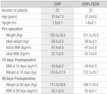
There was no significant statistical difference for any of the variables (p<0.05); CRP=Conventional respiratory physical therapy; CRP+TEDS=CRP associated with trans-cutaneous electrical diaphragmatic stimulation, BMI=Body Mass Index.
Table 1. Anthropometric characteristics and age of the patients under study in the CRP and CRP+TEDS groups, pre-operative and 15 and 30 days post-operative.
between these values was considered the thoracic or abdomi-nal mobility value for each one of the three lines17.
To apply TEDS, the Phrenix Dualpex equipment was used with the following parameters: pulse width of 1.2 m, rise time (ramp) of 0.7 second, respiratory frequency of 14 rpm, pulse frequency of 30 Hz, and sufficient intensity to promote a palpable contraction of the diaphragm muscle3,14. Two
pairs of carbon electrodes were used. One pair was placed on the parasternal region, next to the xiphoid process, and the other pair between the sixth and seventh intercostal spaces, bilaterally on the anterior axillary lines18. The
elec-trodes were fixed with micropore tape to the skin, which was previously cleaned with alcohol. The TEDS application time was 30 minutes per session. For the application, the participants were in supine position, with the headboard tilted to 30º, knees semiflexed, feet supported, arms along the body and with the head on a pillow.
CRP consisted of diaphragm respiratory exercises, deep inspiration, interval-based (two and three) inspiratory training and respiratory exercises associated with shoulder lexion and upper limb extension movements. One set of 10 repetitions of each exercise was carried out. Exercises for the prevention of deep vein thrombosis and ambulation were also carried out. he CRP and CRP+TEDS sessions were applied from the irst to the third day postoperative, morning and afternoon, with a total of ive sessions. All evaluated participants spent four days
in the hospital and received physical therapy treatment until hospital discharge.
he software GraphPad InStat for Windows version 3.05 was used for statistical analysis. Initially, the Kolmogorov-Smirnov normality test was used, followed by the Friedman non-parametric test to compare repeated measures within groups, and the Mann-Whitney test for comparisons between groups. he α level considered for all analyses was set at 0.05. he software GraphPad StatMate version 1.01i was used for the sample size calculation. he variable axillary cyrtometry was considered for this calculation as it is a reliable parameter for determining thoracoabdominal mobility. he conidence level was set at 95%, and the power was set at 95% for a total of 44 individuals. he Student t test was used to compare the
anthro-pometric characteristics and age between groups at the three moments of evaluation.
Results
As there was no sample loss, the 44 participants had a mean age of 37.4±8.1 yrs, mean height of 1.6±0.1 m, initial weight of 121.9±16.9 Kg and BMI of 47.4±6.1 Kg/m2. he
char-acteristics which composed the proile of the studied sample were not signiicantly diferent (p<0.05) for the anthropomet-ric variables (weigh, height and BMI), as well as age, when comparing the three moments of evaluation both in the CRP group and the CRP+TEDS group. his indicates a homoge-neous distribution of the groups as described in Table 1. None of the participants had changes in the preoperative pulmo-nary function tests, therefore no presence of restrictive or obstructive pulmonary disease.
Given the absence of any kind of change in pulmonary vol-umes and low, except in the compartments which compose the vital capacity (VC), only the following spirometry vari-ables were analyzed in this study: inspiratory reserve volume (IRV), expiratory reserve volume (ERV) and tidal volume, through inspiratory capacity (IC). In the CRP+TEDS group, there were signiicant increases in the IRV values when com-paring the irst evaluation to the second evaluation (p<0.05) and in the ERV values when comparing the irst evaluation to the third evaluation (p<0.05). In the CRP group, there were no signiicant diferences in the IRV values or the EVR values, as shown in Table 2.
Pre-operative 15 days 30 days
RIV (L) CRP 1.71±0.3 1.73±0.5 1.65±0.4
CRP+TEDS 1.6±0.3 1.92±0.5* 1.76±0.4
REV (L) CRP 0.67±0.3 0.72±0.3 0.82±0.4
CRP+TEDS 0.67±0.3 0.81±0.3 0.88±0.4#
IC (L) CRP 2.4±0.3
# 2.34±0.4 2.21±0.5
CRP+TEDS 2.59±0.5*# 2.32±0.4 2.41±0.4
Table 2. Mean and standard deviation and statistical results for the
spirometry variables: RIV, REV, IC, pre-operative and 15 and 30 days post-operative for the CRP and CRP+TEDS groups.
*significant difference between the first and second evaluations (p<0.05); # significant difference between the first and third evaluations (p<0.05); CRP=Conventional respiratory physical therapy; CRP+TEDS=CRP combined with transcutaneous electrical diaphrag-matic stimulation; RIV=reserve inspiratory volume; REV=reserve expiratory volume; IC=inspiratory capacity.
Cyrtometry
Thoracic-abdominal (cm) Pre 15 days 30 days
CRP
Axillary 8.4±2.1 7.9±2.0 8.8±1.6
Xiphoid 4.9±2.0 5.8 ±1.8 6.2±1.5
Abdominal 1.2±4.9 3.7±1.8 3.9±3.1
CRP+TEDS
Axillary 6.1±1.7 7.5±2.0* 7.8±2.0#
Xiphoid 4.0±1.7 5.1±2.3 6.5±1.6#
Abdominal 0.72±4.1 1.8±3.9 4.0±3.1#
Table 3. Means, standard deviations and statistical results for the
differences in value in axillary, xiphoid and abdominal cyrtometry for the conventional respiratory physical therapy (CRP) and conventional respiratory physical therapy combined with TEDS (CRP + TEDS) groups in the three evaluations.
*significant difference between the first and second evaluations (p<0.05); # significant difference between the first and third evaluations (p<0.05).
groups (Table 2). With regard to thoracoabdominal mobil-ity, the CRP group had no postoperative diferences (p<0.05) in any of the variables after 15 and 30 days. However, in the CRP+TEDS group, the diferences were signiicant between the evaluations in the three lines (axillary, xiphoid and ab-dominal), showing a signiicant increase in thoracoabdomi-nal mobility in the participants after the bariatric surgery as demonstrated in Table 3.
Discussion
According to the literature, obese individuals have de-creased ERV and functional residual capacity (FRC) espe-cially in the vertical position, and tidal volume can decrease according to the occlusion capacity of the airways, causing changes in pulmonary ventilation and perfusion or even areas of pulmonary shunt with subsequent hypoxemia19.
Therefore, the application of TEDS in the participants of this study may have prevented postoperative IRV and ERV reduction because this technique increases diaphragm contraction. This increase, combined with postoperative abdominal decompression due to the loss of excess fat, may have been decisive for such prevention and may have resulted in an increase in some volumes.
Based on the present results, the IRV and ERV increases observed in the participants can be attributed to the respi-ratory physical therapy, especially to the CRP+TEDS treat-ment. Although it was not possible to evaluate the variables in a group without physical therapy intervention, such as a control group, these results can be attributed to the physical therapy intervention because, in most of the literature refer-ences, there is a postoperative reduction in the variables6-8,11.
he changes in volume also indicate a relationship with the thoracic and abdominal mobility changes veriied by cyr-tometry, although it is not an appropriate form of measuring pulmonary volumes20.
Cyrtometry or thoracoabdominal perimetry, which consists of a group of chest and abdominal circumference measurements, has the purpose of evaluating thoracic ex-pandability and can be carried out in a simple and acces-sible way21. This technique is considered a valid measure
for analyzing the dimensions and widths of thoracic and abdominal movements20. Although seldom referred to in
the literature, this measuring technique is widely used in clinical physical therapy practice to evaluate abdominal and thoracic mobility during respiratory movements22. The
results of the cyrtometry for the thoracoabdominal mobility of both groups, 15 and 30 days postoperative, demonstrated
especially the diaphragm and the abdominal wall muscles which were stimulated by TEDS.
With regard to the normality values of cyrtometry or thoracoabdominal mobility, there is no consensus in the lit-erature, especially for morbidly obese individuals. According to Jamami17, in spite of the importance of thoracoabdominal
mobility for good respiratory movements, it is necessary to consider the relative values, i.e. the differences related to each individual’s physical structure. Nevertheless, there was a considerable increase in thoracoabdominal mobil-ity in the participants, especially those who composed the CRP+TEDS group. It is possible that the respiratory physical therapy, especially with the electrical stimulus, contributed to this result.
It is worth noting that abdominal mobility varied the most between the three studied lines. According to Tribastone23, thoracoabdominal mobility varies
accord-ing to the anatomy of the individual’s ribs. Physiologically, however, the lower ribs are more oblique than the upper ribs and, the more oblique they are, the greater the move-ment that the individual can accomplish. In addition to this physiological aspect, which is common in individuals with normal BMI, it should be taken into account that the participants lost weight, and this resulted in a decrease in abdominal fat. According to Sue24, the abdominal content of
obese individuals compresses the thoracic and abdominal areas, restricting chest movement.
Obese patients can also have signiicant changes in venti-latory mechanics. here is a general notion that total respira-tory compliance is reduced due to compromised thoracic and pulmonary function, especially thoracic function given the presence of fat around the ribs and thorax19. Pelosi et al.2
inves-tigated the efects of BMI on ventilatory mechanics (compli-ance and resist(compli-ance) in a group of anesthetized obese patients, and they found that the reduction in respiratory compliance related to BMI increase is caused mainly by the pulmonary component, while thoracic wall compliance was only weakly dependent on BMI and had a minimal contribution to the variation in total pulmonary compliance.
he decrease in pulmonary compliance is also attributed to alveolar collapse. his condition is frequent in morbidly obese individuals and makes the lungs more rigid and more diicult to insulate, promoting an increase in respiratory work25. To promote the same percentage of ventilation of a
healthy individual in obese individuals, more diaphragmatic activity is needed to overcome pulmonary elastance which generates a greater need for blood low to the diaphragm. he muscles of obese individuals do twice as much work as those of non-obese individuals4.
Although the objective was not to evaluate pulmonary or thoracic compliance as there were no adequate instruments for such measurement, the increase in thoracic mobility in the CRP+TEDS group was attributed to the preservation of the respiratory muscles, which may have promoted greater chest mobility as a consequence of less respiratory work due to weight loss. This aspect leads to the conclusion that TEDS has an important role in the mechanical recov-ery of thoracic and abdominal movement after bariatric surgery. Auler Jr, Giannini and Saragiotto26, in their study
with anesthetized morbidly obese patients, showed that the main factor in compliance reduction may be the pul-monary factor, as thoracic compliance is little affected in obese patients compared to normal patients and does not show variations during laparotomy. Besides low ventilatory compliance, the patients studied by those authors showed increased airway resistance, which was determined mainly by the pulmonary factor. However, the authors accepted the claim that intra-abdominal pressure can play an important role in the decrease in compliance and the increase in pul-monary resistance. These findings reinforce the theory of cranial displacement of the diaphragm during anesthesia, reducing FRC, pulmonary compliance and, consequently, total compliance26.
Clearly the literature has so far presented a great discrep-ancy in the subject, and the controversies over it remain. In conscious patients, the investigations using different meth-ods report a decrease in thoracic compliance4,27. In contrast,
Suratt et al.28 compared obese and non-obese conscious
patients and did not find any correlation between BMI and thoracic wall compliance. According to Nguyen and Wolfe29,
the decrease in respiratory compliance in the intraopera-tive period of open bariatric surgeries is due to the rigid mechanical retractors placed in the abdominal wall, while in laparoscopic bariatric surgeries, the reduction in compli-ance is even greater and due to increased intra-abdominal pressure.
1. Enzi G, Baggio B, Vianello A. Respiratory disturbances in visceral obesity. Int J Obesity. 1990;14 Suppl 2:26.
2. Pelosi P, Croci M, Ravagnan I, Tredici S, Pedoto A, Lissoni A, et al. The effects of body mass on lung volumes, respiratory mechanics, and gas exchange during general anesthesia. Anesth Analg. 1998;87(3):654-60.
3. Gibson GJ. Obesity, respiratory function and breathlessness. Thorax. 2000;55 Suppl 1:S41-4.
4. Naimark A, Cherniack RM. Compliance of the respiratory system and its components in health and obesity. J Appl Physiol. 1960;15: 377-82.
5. Wadström C, Muller-Suur R, Backman L. Influence of excessive weight loss on respiratory function. A study of obese patients following gastroplasty. Eur J Surg. 1991;157(5):341-6.
6. Weiner P, Waizman J, Weiner M, Rabner M, Magadle R, Zamir D. Influence of excessive weight loss after gastroplasty for morbid obesity on expiratory muscle performance. Thorax. 1998;53(1):39-42.
7. Eichenberger A, Proietti S, Wicky S, Frascarolo P, Suter M, Sapan DR, et al. Morbid obesity and postoperative pulmonary atelectasis: an underestimated problem. Anesth Analg. 2002;95(6):1788-92.
8. Siafakas NM, Mistrouska I, Bouros D, Georgopoulos D. Surgery and the respiratory muscles. Thorax. 1999;54(5):458-65.
9. Lawrence VA, Cornell JE, Smetana GW, American Collage of Physicians. Strategies to reduce postoperative pulmonary complications after noncardiothoracic surgery: systematic review for the american college of physicians. Ann Intern Med. 2006;144(8):596-608.
10. Chuter TA, Weissman C, Mathews DM, Starker PM. Diaphragmatic breathing maneuvers movement of the diaphragm after cholecystectomy. Chest. 1990;97(5):1110-4.
11. Joris JL, Hinque VL, Laurent PE, Desaive CJ, Lamy ML. Pulmonary function and pain after gastroplasty performed via laparotomy or laparoscopy in morbidly obese patients. Br J Anaesth. 1998;80(3):283-8.
12. Nguyen NT, Goldeman C, Rosenquist CJ, Arango A, Cole CJ, Lee SJ, et al. Laparoscopic versus open gastric bypass: a randomized study of outcomes, quality of life, and costs. Ann Surg. 2001;234(3):279-89.
13. Geddes LA, Voorhees WD, Lagler R, Riscili C, Foster K, Bourland JD. Electrically produced artificial ventilation. Med Instrum. 1988;22(5): 263-71.
14. Forti EMP, Pachani GP, Montebelo MIL, Costa D. Eletroestimulação diafragmática transcutânea em indivíduos saudáveis. Fisioter Bras. 2005;6(4):261-4.
15. Miller MR, Hankinson J, Brusasco V. Burgos F, Casaburi R, Coates A, et al. Series ATS\ERS task torce: standardisation of lung function testing. standardisation of spirometry. Eur Respir J. 2005;26(2):319-38.
16. Pereira CAC. Diretrizes para Testes de Função Pulmonar. J Pneumol. 2002;28 Suppl 3:S1-82.
17. Jamami M, Pires VA, Oishi J, Costa D. Efeitos da intervenção fisioterápica na reabilitação pulmonar de pacientes com doença pulmonar obstrutiva crônica (DPOC). Rev Fisioter Univ São Paulo. 1999;6(2):140-53.
18. Geddes LA, Voorhees WD, Boulland JD, Riscili CE. Optimum stimulus frequency for contracting the inspiratory muscle with chest-surface electrodes to produce artificial respiration. Rev Ann Biomed Eng. 1990;18(1):103-8.
19. Luce JM. Respiratory complications of obesity. Chest. 1980;78(4):626-31.
20. Caldeira VS, Starling CCD, Britto RR, Martins JA, Sampaio RF, Parreira VF. Precisão e acurácia da cirtometria em adultos saudáveis. J Bras Pneumol. 2007;33(5):519-26.
21. Maciel SS, Paulo MQ, Souza CO, Silva LG, Tavares RR. Efeito broncodilatador do acanthospermum hispidum DC, nos doentes pulmonares obstrutivos crônicos (DPOC). Rev Bras Cienc Saúde. 1997;1(1/3):23-30.
22. Costa D, Sampaio LMM, Lorenzzo VAP, Jamami M, Damaso AR. Avaliação da força muscular respiratória e amplitudes torácicas e abdominais após a RFR em indivíduos obesos. Rev Latinoam Enferm. 2003;11(2):156-60.
23. Tribastone F. Tratado de exercícios corretivos aplicados à reeducação motora postural. São Paulo: Manole; 2001.
24. Sue DY. Obesity and pulmonary function: more or less? Chest. 1997;111(4):844-5.
Conclusions
Based on the present indings, it can be concluded that the obese women who underwent bariatric surgery and received postoperative respiratory physical therapy did not have a reduc-tion in pulmonary volumes. here were signiicant changes in the
VC compartments and in the dynamics of the respiratory move-ments. hese results may be linked to the natural decompression of the thorax and abdomen due to fat tissue loss after the bariatric surgery, as well as the improved dynamics of the respiratory mus-cles, especially the diaphragm, due to the conventional respiratory physical therapy and particularly to the TEDS.
25. Charlebois D, Wilmoth D. Critical care of patients with obesity. Critical Care Nurse 2004;24(4):19-27.
26. Auler Jr JOC, Giannini CG, Saragiotto DF. Desafios no manuseio peri-operatório de pacientes obesos mórbidos: como prevenir complicações. Rev Bras Anestesiol. 2003;53(2):227-36.
27. Sharp JT, Henry JP, Sweany SK, Meadows WR, Pietras RJ. The total
work of breathing in normal and obese men. J Clin Invest. 1964;43: 728-39.
28. Surat PM, Wilhoit SC, Hsiao HS, Atkinson RL, Rochester DF. Compliance of chest wall in obese subjects. J Appl Physiol. 1984;57(2):403-7.