Fonoaudiologia Baseada
em Evidências
Tainá Soares Ferreira1
Laura Davison Mangilli2
Fernanda Chiarion Sassi3
Talita Fortunato-Tavares2
Suelly Cecília Olivan Limongi4
Claudia Regina Furquim de Andrade4
Descritores
Terapia por exercício Terapêutica Fisiologia Músculo esquelético Revisão Keywords
Exercise therapy Therapeutics Physiology Muscle, skeletal Review
Correspondence address:
Claudia Regina Furquim de Andrade R. Cipotânea, 51, Cidade Universitária, São Paulo (SP), CEP: 05360-160. E-mail: [email protected]
Received: 3/18/2011
Accepted: 6/1/2011
Study conducted at the Department of Physical Therapy, Speech-Language Pathology and Audiology, and Occupational Therapy, School of Medicine, Universidade de São Paulo – USP – São Paulo (SP), Brazil. (1) Enhancement Course in Hospital Speech-Language Pathology and Audiology in Orofacial Functions of the General Hospital, School of Medicine,Universidade de São Paulo – USP – São Paulo (SP), Brazil.
(2) Graduate Program (Doctorate degree) in Rehabilitation Sciences, Department of Physical Therapy, Speech-Language Pathology and Audiology, and Occupational Therapy, School of Medicine, Universidade de São Paulo – USP – São Paulo (SP), Brazil.
(3) Speech-Language Pathology and Audiology Sector of the Central Institute of the General Hospital, School of Medicine, Universidade de São Paulo – USP – São Paulo (SP), Brazil.
(4) Department of Physical Therapy, Speech-Language Pathology and Audiology, and Occupational Therapy, School of Medicine, Universidade de São Paulo – USP – São Paulo (SP), Brazil.
review of the literature
Fisiologia do exercício fonoaudiológico: uma revisão crítica
da literatura
ABSTRACT
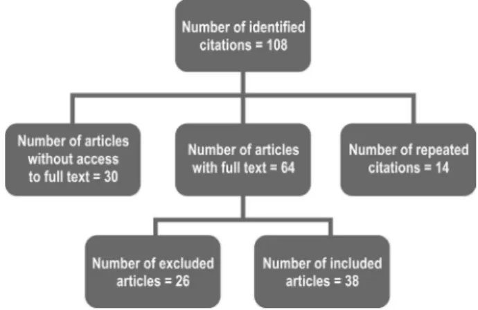
Purpose: To analyze the scientiic literature about the physiology and effects of exercises used in the treat-ment of oral myofunctional disorders. Research strategy: The methodology used followed the concepts of the Cochrane Handbook and involved question formulation related to the topic of investigation, identiication and selection of the studies, and a critical evaluation of the selected articles. Selection criteria: Papers were selected on PubMed database using the following keyword combinations: “physiology exercise AND speech, language and hearing science”, “exercise physiology AND speech therapy”, “exercise physiology AND myo-functional therapy”, and “physiology exercise AND swallowing therapy”. Only papers written in English and published between the years of 2000 and 2010 were included in the analysis. Data analysis: Manuscripts were analyzed according to their objectives, research design, participants, inclusion of a control group, assessment criteria, therapeutic proposal, results and existence about physiology of the chosen exercises. Results: One hundred and eightstudies were identiied, out of which 38 had access to the full text and were directly related to the investigated topic. The articles were classiied as clinical trials and experimental research, case studies, literature reviews and theoretical articles, letters to the editor and critical analyses. Conclusion: This review concluded that there is a lack of knowledge about the effects of the myofunctional exercises used by clinicians. Also there is a lack of scientiic evidence to determine the frequency at which they should be performed. Generally, the articles investigate the eficacy of treatment programs without inquiring whether the included exercises are individually effective.
RESUMO
INTRODUCTION
The Speech-Language and Hearing Science is the ield of knowledge that deals with the rehabilitation of deicits involving the oral/facial myofunctional system, speech and swallowing, including the temporomandibular articulation, obstructive sleep apnea, facial paralysis, dysphagia, hypernasality, speech articulation disorders, among others. In order to do this, the-rapists generally use exercises involving muscles of the lips, cheeks, tongue, soft palate, pharynx and larynx. Exercises, however, are chosen based on a reduced number of researches and case studies, in non-replicable results or are based on the clinician’s intuition(1). Recently, heightened discussions about
the relationship between myofunctional exercises and principals of neuroplasticity, muscle adaptation and overall principles of exercise training have raised more questions than answers(1).
When using training exercises it is important to know that modiications in strength are generally the result of modiications in how the nervous system activates the muscle rather than a structural alteration in the muscle itself. Improved performance may be the result of an increased number of motor units recruited or improved speed and coordination of motor unit recruitment. Studies indicate cortical reorganization during the motor reha-bilitation process(1). This fact gives support to the idea that the
neuromotor system can suffer modiications due to experiences and that restructuring can either occur at a central or peripheral level. Also, these studies offer a neuromotor organization model that can be used to investigate the best training intensity and endu-rance to maximize central and peripheral adaptation. It is already known that in order to promote adaptations in the neuromotor system, exercises need to recruit more of the neuromuscular system than ordinary activities. For this reason, it is important to determine the adequate manner to perform oral motor exercises in order to achieve treatment goals(1).
PURPOSE
The aim of this study was to conduct a systematic review of studies in the ield of the Speech-Language and Hearing Sciences related to the physiology and the effects of exerci-ses used in the treatment of oral/facial motricity, speech and swallowing alterations.
RESEARCH STRATEGY
We followed the precepts of the Cochrane Handbook for the establishment of the research method(2):
1. Formulation of the research question: analysis of texts about the physiology of oral motor exercises used in the treatment of oral facial motricity, speech and swallowing disorders; 2. Location and selection of studies: research of studies
pu-blished on the topic;
3. Critical analyses of the studies: studies were analyzed in terms of their purpose, research design, studied population, existence of control groups, assessment criteria, therapeu-tic proposal, overall results, discussion about the muscle physiology of the exercises.
SELECTION CRITERIA
The manuscripts were selected through a PubMed database using the keywords “physiology exercise AND speech, langua-ge and hearing science”, “exercise physiology AND speech therapy”, “exercise physiology AND myofunctional therapy”, “physiology exercise AND swallowing therapy”. Only articles published in English and between the years of 2000 and 2010 were included for the analyses.
The search in the database was performed independently by the two authors in order to minimize possible losses of cita-tions. Each citation retrieved in the database was independently analyzed by the two authors, who judged the relevance of its inclusion or exclusion. We excluded citations in languages other than English. Articles that did not allow access to full text (obtained in the CAPES Journal Portal), or which were repeated by the overlapping keywords were also excluded.
Full texts that were not directly related to the theme of the review were excluded from the analyses. All phases of the study were independently conducted by each of the authors. When disagreement occurred, we only included the texts for which the inal position was consensual. All of the studies related to the theme of the review were included in the analyses inde-pendently of their design.
Figure 1 displays the route for the inal selection of the analyzed articles.
DATA ANALYSIS
The following indicators were considered for analysis of the 38 selected articles: type of study (clinical trials and experimen-tal research, case studies, review of the literature, letters to the editor); purpose, number of therapeutic sessions; description of exercises and their physiology, main results.
RESULTS
Clinical trials and experimental research
A study was conducted(3) with the purpose of verifying
poetry) on heart rate, during the irst 15 minutes following exercise. Seven healthy individuals, mean age of 44 years, underwent 15 one hour weekly sessions that involved speech or control exercises (walk slowly around the room maintaining a conversation with the therapist). Measurements of the heart rate were taken 15 minutes prior and post therapy. The results indicate that the proposed therapy promotes changes in the dynamics of heart rate and in the rhythm of heart beat that are different from those observed for the control exercises. Also, it was observed that these changes are maintained for at least 15 minutes post exercises. The breathing rate also increased after speech exercises and decreased after the control exercises. Rhythmic speech with poetry instantly modulates the heart rate through breathing. At the end of the text, the author discusses the limitations of the study and concludes that it is not pos-sible to differentiate if the obtained results were caused by the artistic speech form our by the coordination between breathing and speaking.
Other authors(4) developed a study in order to compare the
eficacy of the McNeill Dysphagia Therapy Program (a sys-tematic program based on exercises to rehabilitate swallowing) to the eficacy of a traditional therapy program for swallow-ing, involving biofeedback with surface electromyography (sEMG). The research group was composed by eight patients and the control group by 16 individuals (all participants pre-sented ages below 90 years). The proposed program focuses on the strengthening and coordination of swallowing during functional activities and in the development of movements and reinement of coordination of the muscle components involved in swallowing. The program uses the effortful swallowing maneuver during hierarchical feeding tasks. As the program advances, demand increases – i.e. individuals are requested to use more strength and to increase movement rate. Results of the study indicate that 69% of the patients improved. Both therapy programs improved the swallowing function. However, the McNeill Dysphagia Therapy Program presented better results when considering the removal of alternate feeding and laryngotracheal aspiration.
In another study, researchers(5) investigated the adhesion of
healthy elderly individuals to the Shaker exercise, the number of days to reach isometric and isokinetic goals, frequency and reasons to give up, as well as complaints associated to the given exercises. Twenty six elderly individuals without the diagnosis of dysphagia (ages between 66 and 93 years) performed the Shaker exercise three times a day, during six weeks, and completed a questionnaire about their performance and possible dificulties. Four individuals were submitted to a videodeglutogram pre and post therapeutic intervention. For these individuals, an increase in the laryngeal excursions and in the opening of the upper esophageal sphincter was observed. The time needed to reach the purposes of the exercise varied among the participants; the isokinetic goal was the easiest to accomplish. The results indicate that the Shaker exercise was effective regarding the proposed objectives. The authors, how-ever, suggest that when treating dysphagia it might be necessary to include this exercise in a structured progressive therapeutic program in order to reach safe and eficient swallowing.
Another author(6) used the Shaker exercise three times a
day, during six weeks, with 11 healthy women and ten healthy men (mean age 70 years), who had not been diagnosed with dysphagia or dyphonia, and compared them to a control group of ive individuals. The purpose of the study was to determine if the exercise has an effect on voice and swallowing related to age. Biomechanical measurements of voice and swallow-ing improved in 10 out of the 21 participants. Changes were not observed for participants in the control group. The author concludes that a randomized controlled study, with a larger number of individuals, including periodic monitoring of the heath state and accuracy when performing the exercise is nec-essary to determine the effect of the Shaker exercise on voice and swallowing. The author points that this exercise could be recommended, as a preventive measure, to decrease the ef-fects of sarcopenia on the muscles involved in swallowing and phonation, and could delay the progression of presbyphonia and presbyphagia.
A few authors(7) used sEMG to analyze the masseter muscle
with the purpose of testing an exercise protocol for mastica-tion (to increase muscle strength, improve muscle funcmastica-tion and to decrease pain at rest). Participants of the study were 20 patients, with ages ranging from 30 to 45 years, with the diagnosis of myofascial pain and with a slight increase in the masseter muscle volume during maximal dental clenching, among other criteria. Half of the group was submitted to the protocol that involved the mastication of chewing gums three times, progressively from 10 to 30 minutes, during eight weeks. The results of the sEMG indicate that the exercise produced objective physiological results, with a signiicant increase of the electric activity of the masseter muscle during maximal dental clenching. Also, it was observed a decrease in pain at rest and during testing.
A research was developed(8) with the purpose of testing
the effects of an exercise to improve tongue pressure against the palate during swallowing and of a head maneuver (tilting the head backwards) in patients with presbyphagia. In order to control the results, sEMG was performed on the submental muscles. Participants of the study were 53 healthy volunteers (mean age 35.3 years). Differences in sEMG results were not observed between the tongue isometric (movement repetition) exercises and the head maneuver. However, when considering the tongue isotonic (strength endurance) exercises and the head maneuver, the latter produced better results. Overall, the results suggested that the tongue exercise was more effective for the submental muscles and that the beneits reached could improve the swallowing function.
A different study(9) veriied the impact of oropharyngeal
of sleep quality and complete polysomnography. The authors conclude that the oropharyngeal exercises signiicantly reduced the severity and symptoms of sleep apnea.
A retrospective study(10) with post stroke dysphagic patients
investigated if training using an oral acrylic plate positioned between the teeth and lips could improve labial strength and the swallowing of these individuals. Thirty patients (mean age of 70 years) were investigated using a labial strength measur-ing device (Lip Force Meter – LF100). This device veriies the ability of the lips to support pressure exerted by an oral plate. A swallowing test was also performed pre and post training (training was done three times a day, using the oral plate, for at least ive weeks). The results indicate that training with the oral plate improves labial strength and the swallowing function of the tested patients. The authors conclude that the results obtained with the treatment are probably related to the sensory motor stimulation and to the plasticity of the central nervous system that was stimulated through the exercises and not simply by the increase in strength of the lip muscles.
Other authors(11) published a study that had as a purpose to
compare two equipments that measure tongue pressure - the MOST(Madison Oral Strengthening Therapeutic)and theIOPI (Iowa Oral Performance Instrument) that is already commer-cialized. The authors also tried to identify what would be the adequate tongue pressure when performing isometric exercises with both equipments. Participants were 30 healthy individuals (with ages between 19 and 71 years), who were divided in two groups: one group performed the exercises with the IOPI and the other group with the MOST (participants were instructed to press the tongue against the palate as hard as possible for three seconds, three times). The maximal tongue pressure was very similar with both equipments. The authors conclude that the MOST, not yet available for commercialization, was well adjusted to the mouth in a replicable way, besides presenting a simple interface to the user.
In a multicenter study, a group of researchers(12) developed
a research to determine if the hypernasality of individuals born with a cleft palate could be reduced through velopharyngeal exercises performed with a continues positive airway pressure (CPAP). Participants were 43 subjects, with ages ranging from 3 to 24 years. Participants performed exercises that involved the production of selected words and sentences (considering consonants) using the mask for a period of eight weeks. Dura-tion of the sessions as well as the pressure exerted by the CPAP increased gradually, taking into consideration the principles of progressive resistance training. The results indicate that patients presented a reduction in hypernasality after intervention, but the results varied among the patients and the centers involved in the study. The use of the CPAP during oral training seems to be capable of substantially reducing speech hypernasality in a few subjects with cleft palateA multicenter clinical trial(13) was
developed with the purpose of comparing the Shaker exercise with traditional dysphagia therapy in order to determine the better strategy to reduce laringotracheal aspiration and to im-prove swallowing. Seven institutions took part in the research. Participants were 19 patients with a three months history of aspiration and oropharyngeal dysphagia involving the upper
esophageal opening. Traditional treatment involved a series of exercises that includes the super supraglotic swallowing, the Mendelsohn maneuver and exercises for the base of tongue performed during ive minutes, ten times a day, for six weeks. The results indicate that traditional therapy and the Shaker exercise have very different effects and that both approaches promote signiicant changes in swallowing. The Shaker exercise signiicantly decreased post swallowing aspiration. Traditional therapy, however, also resulted in the improvement of several swallowing parameters, especially regarding the swallowing of pasty food.
In another study, researchers(14) investigated the effect of the
Shaker exercise in the shortening of the thyrohyoid muscle of 11 dysphagic patients, with a dysfunction of the upper esopha-geal sphincter. Six patients performed traditional swallowing therapy (including laryngeal elevation and tongue exercises, the Mendelsohn and super supraglotic maneuvers, and effortful swallowing) and ive patients performed the Shaker exercise. The shortening of the thyrohyoid muscle was measured through videoluoroscopy pre and after six weeks of exercise (exercises were performed twice a week for 45 minutes). After therapy, changes in the thyrohyoid muscle were signiicantly greater for patients in the research group. The conclusion of the research is that the Shaker exercise improves the shortening of the thyro-hyoid muscle besides strengthening the suprathyro-hyoid muscles, as already observed in other studies. Both effects contribute for the improvement in the opening of the upper esophageal sphincter. Researchers(15) also investigated the effects of an exercise
program (15 minutes, twice a day, for 18 weeks) on the reduced oral opening of a group of 35 patients (mean age of 61 years). Patients presented systemic scleroderma and severe microsto-mia. The results indicate that maximal oral opening increased signiicantly in all subjects. Besides that, patients indicated that activities such as eating, speaking and brushing the teeth were more easily done.
Another study(16) investigated the effects of an eight week
therapy program involving progressive tongue resistance exercises during swallowing. Participants were ten healthy elderly individuals (mean age 80 years). Subjects improved isometric maximal tongue pressure and tongue pressure dur-ing swallowdur-ing, even though they did not perform exercises directly involved with this function. Magnetic resonance was performed in four of the participants and indicated that there was an increase in tongue volume.
In another study(17), the same group of researchers
transit time and increase of the swallowing pharyngeal phase. The penetration-aspiration index also decreased signiicantly, making swallowing safer. The conclusion of the research was that tongue isometric exercises increase tongue strength asso-ciated to the improvement of swallowing pressures and in the protection of the inferior airway.
Other authors(18) evaluated the effect of the Shaker exercise
in the swallowing rehabilitation of patients with dysphagia, caused by the abnormal opening of the upper esophageal sphincter (patients presented post swallowing food residue and aspiration, needing to be fed with an alternative method). Participants of the research were 11 patients (mean age of 72 years). Videoluoroscopy and functional assessment of swallowing was performed pre and post the execution of the Shaker exercise (exercise was practiced during six weeks). Seven randomly selected patients performed control exercises (15 repetitions of passive tongue lateralization, three times a day, during six weeks) before being submitted to the tested exercise program. Changes were not observed after the per-formance of the control exercises. However, after the Shaker, exercise all of the patients presented a signiicant improvement in the opening of the upper esophageal sphincter and in the laryngeal excursions. Also it was observed absence of post swallowing aspiration, making it possible to remove alternate feeding. The authors conclude that the exercise proposed for the strengthening of the suprahyoid muscles was effective for the investigated patients.
A study(19) was developed in order to investigate if muscle
warming up decreases muscle rigidity during speech production or if it has adverse effects due to fatigue and exhaustion caused by the intensive speech activity. Participants were 30 subjects (mean age of 40 years) with myotonic dystrophy (onset during adulthood), and ten control subjects (mean age of 34 years). For the patients with myotonic dystrophy, muscle warming up increased speech rate and decreased variability. No signs of fatigue or exhaustion due to the prolonged and intensive use of the muscles were observed. Signiicant changes were not observed for the control group. After muscle warming up, the research group achieved normal speech rate during reading and speaking, as observed for the control group. The study suggests that the speech production of individuals with myo-tonic dystrophy improves with muscle activity and that muscle rigidity during speech can be reduced with muscle warming up (i.e. with the execution of repetitive movements). The authors conclude that muscle warming up is a valid intervention since it promotes the increase of speech rate and of speech luency, without aggravating the signs of laccid dysarthria, commonly observed in patients with myotonic dystrophy.
A prospective study(20) was developed with the purpose
of investigating the presence of signs and symptoms of tem-poromandibular disorder (TMD) in individuals with tinnitus. The study also evaluated the long term effect of treating TMD on tinnitus. Seventy-three patients with TMD, focal pain and tinnitus were submitted to treatment. Fifty other patients who awaited treatment composed the control group. According to the diagnosis and clinical indings, individual therapeutic plans were elaborated involving mandible isotonic and isometric
exercises. Subjects were asked to ill in a questionnaire two years after treatment. Results indicate that for the research group 43% of the patients presented improvement of their tin-nitus, 39% did not refer changes and 17% reported worsening of the tinnitus when compared to the beginning of treatment. The authors of the research state that signs and symptoms of TMD are common in patients with tinnitus and that the treat-ment of TMD has a positive effect over tinnitus in the long run, especially for patients with luctuating tinnitus.
Case studies
A few authors(21) reported an uncommon case of late
neuro-logic recovery. The case study involves a 26-year-old woman with a deiciency in the production of the growth hormone caused by a brain surgery (bulbar astrocytoma) performed at 11 years of age. This surgery caused signiicant neurologic déicits, including paralysis of the oropharyngeal structures and of the vocal folds, absence of primary esophageal peristalsis) secondary to iatrogenic paralysis of the cranial nerves IX, X and XII. Most of the sequels that were identiied during assess-ment were minimized after eight months of associated drug therapy (growth hormone), physiotherapy and speech-language therapy. Authors observed an increase in the size of the tongue and in its mobility, improvement in the quality and quantity of saliva, improvement in phonation, recovery of the esophageal peristalsis, and recovery from sleep apnea.
Other authors(22) described the therapeutic development of
a 49-year-old man with temporomandibular disorder and oral facial myofunctional deicits. Therapy involved oral myofunc-tional therapy and the use of an occlusion plate. Myofuncmyofunc-tional therapy began 60 days after using the occlusion plate and involved 50 minutes sessions, every ifteen days. Therapy ses-sions included counseling about the disorder, instructions about how to perform the exercises at home and counseling about how to avoid dislocation. The authors described all exercises and the frequency at which they should be performed. The authors conclude that the combination between oral myofunctional therapy and the occlusion plate can beneit patients with hy-permobility temporomandibular disorder.
In another case study(23) an intraoral device was developed
to correct abnormal neuromuscular patterns and to improve the facial aesthetic of individuals with facial paralysis. The participant of the study was an 18-year-old woman with a two year and eight months facial paralysis (facial trauma and brain edema). sEMG was used during the performance of exercises for the coordination between the action of the device and the smiling movement on the affected side of the face. Exercises were performed for 20 minutes, four times a day. The results of the study indicated that the device improved facial symmetry at rest, due to the contralateral muscle traction, and improved the positioning of the labial iltrum.
The literature also describes a case study of a 54-year-old man, who was admitted to the hospital due to subarachnoid hemorrhage and brainstem stroke(24). Although scintigraphy
cough. During the swallowing training, the patient was guided to hold his breath for two seconds, to contract his abdominal muscles and to cough with his throat contracted. Initially, two series of three voluntary coughs were performed twice a day and latter, when the patient presented good laryngeal elevation and absence of fatigue, training was increased to three times a day. After the reintroduction of an oral diet, the patient contin-ued to perform training at all meal times and was instructed to maintain training until hospital discharge. Although the patient still presented a few events of penetration or laryngotracheal aspiration, it was possible to avoid pulmonary infection. The authors conclude that the use of voluntary cough seems to be effective in avoiding aspiration pneumonia. However, this exercise is not directly related to the improvement of the swal-lowing function. The scintigraphy of swalswal-lowing is a useful instrument to evaluate airway clearing and to determine if the reintroduction of an oral diet is possible.
A group of authors(25) described the case of a 28-year-old
man who was injured by a ire weapon. The bullet fractured the left coronoid process and the pyterygoid processes bilater-ally, resulting in a mouth opening restriction and interfering in mastication. Clinical assessment of the extra and intraoral aspects was performed, as well as computerized tomography that identiied a parotid lesion. The patient began oral motor therapy ten days after the accident. Three one-hour weekly sessions were performed during a month. The patient was also guided to perform the exercises at home and after the therapy program had ended to continue with the same exercises for two other months. The therapy protocol involved the relaxation of the masticatory muscles using a hot stimulus and manual mas-sage to increase blood low, eliminating metabolic residues; mandibular exercises for mouth opening with the tongue against the palate and using a lever of spatulas; manual traction to avoid mandibular deviation to the right and to promote lateralization to the left; mandibular protrusion movements with the purpose of relaxing antagonist muscles and to coordinate oral facial movements. The therapeutic procedure also involved functional therapy to rehabilitate chewing. In this case, the patient was guided to chew on the contralateral side to the fracture. After nine months of treatment, the patient’s maximal oral opening increased from 28 mm to 40 mm; mandibular deviation during mouth opening and mandibular protrusion was not observed. Also, a limited lateralization movement to the left was observed. The authors concluded that the adopted conservative treatment was successful.
Other authors(26) performed a study involving six individuals
(three with mean age of 26 years and three with mean age of 66 years) in order to verify if the Doppler ultrasound could be used to identify changes in the blood low of the tongue during selected articulatory gestures (emission of the phonemes /t/ and /k/). The study also tried to verify if movements produced with greater strength increased the blood low of the tongue. The results indicated an increase in the blood low of the tongue after the articulatory gestures. However, no difference was observed between the tested phonemes or between the differences in muscle strength. Younger individuals presented a higher blood low when compared to the older ones.
A group of authors(27) described the case of three
50-year-old men, who were diagnosed with neurologic dysphagia and who were submitted to a experimental therapy protocol. The therapeutic program involved two to three weekly 45 minute sessions using isometric tongue exercises (ten series of pressing the tongue against the palate per session) and accuracy tasks for pressing the tongue against the palate using biofeedback equipment. The patients only performed the exercises during therapy sessions. After treatment, an increase in the isometric strength of the tongue was observed, as well as improvement in accuracy to generate tongue pressure, improvement in food bolus control during videoluoroscopy and functional improvement in oral intake. The proposed training program was effective for all three patients, improving functional and instrumental aspects of swallowing.
Reviews of the literature and theoretical texts
One of the consulted reviews of the literature(1) discusses the
physiological impact of neuromuscular treatments on deicits commonly related to dysarthria and dysphagia. The authors of this review also describe muscle physiology, relevant pa-rameters used to determine the exercises (strenght/endurance, rate, duration, dynamics, frequency and progression) that will be used and discuss the differences between passive and active exercises. The uses of thermal, tactile and electric stimuli are largely discussed. The authors conclude that the physiological precepts are important parameters that should be considered when developing a muscle rehabilitation program.
A group of authors(28) presented a review about swallowing,
describing its physiology, discussing the impacts of neurologic disorders in swallowing and presenting existing assessment methods and therapeutic proposals. The authors conclude that dysphagia is a frequent neurologic problem in the population. For this reason, appropriate treatment should include clinical indings, individual results of patients when using compensa-tory maneuvers and should also consider which deicits can be rehabilitated taking into consideration the presented neurologic disease and its natural progression.
In another review of the literature, two authors(29) discuss
the impact of aging in swallowing, the physiology of normal swallowing and the neural control involved in this function. The authors also present the existing intervention proposals. Considering therapy, the authors of the review point that more researches are necessary in order to determine the eficacy of combining exercise and diet modiication. Although a few stud-ies have already indicated that the use of swallowing maneuvers are more effective in rehabilitation than diet modiication alone, especially when considering elderly individuals with dysphagia, these maneuvers seem to offer little resource to patients for the functional rehabilitation of swallowing. Motor exercises that aim to increase muscle strength of the tongue and to increase oropharyngeal movements seem to be more effective to improve swallowing in healthy and dysphagic elderly individuals.
Another author(30) presented a review of the literature with
to comment about individual variability regarding time and type of swallowing, physiological modiications related to different food consistencies, voluntary control, swallowing disorders, deicit assessment, treatment and multidisciplinary approach. In this manuscript, the author describes the existing therapy approaches without making any criticism and without presenting any data about the duration tof treatment programs.
In another manuscript, the same author(31) makes a critical
analyses about therapists who assess patients only at predeter-mined time intervals to follow-up dysphagia and to determine the adequate moment to reintroduce or to suspend oral diet. The author also describes the existing treatments to rehabilitate swallowing. According to him, current treatments include active exercises and compensatory strategies, surgical procedures, drugs and oral prosthesis that were developed to make swal-lowing more secure and eficient. Two treatment possibilities exist: compensations that allow the patient to eat a few food consistencies without presenting aspiration; and exercise that improve muscle strength and coordination during swallowing, so that patients do not need to use compensations and can have an oral diet completely. In this review, the author approaches all treatment options and presents available scientiic evidence about the eficacy of each one of them.
Other authors(32) present a review in which they discuss
the impact of aging on swallowing and also discuss possible intervention strategies to be adopted in these cases. The review differentiates presbyphagia and dysphagia, it discusses the ef-fects of comorbidity in swallowing, it presents issues related to nutrition and hydration, it describes possible alternate feeding methods and therapeutic interventions (including maneuvers, modiication of food consistencies and exercise). The authors did not present data regarding the duration of treatments.
An author(33) proposed a discussion about head and neck
cancer treatments and their negative impact on swallowing. A long the review, several treatment procedures are described, as well as their eficacy for post treatment head and neck cancer patients. The author also describes the possible physiological changes caused by maneuvers and exercises commonly used during the swallowing rehabilitation process of these patients. Other authors(34) describe the effects of brain stroke on the
neural control of swallowing. The authors mention the impacts of dysphagia, the assessment methods, the mechanisms of recovery in post stroke dysphagic patients, the use of compen-satory strategies and rehabilitation programs.
Finally, a review(35) discussed the use of neuroprothesis
as a treatment for dysphagia. The review details the swal-lowing process, as well as the muscles and nerves involved, the effects of dysphagia and other treatment options besides the neuroprothesis. The neuroprotheis interferes in laryngeal elevation, stimulating the muscles related to this movement (especially the geniohyoid, mylohyoid and thyrohyoid), that are supericial and therefore susceptible to minimally invasive stimulation – i.e. through supericial and intramuscle electrodes. The author points that it already has been demonstrate, through clinical trials, that the neuroprotheis potentially reduces bron-choaspiration.
Letters to the editor and critical reviews
In a letter to the editor, an author(36) comments about the
indings of a group of researchers on an exercise program for swallowing disorders. The author also makes comments about other groups of researchers: a group who developed and studied the Shaker exercise; a group who studied resistance exercises to improve tongue strength using an instrument to measure the pressure of the tongue against the palate – the IOPI; and a third group who studied tongue strength and mobility post supraglotic laryngectomy.
Other two authors(37) summarized and criticized eight
articles on dysphagia published between 2001 and 2002. The researches that were better evaluated were: a) the proposal of a rehabilitation program for patients with upper esophageal sincter disfunction using the Shaker exercise(18); b) the
inves-tigation of oropharyngeal electro stimulation in rats to trigger swallowing(38). The study concluded that the palatine pillars, the
posterior pharyngeal wall and the soft palate, when stimulated demonstrated to trigger swallowing. The study also concluded that when the glossopharyngeal nerve is injured, the swallowing relex becomes absent when stimulating the above areas, i.e. the pharyngeal portion if primordial to elicit the swallowing relex; c) a longitudinal investigation related to the quality of life of patients with oral cancer who were treated surgically(39).
The study discusses the functional implications of treatments based on the patient’s perspective.
The literature(40) also presents a critical review of a research(9)
that investigated the effects of oral motor exercises in patients with moderate obstructive sleep apnea. The author of the criti-. The author of the criti-The author of the criti-cal review discusses each one of the exercises proposed in the therapeutic program, pointing that it is important to determine if the purpose of an exercise is to strengthen the muscles, improve resistance or to improve movement rate and endurance. Accord-ing to the author, only when the purpose of each exercise is clear, it is possible to determine exercise load, intensity, frequency and endurance. Based on these considerations, the use of a few exercises proposed in the original article (especially those related to the lips and cheeks) are not justiied. Besides that, the author also points that a few of the used exercises do not have enough evidence about their effects. In the author’s opinion, only two tongue exercises were responsible for the changes observed in the original research. The author concludes that there is a lack of researches that conirm if myofunctional exercises can produce changes in the upper airways and avoid their collapse during sleep, that investigate exercise variables and that conirm if there is a real need to perform them continuosly to maintain results.
Three authors(41) published the records of their lectures and
point the lack of literature about the effectiveness of singing and blow instruments on the occurrence of cough. The authors conclude that there is a lack of evidence about the physiology related to the onset of couth, its modulation, intensiication or excitement, and if the onset of cough occurs at a peripheral level (airways relex), cortical level or both. Also it is not clear if the same results would be obtained for provoked and spontaneous cough. The authors point that exercise, speech and music can modify the sensibility characteristics of cough and that these should be explored in future studies.
CONCLUSION
Overall, experimental studies describe the exercises used in therapeutic programs; they describe how many times a week the program was performed and for how long; however the number of repetitions of each exercise is not stated. Besides that, only a few of the studies describe the purpose of the exercises that were used. It was observed that the authors do not describe which muscles are activated during exercise practice, the physiology of this activation and its relationship with the physiological goals. It is important to highlight that most of the studies are related to dysphagia. The purpose of the present review was to verify, within a pre established period, all of the articles related to exercise physiology. Given this, articles were categorized by variables such as pathology, association with objective exams, type of exercise that was used, etc.
Regarding the design of the consulted articles, it is possible to conclude that there is deinitely a lack of clinical trials in the Field of the Speech-Language and Hearing Science, and that the existing studies involve a very limited number of partici-pants. Almost half of the case studies describe single cases of patients with oral motor alterations that present improvement after therapy. Most of these studies focused mainly on the modiications observed in pre and post treatment assessments and do not describe the applied therapy techniques.
The other analyzed studies have experimental designs that correspond to a pilot study. These articles focused mainly on the description of a therapeutic proposal, indicating which exercises were used and the frequency at which they were performed. A few also present discussions about the muscle physiology that would justify the results that were obtained.
Theoretical texts that discuss the concepts related to dys-phagia, although with different approaches, were also found. Most of these texts explain the physiology of normal/healthy swallowing and then discusses more deeply some type of pa-thology that has dysphagia as a symptom. All of them discuss the existing therapy methods, making reference to maneuvers and rehabilitation exercises.
Regarding the purposes of the present review, only one of the consulted articles reached expectations, discussing in details muscle physiology, varibles that should be taken into consideration when chosing exercises (strength/endurance, rate, duration, dynamics, frequency and progression), and expected physiological responses when executing passive and active exercises, thermal stimulation and when receiving tactile and electric stimulus.
This review indicates that the knowledge about the effects of muscle exercises used by clinicians is insuficient and that there is a lack of scientiic evidence to determine the frequency at which these should be performed. Overall, researchers verify the eficacy of therapeutic programs, considering their effects. However, it is not possible to know if the selected exercises are effective when applied individually and at which frequency whey should be performed in order to achieve the proposed goals. Based on the data obtained in this review, it is possible to suggest that researches developed in the ield of the Speech-Language and Hearing Science involving oral myofunctional exercises, should primarily investigate: exercise physiology; muscle fatigue; necessary muscle activation when performing isotonic and/or isometric exercises.
REFERENCES
1. Burkhead LM, Sapienza CM, Rosenbek JC. Strength-training exercise in dysphagia rehabilitation: principles, procedures, and directions for future research. Dysphagia. 2007;22(3):251-65.
2. The Cochrane Collaboration. Cochrane handbook for systematic reviews of interventions [Internet]. 2011 [cited 2011 May 11]. Available from: www.cochrane.org/training/cochrane-handbook.
3. Clark HM. Neuromuscular treatments for speech and swallowing: a tutorial. Am J Speech Lang Pathol. 2003;12(4):400-15.
4. Carnaby-Mann GD, Crary MA. McNeill dysphagia therapy program: a case-control study. Arch Phys Med Rehabil. 2010;91(5):743-9. 5. Easterling C, Grande B, Kern M, Sears K, Shaker R. Attaining and
maintaining isometric and isokinetic goals of the Shaker exercise. Dysphagia. 2005;20(2):133-8.
6. Easterling C. Does an exercise aimed at improving swallow function have an effect on vocal function in the healthy elderly? Dysphagia. 2008;23(3):317–26.
7. Gavish A, Winocur E, Astandzelov-Nachmias T, Gazit E. Effect of controlled masticatory exercise on pain and muscle performance in myofascial pain patients: a pilot study. Cranio. 2006;24(3):184-90. 8. Guimarães KC, Drager LF, Genta PR, Marcondes BF, Lorenzi-Filho G.
Effects of oropharyngeal exercises on patients with moderate obstructive sleep apnea syndrome. Am J Resp Crit Care Med. 2009;179(10):962-6. 9. Hägg M, Anniko M. Lip muscle training in stroke patients with
dysphagia. Acta Otolaryngol. 2008;128(9):1027-33.
10. Hewitt A, Hind J, Kays S, Nicosia M, Doyle J, Tompkins W, et al. Standardized instrument for lingual pressure measurement. Dysphagia. 2008;23(1):16-25.
11. Kuehn DP, Imrey PB, Tomes L, Jones DL, O’Gara MM, Seaver EJ, et al. Eficacy of continuous positive airway pressure for treatment of hypernasality. Cleft Palate Craniofac J. 2002;39(3):267-76.
12. Logemann JA, Rademaker A, Pauloski BR, Kelly A, Stangl-McBreen C, Antinoja J, et al. A randomized study comparing the Shaker exercise with traditional therapy: a preliminary study. Dysphagia. 2009;24(4):403-11. 13. Mepani R, Antonik S, Massey B, Kern M, Logemann J, Pauloski B, et al. Augmentation of deglutitive thyrohyoid muscle shortening by the Shaker Exercise. Dysphagia. 2009;24(1):26-31.
14. Pizzo G, Scardina GA, Messina P. Effects of a nonsurgical exercise program on the decreased mouth opening in patients with systemic scleroderma. Clin Oral Investig. 2003;7(3):175-8.
15. Robbins J, Gangnon RE, Theis SM, Kays SA, Hewitt AL, Hind JA. The effects of lingual exercise on swallowing in older adults. J Am Geriatr Soc. 2005;53(9):1483-9.
16. Robbins J, Kays SA, Gangnon RE, Hewitt A, Hind JA, Hewitt AL, et al. The effects of lingual exercise in stroke patients with dysphagia. Arch Phys Med Rehabil. 2007;88(2):150-8.
Gastroenterology. 2002;122(5):1314-21.
18. de Swart BJ, Engelen BG, Maassen BA. Warming up improves speech production in patients with adult onset myotonic dystrophy. J Commun Disord. 2007;40(3):185-95.
19. Tullberg M, Ernberg M. Long-term effect on tinnitus by treatment of temporomandibular disorders: a two-year follow-up by questionnaire. Acta Odontol Scand. 2006;64(2):89-96.
20. Devesa J, Reimunde P, Devesa A, Souto S, Lopez-Amado M, Devesa P, et al. Recovery from neurological sequelae secondary to oncological brain surgery in an adult growth hormone-deicient patient after growth hormone treatment. J Rehabil Med. 2009;41(9):775-7.
21. de Félício CM, Freitas RL, Bataglion C. The effects of orofacial myofunctional therapy combined with an occlusal splint on signs and symptoms in a man with TMD-hypermobility: case study. Int J Orofacial Myology. 2007;33:21-9.
22. Grisolía FM, Ferrary T. Development of an intraoral device for facial muscle retraining and its clinical application. Acta Odontol Latinoam. 2007;20(1):49-54.
23. Kanai N, Kurabayashi H, Nakamata N, Yamamoto E, Hishinuma A, Suzuki E, et al. Successful treatment of pulmonary aspiration due to brain stem infarction by using cough exercise based on swallowing scintigraphy: preliminary observations. Dysphagia. 2009;24(4):434-7. 24. de Oliveira DM, Vasconcellos RJ, Laureano Filho JR, Cypriano RV.
Fracture of the coronoid and pterygoid processes by irearms: case report. Braz Dent J. 2007;18(2):168-70.
25. Watkin KL, Gallagher TM, Logemann JA, Rademaker AW. Effects of lingual gestures on blood low into the tongue: a pilot study. Head Neck. 2001;23(5):404-8.
26. Yeates EM, Molfenter SM, Steele CM. Improvements in tongue strength and pressure-generation precision following a tongue-pressure training protocol in older individuals with dysphagia: three case reports. Clin Interv Aging. 2008;3(4):735-47.
27. Yoshida M, Groher ME, Crary MA, Mann GC, Akagawa Y. Comparison of surface electromyographic (sEMG) activity of submental muscles between the head lift and tongue press exercises as a therapeutic exercise for pharyngeal dysphagia. Gerodontology. 2007;24(2):111-6.
28. González-Fernández M, Daniels SK. Dysphagia in stroke and neurologic disease. Phys Med Rehabil Clin N Am. 2008;19(4):867-88, x. 29. Humbert IA, Robbins J. Dysphagia in the elderly. Phys Med Rehabil Clin
N Am 2008;19(4):853-66, ix-x.
30. Logemann JA. Swallowing disorders. Best Pract Res Clinical Gastroenterol. 2007;21(4):563-73.
31. Logemann JA. Treatment of oral and pharyngeal dysphagia. Phys Med Rehabil Clin N Am. 2008;19(4):803-16.
32. Ney D, Weiss JM, Kind AJ, Robbins J. Senescent swallowing: impact, strategies and interventions. Nutr Clin Pract. 2009;24(3):395-413. 33. Pauloski BR. Rehabilitation of dysphagia following head and neck
cancer. Phys Med Rehabil Clin N Am. 2008;19(4):889-928, x. 34. Singh S, Hamdy S. Dysphagia in stroke patients. Postgrad Med J.
2006;82(968):383-91.
35. Tyler DJ. Neuroprostheses for management of dysphagia resulting from cerebrovascular disorders. Acta Neurochir Suppl. 2007;97(Pt 1):293-304. 36. Logemann JA. The role of exercise programs for dysphagia patients.
Dysphagia. 2005;20(2):139-40.
37. Sasaki CT, Leder SB. Comments on selected recent dysphagia literature. Dysphagia. 2003;18(1):64-8.
38. Kitagawa J, Shingai T, Takahashi Y, Yamada Y. Pharyngeal branch of the glossopharyngeal nerve plays a major role in relex swallowing from the pharynx. Am J Physiol Regul Integr Comp Physiol. 2002;282(5):1342-7. 39. Schliephake H, Jamil MU. Impact of intraoral soft-tissue reconstruction
on the development of quality of life after ablative surgery in patients with oral cancer. Plast Reconstr Surg. 2002;109(2):421-30.
40. Steele CM. On the plausibility of upper airway remodeling as an outcome of orofacial exercise. Am J Respir Crit Care Med. 2009;179(10):858-9. 41. Widdicombe J, Fontana G, Gibson P. Workshop – cough: exercise, speech