Article
Printed in Brazil - ©2016 Sociedade Brasileira de Química 0103 - 5053 $6.00+0.00
*e-mail: [email protected]
Chemical Proile and Cytotoxic Activity of Leaf Extracts from
Senna
spp. from
Northeast of Brazil
Juliana G. A. Silva,a Alexander A. Silva,b Isabel D. Coutinho,b Claudia O. Pessoa,c Alberto J. Cavalheirob and Maria G. V. Silva*,a
aDepartamento de Química Orgânica e Inorgânica and cDepartamento de Fisiologia e
Farmacologia, Universidade Federal do Ceará, 60440-900 Fortaleza-CE, Brazil
bInstituto de Química, Universidade Estadual de São Paulo, 14800-060 Araraquara-SP, Brazil
Methanolic, diethyl ether and n-hexane extracts of leaves of four species of Senna (S. gardneri, S. macranthera, S. splendida and S. trachypus) were analyzed by gas chromatography coupled to mass spectrometry (GC-MS). Using linear retention indices and mass spectral data, 34 compounds were identified, including fatty acids, flavonoids, terpenoids and steroids that were not reported previously for these species. Additionally, the cytotoxicity of the extracts against different tumor cell lines was determined. The cytotoxicity was then correlated with the chemical composition of the extracts by partial least squares-discriminant analysis (PLS-DA). The n-hexane extract of S. gardneri and the ethyl ether extract of S. splendida were the most active against human colon (59.75 and 31.37%, respectively) and human glioblastoma (52.85 and 48.28%, respectively) cell lines.
Keywords: Senna spp., GC-MS, dereplication, cytotoxicity, multivariate analysis
Introduction
T h e g e n u s S e n n a ( L eg u m i n o s e a e ) c o n t a i n s approximately 260 species found in tropical and subtropical regions worldwide and is widely distributed in northeastern, southeastern and southern Brazil.1-3 Senna species are
also reported in India, Australia and Africa.4 The genus
is within the tribe Cassieae Bronn and subtribe Cassinae Irwin &Barneby, which also include the genera Cassia L. and Chamaecrista Moench. The species of Senna and
Chamaecrista were included in the genus Cassia before the taxonomic revision of Irwin and Barneby, 1981.5
Senna species produce flavonoids, polysaccharides, steroids, chromones, lactones and triterpenes but the most common classes of secondary metabolites are anthraquinones and piperidine alkaloids.6-8 Based on
literature reports, the leaves of Senna species have a variety of pharmacological activities. The laxative propriety of anthrone rhein is well known, which is obtained as sennosides primarily from Cassia acutifolia and
C. angustifolia and is metabolized by bacteria from the intestinal tract of humans.9 n-Hexane and methanol extracts
of the leaves of S. macranthera have strong laxative activity,
comparable with that of the positive control bisacodyl, and anti-inflammatory activity, similar to that of diclofenac sodium.10
The dimeric indole alkaloid cassiaindoline in the leaves of S. alata has significant analgesic and anti-inflammatory activities, and the anthraquinones in the leaves of this species has effective antifungal and bactericidal activities.11
Lipophilic extracts and anthraquinones obtained from seeds of C. tora inhibit EBV-EA activation induced by teleocidin B-4, which may indicate a chemopreventive action.12 Extracts and the alkaloids cassine and spectaline
of S. spectabilis have no cytotoxic effect on murine macrophages (J774 cell line),8 and the extract of S. sophera
is not cytotoxicity.13
Barakol, an anxiolytic agent isolated by acid hydrolysis of S. siamea leaves, is toxic to P19 cells and induces apoptosis,14 but the ethanolic and aqueous extracts of
leaves from C. siamea (syn. S. siamea) were not cytotoxic to KB and Vero cells.15 Esakkirajan et al.16 obtained
spiro[piperidine-4,2’(1’H)-quinazolin]-4’(3’H)-one from the leaves of C. auriculata with an half maximal inhibitory concentration (IC50) value of 25 µg mL-1 for human colon
cancer cell line HCT15.
Silva et al. 1873 Vol. 27, No. 10, 2016
because of the long chromatographic process required, the waste of organic solvents, and often, the isolation of well-known compounds in various species.17 Therefore,
to avoid the re-isolation of known compounds, qualitative analysis on hyphenated systems (dereplication) has a fundamental role in the analysis of metabolic profiles of the most diverse species of plants.18,19 Additionally, the
comparisons of chemical profiles of extracts with different biological activities can be used to indicate hit compounds related to the observed activity. Then, to correlate chemical compounds and biological activity, data can be analyzed by multivariate statistics; typically combining supervised techniques like principal component analysis (PCA) or hierarchical cluster analysis (HCA), and unsupervised techniques, partial least squares (PLS) and related regression techniques.20,21
Based on these considerations and the positive and negative results previously reported for the cytotoxicity of extracts from Senna and Cassia species, 12 extracts from the leaves of four species of Senna (S. gardneri, S. macranthera, S. splendida and S. trachypus) were evaluated for cytotoxic effects on three human cancer cell lines, i.e., OVCAR-8, HCT-116, and SF-295. The chemical profiles of the extracts obtained by gas chromatography coupled to mass spectrometry (GC-MS) were then compared to identify active compounds.
Experimental
Plant material
The leaves of S. gardneri, S. macranthera, S. splendida
and S. trachypus were collected in Chapada Ibiapaba, Ceará, Brazil, between August 2010 and March 2012. Plants were identified by Prof Edson de Paula Nunes, Departamento de Biologia of Universidade Federal do Ceará (UFC), and the samples were registered as numbers 47.385, 47.384, 47.387 and 47.377, respectively, and stored in the Herbarium Prisco Bezerra at UFC. After drying at room temperature (ca. 25 °C), 3.0 g of leaves from each species of Senna
was extracted with 40 mL of organic solvents (n-hexane, diethyl ether and methanol, consecutively) for 12 min in an ultrasound bath. The extracts were filtered and then concentrated under vacuum on a rotary evaporator.
Reagents and equipment
The homologous series of C12-C40 alkanes, pyridin,
MSTFA (N-trimethylsilyl-N-methyl trifluoroacetamide) and methoxyamine hydrochloride were purchased from Sigma Chemical Co. (St. Louis, MO, USA). All other
chemicals were analytical grade and were purchased from Merck (Darmstadt, Germany). All solvents used for GC-MS analyses were analytical grade. Methanol,
n-hexane and diethyl ether were purchased from Tedia (Fairfield, USA). The samples were analyzed on a Shimadzu GC-MS QP2010 (Tokyo, Japan) equipped with an automatic sampler AOC-20Si using an ionization source of 70 eV and fragmentation by electron ionization (EI), GC-MS Solutions software version 1.02 (Tokyo, Japan) and a fused silica capillary column SULPECO DB-5 (5% phenyl-methylpolysiloxane, 30 m × 0.25 mm × 0.25 µm). For statistical analyses, HCA and PLS-DA were performed using Matlab 7.12.0 (MathWorks, Natick, MA, USA) and PLS_Toolbox (Eigenvector Research, Wenatche, USA), respectively.
Extract derivatization
After drying at room temperature (ca. 25 °C), 3.0 g of leaves from each species of Senna was extracted with 40 mL of solvents (n-hexane, diethyl ether and methanol, consecutively) for 12 min in a sonicator. The extracts were filtered and then concentrated under vacuum. All extracts were treated with the trimethylsilylation reaction. The methanolic extracts were subjected to methoxymation with modifications.22 Extracts (20 mg each) were added to vials
and dissolved in 300 µL of pyridine. Shortly thereafter, 100 µL of methoxyamine hydrochloride (20 mg mL-1) was
added to the vials and the derivatization was performed at 30 °C for 90 min. Following derivatization, 150 µL of MSTFA was added and the final solution was placed in a water bath at 37 ºC for 30 min. The n-hexane and ethyl ether extracts (20 mg) were dissolved in 300 µL of pyridine. Subsequently, 150 µL of MSTFA was added and the above procedure was repeated. After termination of the reaction, the samples were filtered through membranes (Chromafil®
Xtra RC-20/25, with 0.20 µm pores) and stored in 2 mL vials for 24 h at 4 °C before GC-MS analysis.
Gas chromatography coupled to mass spectrometry analyses (GC-MS) of Senna spp. extracts and identification
of compounds
The samples were analyzed by GC-MS with injector temperature was adjusted to 260 °C. Helium (1 mL min-1)
was the carrier gas and injections of 1 µL occurred in split mode (1:10). The oven temperature was kept at 120 °C for 3 min and then programmed to 320 °C at 3 °C min-1.
derivatives were identified by comparison of their mass spectra with those in National Institute of Standards and Technology (NIST) or Wiley libraries and Golm Metabolome Database (GMD), requiring at least 90% similarity and experimental linear retention indices (RI) within literature RI values ± 10.
3-(4,5-Dimethylthiazol-2-yl)-2,5-diphenyltetrazolium bromide (MTT) assay for cell viability
Tumor cell lines OVCAR-8 (ovarian carcinoma), HCT-116 (human colon) and SF-295 (human glioblastoma) were provided by the National Cancer Institute (USA). The extracts from the leaves of Senna species were diluted in dimethyl sulfoxide (DMSO) at a concentration of 125 mg mL-1 and then plated. The cells were added
shortly thereafter and were plated at concentrations of 0.1 × 10⁶ cells mL-1 for OVCAR-8 and 0.7 × 10⁵ cells mL-1
for HCT-116 and SF-295 lineages. The cells were incubated with the extracts for 72 h in an incubator at 5% CO2 at
37 °C. Following treatment, cells were washed and fresh medium was prepared. The MTT dye solution (150 µL) was added to each well for 3 h. The absorbance was measured after dissolving the precipitate with 150 µL of pure DMSO on a plate spectrophotometer at 595 nm.23 Doxorubicin was
purchased from Sigma Aldrich (San Diego, USA) and was used as the positive control. Experiments were repeated independently three times. The results are expressed as percentage of cell viability.
Statistical analyses
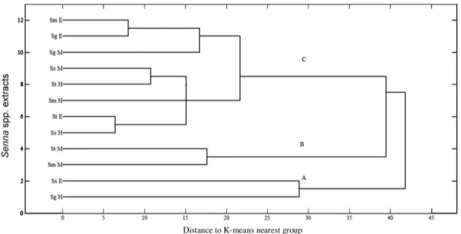
HCA was used for an initial, exploratory analysis of the cytotoxicity data for the 12 extracts on the three tumor cell line lineages (12 × 3), using preprocessing mean center data and distance to k-nearest neighbor. Then, partial least squares-discriminant analysis (PLS-DA) was used to model the three clusters obtained in the HCA. The data set (12 samples and 110 compound areas) was normalized, and five latent variables were selected to build the model.
Results and Discussion
The extracts obtained by sonication with n-hexane, diethyl ether and methanol from the leaves of S. gardneri, S. macranthera, S. splendida and S. trachypus were submitted to silylation and further analyzed by GC-MS to obtain linear retention indices (RIs) and mass spectra of the compounds, which were compared with the mass spectra in three mass spectra (MS) libraries (i.e., NIST, GMD and Wiley). The RIs were calculated from the retention times
obtained from the chromatograms of each compound and of a standard mixture of alkanes (C12-C40), according to
Van den Dool and Kratz equation.24 From the extracts of
the four species obtained with the three different solvents, 34 compounds were identified, including carboxylic acids, fatty acids, fatty alcohols, long-chain alkanes, diterpenes, triterpenes and sterols, in addition to the flavonoids chrysin and quercetin. The compounds identified by GC-MS in the extracts of Senna species are presented in Table 1.
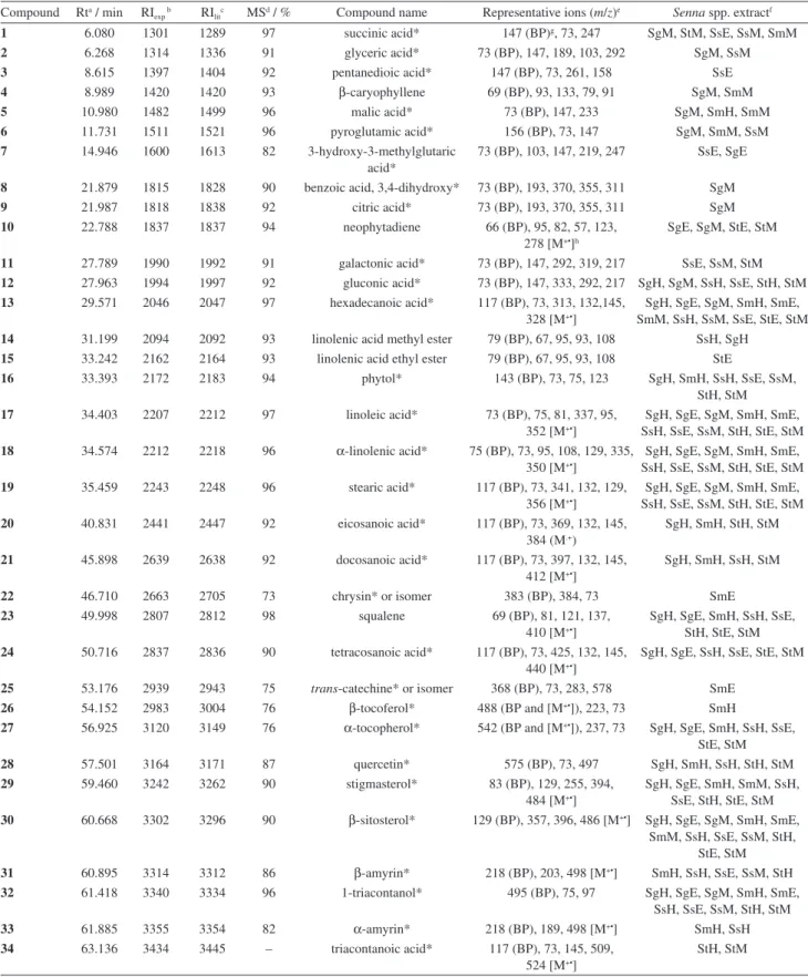
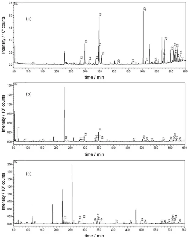
Each extract was prepared and analyzed in triplicate. With the extracts obtained with the different solvents, the metabolite diversity of the samples was demonstrated. As expected, the more polar extracts obtained with methanol contained high levels of mono-, disaccharides and polyols, whereas the n-hexane extracts were rich in terpenoids and fatty acids and other lipophilic compounds. However, the extracts obtained with diethyl ether were less complex. The compounds identified in the GC-MS chemical profiles are shown in Figure 1, and three representative GC-MS chromatograms are shown in Figure 2.
A multivariate analysis using HCA and PLS-DA was conducted of the chemical composition and the cytotoxicity of the extracts against human tumor cells to identify the compounds potentially correlated with this activity. The results of the cytotoxicity assays are shown in Table 2.
The n-hexane extract of S. gardneri had the highest activity against HTC-116 and SF-295 cancer cell lines with cell growth inhibited by 59.75 and 52.85%, respectively, whereas the highest inhibition of OVCAR-8 cells occurred with the methanol extract of S. macranthera. Although the cytotoxicity of plant extracts was generally weak (< 75% inhibition), the cytotoxic potential of these extracts was significantly different. In addition to differences in cytotoxicity, the chemical composition of these extracts was also very different; therefore, we used multivariate analysis of the two data sets (i.e., cytotoxicity and chemical composition) to identify the compounds likely responsible for the biological activity. Thus, the 12 extracts were initially analyzed by HCA, and using cytotoxicity as the dependent variable, the samples were classified into three clusters: (A) the extracts of SgH and SsE with the most activity on HTC-116 and SF-295 cells; (B) the extracts of StM and SmM with the most activity on OVCAR-8 cells; and (C) the extracts with low or no cytotoxicity (Figure 3).
Silva et al. 1875 Vol. 27, No. 10, 2016
Table 1. Compounds and respective peak areas found in the extracts from leaves of Senna spp. as trimethylsilyl derivatives in GC-MS analyses
Compound Rta / min RI
exp b RIlitc MSd / % Compound name Representative ions (m/z)e Senna spp. extractf
1 6.080 1301 1289 97 succinic acid* 147 (BP)g, 73, 247 SgM, StM, SsE, SsM, SmM
2 6.268 1314 1336 91 glyceric acid* 73 (BP), 147, 189, 103, 292 SgM, SsM
3 8.615 1397 1404 92 pentanedioic acid* 147 (BP), 73, 261, 158 SsE
4 8.989 1420 1420 93 β-caryophyllene 69 (BP), 93, 133, 79, 91 SgM, SmM
5 10.980 1482 1499 96 malic acid* 73 (BP), 147, 233 SgM, SmH, SmM
6 11.731 1511 1521 96 pyroglutamic acid* 156 (BP), 73, 147 SgM, SmM, SsM
7 14.946 1600 1613 82 3-hydroxy-3-methylglutaric
acid*
73 (BP), 103, 147, 219, 247 SsE, SgE
8 21.879 1815 1828 90 benzoic acid, 3,4-dihydroxy* 73 (BP), 193, 370, 355, 311 SgM
9 21.987 1818 1838 92 citric acid* 73 (BP), 193, 370, 355, 311 SgM
10 22.788 1837 1837 94 neophytadiene 66 (BP), 95, 82, 57, 123,
278 [M+•]h
SgE, SgM, StE, StM
11 27.789 1990 1992 91 galactonic acid* 73 (BP), 147, 292, 319, 217 SsE, SsM, StM
12 27.963 1994 1997 92 gluconic acid* 73 (BP), 147, 333, 292, 217 SgH, SgM, SsH, SsE, StH, StM
13 29.571 2046 2047 97 hexadecanoic acid* 117 (BP), 73, 313, 132,145,
328 [M+•]
SgH, SgE, SgM, SmH, SmE, SmM, SsH, SsM, SsE, StE, StM
14 31.199 2094 2092 93 linolenic acid methyl ester 79 (BP), 67, 95, 93, 108 SsH, SgH
15 33.242 2162 2164 93 linolenic acid ethyl ester 79 (BP), 67, 95, 93, 108 StE
16 33.393 2172 2183 94 phytol* 143 (BP), 73, 75, 123 SgH, SmH, SsH, SsE, SsM,
StH, StM
17 34.403 2207 2212 97 linoleic acid* 73 (BP), 75, 81, 337, 95,
352 [M+•]
SgH, SgE, SgM, SmH, SmE, SsH, SsE, SsM, StH, StE, StM 18 34.574 2212 2218 96 α-linolenic acid* 75 (BP), 73, 95, 108, 129, 335,
350 [M+•]
SgH, SgE, SgM, SmH, SmE, SsH, SsE, SsM, StH, StE, StM
19 35.459 2243 2248 96 stearic acid* 117 (BP), 73, 341, 132, 129,
356 [M+•]
SgH, SgE, SgM, SmH, SmE, SsH, SsE, SsM, StH, StE, StM
20 40.831 2441 2447 92 eicosanoic acid* 117 (BP), 73, 369, 132, 145,
384 (M.+)
SgH, SmH, StH, StM
21 45.898 2639 2638 92 docosanoic acid* 117 (BP), 73, 397, 132, 145,
412 [M+•]
SgH, SmH, SsH, StM
22 46.710 2663 2705 73 chrysin* or isomer 383 (BP), 384, 73 SmE
23 49.998 2807 2812 98 squalene 69 (BP), 81, 121, 137,
410 [M+•]
SgH, SgE, SmH, SsH, SsE, StH, StE, StM 24 50.716 2837 2836 90 tetracosanoic acid* 117 (BP), 73, 425, 132, 145,
440 [M+•]
SgH, SgE, SsH, SsE, StE, StM
25 53.176 2939 2943 75 trans-catechine* or isomer 368 (BP), 73, 283, 578 SmE
26 54.152 2983 3004 76 β-tocoferol* 488 (BP and [M+•]), 223, 73 SmH
27 56.925 3120 3149 76 α-tocopherol* 542 (BP and [M+•]), 237, 73 SgH, SgE, SmH, SsH, SsE, StE, StM
28 57.501 3164 3171 87 quercetin* 575 (BP), 73, 497 SgH, SmH, SsH, StH, StM
29 59.460 3242 3262 90 stigmasterol* 83 (BP), 129, 255, 394,
484 [M+•]
SgH, SgE, SmH, SmM, SsH, SsE, StH, StE, StM 30 60.668 3302 3296 90 β-sitosterol* 129 (BP), 357, 396, 486 [M+•] SgH, SgE, SgM, SmH, SmE,
SmM, SsH, SsE, SsM, StH, StE, StM
31 60.895 3314 3312 86 β-amyrin* 218 (BP), 203, 498 [M+•] SmH, SsH, SsE, SsM, StH
32 61.418 3340 3334 96 1-triacontanol* 495 (BP), 75, 97 SgH, SgE, SgM, SmH, SmE,
SsH, SsE, SsM, StH, StM
33 61.885 3355 3354 82 α-amyrin* 218 (BP), 189, 498 [M+•] SmH, SsH
34 63.136 3434 3445 – triacontanoic acid* 117 (BP), 73, 145, 509,
524 [M+•]
StH, StM
aRt = retention time; bRI
Chemical Profile and Cytotoxic
Acti
vity of Leaf Extracts from
Senna
spp.
J. Br
az. Chem. Soc.
0 5 10 15 20 25 30 35
Malic acid
Gluconic acid
Hexadecanoic acid
γ-Linolenicacid
Linolenic ca id ethyl ester
Phytol
Linoleic acid
α-Linolenic acid
Stearic acid
Eicosanoic acid
Docosanoic acid
Chrysin
Normalized area / %
Compound Squalene Tetracosanoic acid trans-Catechine β-Tocoferol α-Tocopherol Quercetin Stigmasterol β-Sitosterol β-Amyrin 1-Triacontanol α-Amyrin Cycloartenol acetate Triacontanoic acid 51 21 31 41 51 61 71 81 92 02 12 22 32 42 52 62 72 82 93 03 13 23 33 53 6 (a ) Ss H Sm H Sg H St H
0 5 10 15 20 25 30 35
Succinic acid Pentanedioic acid 3-Hydroxy-3-methylglutaric acid Neophytadiene Galactonic acid Gluconic acid Hexadecanoic acid
Linolenic acid ethyl ester
Phytol
Linoleic acid
α-Linolenic acid
Stearic acid Chrysin Squalene Tetracosanoic acid trans-Catechine α-Tocopherol Stigmasterol β-Sitosterol β-Amyrin 1-Triacontanol Cycloartenol acetate 13 70 11 11 23 15 11 67 18 19 12 22 34 25 27 29 23 01 32 33 5 (b) Ss E Sm E SgE St E Compound
Normalized area / %
0 10 20 30 40 50 60
Succinic acid
Glyceric acid
β-Caryophyllene
Malic acid
Pyroglutamic acid
Benzoic acid, 3,4-dihydroxy
Citric acid Neophytadiene Galactonic acid Gluconic acid Hexadecanoic acid Phytol Linoleic acid
α-Linolenic acid
Stearic acid Eicosanoic acid Docosanoic acid Squalene Tetracosanoic acid α-Tocopherol Quercetin Stigmasterol β-Sitosterol β-Amyrin 1-Triacontanol Lupeol acetate Triacontanoic acid 1245689 10 11 12 13 16 17 18 19 20 21 23 24 27 28 29 30 31 32 34 36 (c ) Ss M SmM Sg M StM Compound
Normalized area / %
e 1
. Chemical profiles of
Senna
spp. lea
v
es e
xtracted with (a) he
xane; (b) eth
yl ether; and (c) methanol. Compounds are sho
wn with more than 1%
xtract. SgH =
S. gar
dneri
he
xane, SgE =
S. gar
dneri
ether
, SgM
= S. gar
dneri
methanol, SmH =
S. macr anther a he xane, Senna macr anther a ether
, SmM =
S. macr
anther
a
methanol, SsH =
S. splendida
he
xane, SsE =
S. splendida
ether
, SsM =
S. splendida methanol, ac hypus he xane, StE
= S. tr
ac
hypus
ether
, StM =
S. tr
ac
hypus
methanol e
xtract.
0 5 10 15 20 25 30 35
Malic acid
Gluconic acid
Hexadecanoic acid
γ-Linolenicacid
Linolenic ca id ethyl ester
Phytol
Linoleic acid
α-Linolenic acid
Stearic acid
Eicosanoic acid
Docosanoic acid
Chrysin
Normalized area / %
Compound Squalene Tetracosanoic acid trans-Catechine β-Tocoferol α-Tocopherol Quercetin Stigmasterol β-Sitosterol β-Amyrin 1-Triacontanol α-Amyrin Cycloartenol acetate Triacontanoic acid 51 21 31 41 51 61 71 81 92 02 12 22 32 42 52 62 72 82 93 03 13 23 33 53 6 (a ) Ss H Sm H Sg H St H
0 5 10 15 20 25 30 35
Succinic acid Pentanedioic acid 3-Hydroxy-3-methylglutaric acid Neophytadiene Galactonic acid Gluconic acid Hexadecanoic acid
Linolenic acid ethyl ester
Phytol
Linoleic acid
α-Linolenic acid
Stearic acid Chrysin Squalene Tetracosanoic acid trans-Catechine α-Tocopherol Stigmasterol β-Sitosterol β-Amyrin 1-Triacontanol Cycloartenol acetate 13 70 11 11 23 15 11 67 18 19 12 22 34 25 27 29 23 01 32 33 5 (b) Ss E Sm E SgE St E Compound
Normalized area / %
0 10 20 30 40 50 60
Succinic acid
Glyceric acid
β-Caryophyllene
Malic acid
Pyroglutamic acid
Benzoic acid, 3,4-dihydroxy
Citric acid Neophytadiene Galactonic acid Gluconic acid Hexadecanoic acid Phytol Linoleic acid
α-Linolenic acid
Stearic acid Eicosanoic acid Docosanoic acid Squalene Tetracosanoic acid α-Tocopherol Quercetin Stigmasterol β-Sitosterol β-Amyrin 1-Triacontanol Lupeol acetate Triacontanoic acid 1245689 10 11 12 13 16 17 18 19 20 21 23 24 27 28 29 30 31 32 34 36 (c ) Ss M SmM Sg M StM Compound
Silva et al. 1877 Vol. 27, No. 10, 2016
Figure 2. Representative GC-MS chromatograms from (a) hexane extract of leaves from Senna splendida; (b) ethyl ether extract of leaves from Senna splendida; and (c) methanol extract of leaves from Senna trachypus.
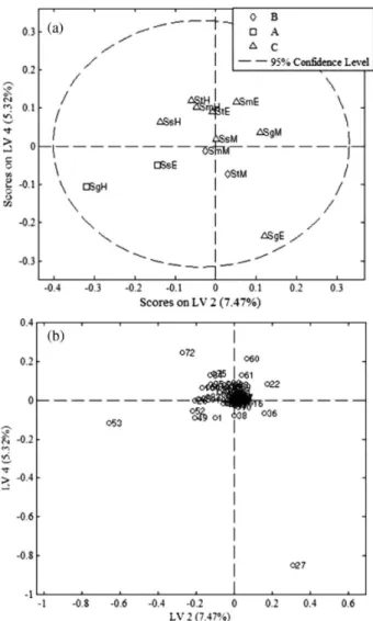
variances of the data, with the residual Q and Hotelling’s T2 showing only one sample (SgM) outside the 95% confidence intervals. The best adjustments were obtained in the score (Figure 4a) and the loading (Figure 4b) plots of LV4 × LV2 in which the cytotoxic extracts from cluster A (SgH and SsE) were in the identical quadrant (Figure 4a) as variable 53 (Figure 4b), which could be related to the cytotoxicity of these extracts. Variable 53 corresponded to
compound 18, identified as α-linolenic acid (ALA). Several studies examined linoleic acid in combination with other compounds in evaluations of anticancer activity.25,26
Dai27 and Sun et al.28 reported that polyunsaturated
the generation of free radicals and lipid peroxidation.29
Dai et al.30 showed that polyunsaturated fatty acids are
cytotoxic to tumor cells, and of the fatty acids tested, linoleic acid (LA) and α-linolenic acid (ALA) were the most effective in suppressing the growth of normal gastric cells (GES1) at 180 and 200 µmol L-1 and those of gastric
carcinoma (MGC and SGC) at 200 µmol L-1. The induction
of apoptosis by α-linolenic acid (18) (ALA) was observed by Vecchini et al.31 and Scheim32 and likely occurred
because of the reduction of nitric oxide, as proposed by Deshpande et al.33 In recent studies with mouse models,
ALA (18) reduced breast tumor growth while increasing the efficacy of chemotherapeutic agents. However, these studies did not confirm whether the effects were caused by ALA or its metabolites.34 No compound was correlated with
the cytotoxicity of the samples in cluster B, which included two methanolic extracts, most likely because several of the polar compounds in these extracts did not elute in the chromatographic conditions used in this study.
Additionally, some compounds were detected in only one species; for example, the flavonoid, chrysin (22), was identified only in S. macranthera, and the flavonoid,
Table 2. Cytotoxic activity of Senna spp. extracts on human cancer cell lines (% inhibition ± SDa)
Sampleb HTC-116c SF-295d OVCAR-8e
SgH 59.75 ± 0.60 52.85 ± 5.21 24.61 ± 13.82
SgE 11.63 ± 2.53 24.29 ± 6.13 0.01 ± 0.00
SgM 6.56 ± 10.65 5.77 ± 2.80 0.01 ± 0.00
SmH 28.27 ± 3.05 39.27 ± 7.14 0.01 ± 0.01
SmE 5.59 ± 6.05 20.54 ± 6.22 0.01 ± 0.00
SmM 1.37 ± 3.82 18.56 ± 5.55 44.64 ± 0.99
SsH 13.85 ± 1.29 36.92 ± 0.14 10.43 ± 41.62
SsE 31.37 ± 6.65 48.28 ± 10.04 21.92 ± 1.67
SsM 19.65 ± 5.28 28.66 ± 11.53 0.01 ± 0.00
StH 27.39 ± 3.52 21.19 ± 3.96 0.01 ± 0.00
StE 20.19 ± 1.67 37.12 ± 5.65 9.41 ± 45.94
StM 7.23 ± 1.29 32.38 ± 2.32 35.4 ± 23.31
DOX, IC50 [µmol L–1]f 0.12 (0.09-0.17) 0.22 (0.16-0.24) 0.34 (0.31-0.36)
aSD =standard deviation;bsample:SgH = S. gardneri hexane, SgE = S. gardneri ether, SgM = S. gardneri methanol, SmH = S. macranthera hexane, SmE = Senna macranthera ether, SmM = S. macranthera methanol, SsH = S. splendida hexane, SsE = S. splendida ether, SsM = S.splendida methanol, StH = S. trachypus hexane, StE = S. trachypus ether, StM = S. trachypus methanol; cHTC-116 = human colon; dOVACAR-8 = ovarian carcinoma; eSF-295 = human glioblastoma; fDOX = doxorubicin was the positive control. IC
50 is the drug concentration that caused 50% inhibition of cell growth, with the corresponding 95% confidence interval (CI 95%) shown below.
Silva et al. 1879 Vol. 27, No. 10, 2016
quercetin (28), was identified only in the methanolic extract of S. trachypus. These compounds have shown excellent potential for chemopreventive and cancer therapy35 and have anti-inflammatory and antioxidant
activities.36 However, eleven compounds were identified in
all the extracts that are typically encountered in different families and genera of higher plants. For example, all extracts contained the triterpene squalene (23). This compound, which is produced by all higher organisms and has beneficial effects on human health and antioxidant activity, has also been isolated from the leaves and roots of
Ramonda serbica and R. nathaliae,37 and from the marine
diatom Pleurosigma strigosum.38 The triterpenes α-amyrin
(33) and β-amyrin (31) were detected only in the n-hexane extracts of leaves of S. macranthera and S. splendida. These compounds, with anti-inflammatory, anti-conceptive, and hepatoprotective pharmacological activities, have also been isolated as a mixture from different natural
Figure 4. Score (a) and loading (b) plots obtained after PLS-DA using 12 extracts from Senna spp. and 110 compound areas (12 × 110). A) extracts cytotoxic to HTC-116 and SF-295 cells; B) extracts cytotoxic to OVCAR-8 cells; and C) extracts with low or no cytotoxicity.
sources, including other species of the genus Senna, i.e.,
S. spectabilis var. excelsa and S. reticulata.7 The steroids β-sitosterol (30) and stigmasterol (29) are common compounds in species of several genera and families, and the properties of β-sitosterol isolated from S. spectabilis
var. excelsa have been reported as antibacterial, anti-inflammatory and analgesic.6
Conclusions
The GC-MS analyses of 12 extracts from the leaves of four species of Senna identified 34 compounds in different groups that included fatty acids, steroids, triterpenes and flavonoids not reported previously for these species. The use of multivariate analyses (HCA and PLS-DA) led us to infer that the cytotoxicity of some Senna extracts to HTC-116 and SF-295 tumor cells lines was attributed to linolenic acid, an inference that was reinforced by literature data. Moreover, with analytical techniques, dereplication and multivariate statistical analysis, this study demonstrated that it was possible to effectively identify hit compounds and by avoiding the steps of extract fractionation and purification of known compounds, to improve the research approach in the prospect for new drug prototypes from biological sources.
Supplementary Information
Supplementary data (MS spectra) are available free of charge at http://jbcs.sbq.org.br.
Acknowledgments
The authors are very thankful to the Federal University of Ceará (UFC), State University Paulista (UNESP), Prof Norberto Peporine Lopes and Izabel Cristina Casanova Turatti from the University of São Paulo (USP) for their support with the GC-MS analyses, to Prof Fernando Batista Costa from the University of São Paulo (USP) for accurate comments and to FAPESP, CNPq and CAPES for all the financial support.
References
1. Irwin, H. S.; Barneby, R. C.; The American Cassiinae: a Synoptical Revision of Leguminoseae Tribe Cassia Subtribe
Cassinae in the New World, New York Bot. Gard: New York, USA, 1982.
2. Rodrigues, R. S.; Flores, A. S.; Miotto, T. S.; Baptista, L. R. S. R.; Acta Bot. Bras. 2005, 19, 1.
4. Rahman, M. O.; Rahman, M. D. Z.; Begun, A.; Bangladesh J. Plant Taxon. (Online) 2013, 20, 77.
5. Irwin, H. S.; Barneby, R. C. In Advances in Legume Systematics; Polhill, R. M.; Raven, P. H., eds.; Kew: The Royal Botanic Gardens: London, UK, 1981, ch. 1.
6. Silva, F. O.; Oliveira, I. R.; Silva, M. G. V.; Braz-Filho, R.; Quim. Nova2010, 33, 1874.
7. Santos, R. N.; Silva, M. G. V.; Quim. Nova2008, 31, 1979. 8. Melo, G. M. A.; Silva, M. C. R.; Guimarães, T. P.; Pinheiro,
K. M.; Matta, C. B. B. Q.; Pivatto, M.; Bolzani, V. S.; Alexandre-Moreira, M. S.; Viegas Jr., C.; Phytomedicine2013, 3, 277. 9. Hardcastle, J. D.; Wilkins, J. L.; Gut1970,11, 1038. 10. Guarize, L.; Costa, J. C.; Dutra, L. B.; Mendes, R. F.; Lima,
I. V. A.; Scio, E.; Nat. Prod. Res.2012, 26, 331.
11. Villaseñor, I. M.; Sanchez, A. C.; Z. Naturforsch. 2009, 64, 335. 12. Yeung-Beom, P.; Seon-Bong, K.; J. Microbiol. Biotechnol.
2011, 21, 1043.
13. Rahman, M. A.; Sultana, R.; Emran, T. B.; Islan, M. S.; Rahman, M. A.; Chakma, S. C.; Rashid, H.; Hasan, C. M. M.; BMC Complementary Altern. Med. 2013, 13, 25.
14. Wongtongtair, S.; Chanvorachote, P.; Hutamekalin, P.; Chaichantipyuth, C.; Lipipun, V.; Tiensiwakul, P.; Meksuriyen, D.; J. Ethnopharmacol. 2011, 137, 971. 15. Nsonde-Ntandou, G. F.; Banzouzib, J. T.; Mbatchia, B.;
Elion-Itoua, R. D. G.; Etou-Ossibia, A. W.; Ramosd, S.; Benoit-Vicale, F.; Abenaa, A. A.; Ouambah, J. M.; J. Ethnopharmacol. 2010, 127, 108.
16. Esakkirajan, M.; Prabhu, N. M.; Arulvasu, C.; Beulaja, M.; Manikandan, R.; Thiagarajan, R.; Govindaraju, K.; Prabhu, D.; Dinesh, D.; Babu, G.; Dhanasekaran, G.; Spectrochim. Acta, Part A2014, 120, 462.
17. Funari, C. S.; Castro-Gamboa, I.; Cavalheiro, A. J.; Bolzani, V. S.; Quim. Nova2013, 10, 1605.
18. Rochfort, S.; J. Nat. Prod. 2005, 68, 1813.
19. Kanani, H.; J. Chromatogr. B: Anal. Technol. Biomed. Life Sci. 2008, 87, 191.
20. Madsen, R.; Lundstedt, T.; Trygg, J.; Anal. Chim. Acta2010, 659, 23.
21. Chagas-Paula, D. A.; Zhang, T.; Costa, F. B.; Edrada-Ebel, R.; Metabolites 2015, 5, 404.
22. Isidorov, V. A.; Szczepaniak, L.; J.Chromatogr. A2009, 1216, 8998.
23. Mossman, T.; J. Immunol. Methods1983, 65, 55.
24. Van den Dool, H.; Kratz, P. D.; J. Chromatogr. A 1963, 11, 463. 25. Tao, X. M.; Wang, J. C.; Wang, J. B.; Feng, Q.; Gao, S. Y.;
Zhang, L. R.; Zhang, Q.; Eur. J. Pharm. Biopharm. 2012, 82, 406.
26. Roz, E.; Bard, J. M.; Huvelin, J. M.; Nazih, H.; Prostaglandins, Leukotrienes Essent. Fatty Acids2013, 88, 267.
27. Dai, U. N.; Med. Sci. Monit.2002, 8, RA79.
28. Sun, Z.; Wang, H.; Ye, S.; Xiao, S.; Liu, J.; Wang, W.; Jiang, D.; Liu, X.; Wang, J.; Prostaglandins Other Lipid Mediators 2012, 99, 1.
29. Menédez, J. A.; Vázquez-Martín, A.; Ropero, S.; Colomer, R.; Lupu, R.; Trueta, J.; Clin. Transl. Oncol. 2006, 8, 812. 30. Dai, J.; Shen, J.; Pan, W.; Shen, S.; Das, U. N.;Lipids Health
Dis. 2013,12, 71.
31. Vecchini, A.; Ceccarelli, V.; Susta, F.; Caligiana, P.; Orvietani, P.; Binaglia, L.; Nocentini, G.; Ricardi, C.; Calviello, G.; Palozza, P.; Maggiano, N.; Di Nardo, P.; J. Lipid Res. 2004, 45, 308.
32. Scheim, D. E.; Lipids Health Dis. 2009, 8, 54.
33. Deshpande, R.; Mansara, P.; Suryavanshi, S.; Kaul-Ghanekar, R.; J. Mol. Biochem. 2013, 2, 6.
34. Mason, J. K.; Klaire, S.; Kharotia, S.; Wiggins, A. K. A.; Thompson, L. U.; Lipids Health Dis. 2015, 14, 90.
35. Kasala, E. R.; Bodduluru, L. N.; Madana, R. M.; V, A. K.; Gogoi, R.; Barua, C. C.; Toxicol. Lett.2015, 4, 214.
36. Joshi, U. J.; Gadge, A. S.; D’Mello, P.; Sinha, R.; Srivastava, S.; Govi, G.; J. Pharm. Pharm. Sci.2011, 2, 1756.
37. Radulović, N. S.; Blagojević, P. D.; Palić, R. W.; Zlatković, B. K.; Stevanović, B. M.; J. Serb. Chem. Soc. 2009, 74, 35. 38. Grossi, V.; Beker, B.; Geenevasen, J. A. J.; Schouten, S.;
Raphel, D.; Fontaine, M.; Damtsé, J. S. S.; Phytochemistry 2004, 65, 3049.
Submitted: November 4, 2015
Published online: March 14, 2016