Fatty Acid and Sterol Composition of Three
Phytomonas
Species
Celso Vataru Nakamura
+, Luciana Waldow, Sandra Regina Pelegrinello,
Tânia Ueda-Nakamura, Benício Alves de Abreu Filho,
Benedito Prado Dias Filho
Laboratório de Microbiologia, Departamento de Análises Clínicas, Centro de Ciências da Saúde, Universidade Estadual de Maringá, Campus Universitário, Av. Colombo 5790, 87020-900 Maringá, PR, Brasil
Fatty acid and sterol analysis were performed on Phytomonas serpens and Phytomonas sp. grown in chemically defined and complex medium, and P. françai cultivated in complex medium. The three species of the genus Phytomonas had qualitatively identical fatty acid patterns. Oleic, linoleic, and linolenic were the major unsaturated fatty acids. Miristic and stearic were the major saturated fatty acids. Ergosterol was the only sterol isolated from Phytmonas sp. and P. serpens grown in a sterol-free medium, indicating that it was synthesized de novo. When P. françai that does not grow in defined medium was cultivated in a complex medium, cholesterol was the only sterol detected. The fatty acids and sterol isolated from Phytomonas sp. and P. serpens grown in a chemically defined lipid-free medium indicated that they were able to biosynthesize fatty acids and ergosterol from acetate or from acetate precursors such as glucose or threonine.
Key words: Phytomonas - fatty acids - sterols - trypanosomatids
Protozoan parasites must interact with the host at a variety of levels: the acquisition of nutrients, evasion or confusion of the host’s response, estab-lishment and maintenance of the infected state. This interface and the interactions are, by necessity, membrane mediated (Fish 1995).
Lipids, which are essential structural compo-nents of biological membranes, also affect cell sur-face recognition, cell interactions, and the expres-sion of antigenic determinants (Yamakawa & Nagai 1978, Cullis & Kruijiff 1979, MacMurchie & Raison 1979, Elbein 1979).
The cell membranes of a variety of biological systems are altered in response to temperature changes, a process for which the term homeovis-cous adaptation has been proposed (Sinensky 1974). In general, the lipid composition is charac-terized by an increase in unsaturated fatty acid with the decrease in environmental temperature ( Roy et al. 1991, Imhoff & Thiemann 1991, Buzzi et al. 1993). Moreover in protozoa, the lipid content and metabolism are often influenced by environmen-tal factors including the composition of the growth
This work was supported by grants from CNPq (no. 520245/93-8)
+Corresponding author. Fax: +55-44 -261.4490. E-mail: [email protected]
Received 21 September 1998 Accepted 3 March 1999
medium (Pinto et al. 1982, Racagni et al. 1995, Ellis et al. 1996) and temperature (Fagundes et al. 1980, Jones et al. 1993, Avery et al. 1995, Florin-Christensen et al. 1997).
Flagellate trypanosomatids of the genus
Phytomonas are etiologic agents of diseases
affect-ing fruits and plants of great economical impor-tance including tomato, cashew, coffee, cassava, coconut and oil palms (Lopez et al. 1975, Dollet & Lopez 1978, Vainstein & Roitman 1985, Conchon et al. 1989) although they also act as parasites of lactiferous without any apparent pathogenicity (Attias & De Souza 1986). Insects have been sus-pected as a vector of plant flagellates. Jankevicius et al. (1989) showed in controlled laboratory cage experiments that P. serpens, the tomato parasite, is
transmitted by the bite of coreid insect Phthia picta.
The presence of trypanosomatids in plants of eco-nomic interest has attracted the attention of sev-eral research groups. A study on the fatty acid and sterol composition of three Phytomonas strains was
undertaken in the present work.
MATERIALS AND METHODS
Microorganisms - P. françai isolated from
cas-sava (Vainstein & Roitman 1985), P. serpens
iso-lated from the salivary glands of the phytophagous insect P. picta (Brasil et al. 1990), and Phytomonas
sp. isolated from the latex of Euphorbia hyssopifolia (Attias & De Souza 1986), were
trypticase 5, yeast extract 5, folic acid 0.002, hemin dissolved in quadrol 25% 0.02; pH 7.0 (Roitman et al. 1972). Cells were grown at 28ºC for 48 hr and thereafter were kept at 4 to 6ºC. For the ex-periments, cells were grown in 1 l flasks contain-ing 500 ml of complex medium. The medium was autoclaved at 121ºC for 20 min. However, in some experiments Phytomonas sp. and P. serpens were
also cultivated in a chemically defined medium (Silva & Roitman 1989) (Table I). The inoculum consisted of 50 ml of a 48 hr culture, correspond-ing to approximately 2 x 10⁸ cells. After 48 hr of incubation the cells were collected by centrifuga-tion (2,000 g for 10 min at 4ºC) and washed four times in cold phosphate-buffered saline (PBS), pH 7.2, 0.01 M.
Extraction of lipids and identification of the fatty acid and sterols - Lipids were extracted from
washed protozoan cells with 10 vols each of chlo-roform-methanol-water mixture (4:8:3 v/v) and chloroform-methanol mixture (1:1 v/v). Combined
extracts were evaporated to dryness. Absolute methanol-diethyl ether (3:1 v/v) was added to the lipid extract followed by saponification with 1 ml of 5N NaOH. Fatty acids were then extracted in n-hexane after adding water and lowering the pH to 1.0. Fatty acids were converted to their correspond-ing methyl esters by treatment with ether-diazomethane and methanol-diethyl ether (1:9, v/v) (Pörschmann 1982). Methyl esters were ana-lyzed by gas-liquid chromatography (GLC) with a temperature programmed and coupled to a mass spectrometer (MS) Hewlett Packard 5992 AGC/ MS System with an ionizing energy of 70 eV. Methyl esters were identified by their retention time relative to methyl esters of known fatty acid stan-dards. The chain lengths of unsaturated fatty acids were also identified by GLC of the products of catalytic hydrogenation of methyl esters carried out at room temperature for 1 hr in ethyl acetate, with 10% palladium on charcoal under a hydrogen pres-sure of 40 psi.
TABLE I
Chemically defined medium for Phytomonas serpens (Silva & Roitman 1989)
Compound g/l Compound mg/l
β-Na glycerophosphate 10 Nicotinamide 2
Glucose 20 Ca pantothenate 2
Inositol 0.04 Na Riboflavine PO4.2H2O 1
Glutamic acid 0.1 Pyridoxamine.2HCl 0.6
L-Serine 0.2 Thiamine HCl 0.6
MgSO4 0.05 Biotin 0.008
KCl 10 Folic acid 2
K2HPO4 1
K3Citrate.H2O 1
Citric acid.H2O 0.5
Malic acid 0.2
Succinic acid 1
MgCO3 1
CaCO3 0.02
Fe(NH4)2(SO4)2.6H2O 0.01
L-arginine HCl 0.4
L-histidine (free base) 0.3
L-isoleucine 0.2
L-leucine 0.2
L-lysine HCl 0.2
L-methionine 0.1
L-phenylalanine 0.2
L-threonine 0.2
L-tryptophan 0.1
L-tyrosine ethyl ester 0.2
L-valine 0.2
Adenine 0.02
Trace elementsa 0.2
Heminb 0.01
Sterols of cells were extracted from total lipids by saponification with 1 ml of 5N NaOH for 5 hr. They were fractionated by thin-layer chromatog-raphy (TLC) on 0.25 mm layers of silica gel GF254 (Merck), using hexane-ethyl acetate (65:35, v/v) as solvent and the spots visualized by u.v. or by spraying with sulfuric acid-ceric acetate (Sthal 1969). After being visualized, the sterols were scraped from the TLC plates, dissolved in metha-nol and analyzed by u.v. spectroscopy (200-400 nm) in a Varian 1E/UV Visible Spectrophotom-eter. Cholesterol and ergosterol (Sigma Chemical Co.) were used as internal standards.
RESULTS
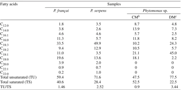
The fatty acid compositions of P. françai, P. serpens and Phytomonas sp. grown in the
com-plex medium are shown in Table II. Lauric (C12:0), miristic (C14:0), palmitoleic (C16:1), palmitic (C16:0), linolenic (C18:3), linoleic (C18:2), oleic (C18:1), stearic (C18:0), eicosanoic (C20:0), erucic (C22:1), and docosanoic (C22:0) acids were detected as components of the total lipid fraction from all parasites. Linolenic, linoleic, oleic, and stearic ac-ids were the major fatty acac-ids of the three
Phytomonas sp. and accounted for more than 60%
of the total fatty acids. P. françai and P. serpens
contained higher levels of linolenic acid (33.5% and 49.9%, respectively) than that observed with
Phytomonas sp.cells (10.2%). In Phytomonas sp.
eicosanoic, euric, and docosanoid acids were ab-sent, oleic (21.1%) and stearic acids (18.1%) were the prominent components and the degree of
unsaturation (47.5%) was lower than those re-corded for P. françai (59.4%) and P. serpens
(71.6%).
When Phytomonas sp. and P. serpens were
grown in chemically defined medium the fatty ac-ids composition showed an increase in the degree of unsaturation. The proportion of the total unsat-urated fatty acids of Phytomonas sp. grown in
chemically defined medium increased by approxi-mately 61%. Changes in the degree of unsaturation were due to variations in the proportion of the un-saturated fatty acids linolenic and oleic. The pres-ence of unsaturated fatty acids was confirmed by catalytic hydrogenation; the characteristic peaks for palmitoleic, oleic, linoleic, and linolenic acids were completely abolished with a corresponding in-crease in the size or the peaks for palmitic and stearic acids (data not shown).
Only one sterol type with a Rf similar to ergos-terol could be detected by TLC of the nonsaponi-fiable component of the lipid extract of Phytmonas
sp. and P. serpens grown in both complex (Fig.
1A) and chemically defined medium (Fig 1B). Its ultraviolet absorption spectrum showed maximum absorption at 293, 283, 271, and 261 nm, which was in good agreement with authentic ergosterol (Fig. 2).
Cholesterol was the only sterol detected in P. françai grown in complex medium as showed in
Fig. 1. This compound was also identified by GLC by comparison with the retention time of the cho-lesterol standard.
TABLE II
Fatty acid composition (%) of three Phytomonas speciesa Fatty acids Samples
P. françai P. serpens Phytomonas sp.
CMb DMc
C12:0 1.8 3.5 8.7 4.8
C14:0 3.8 2.6 13.9 7.3
C16:1 4.6 4.6 5.7 2.5
C16:0 11.3 5.7 11.8 8.2
C18:3 33.5 49.9 10.2 24.3
C18:2 9.4 12.9 10.5 5.7
C18:1 11.0 3.5 21.1 45.0
C18:0 19.6 13.6 18.1 2.2
C20:0 3.9 2.0 0 0
C22:1 0.9 0.7 0 0
C22:0 0.2 1.0 0 0
Total unsaturated (TU) 59.4 71.6 47.5 77.5
Total saturated (TS) 40.6 28.4 52.5 22.5
TU/TS 1.46 2.52 0.9 3.44
DISCUSSION
Even- and odd-numbered, saturated, mono-enoic and polymono-enoic types of fatty acids ranging from C12 to C22 were characterized as components of the total lipid fraction of P. françai, P. serpens,
and Phytomonas sp. In general, this fatty acids
pattern resembles that observed for Herpetomonas
(Pinto et al. 1982), Leishmania donovani (Glew
et al. 1988), and Trypanosoma cruzi (Racagni et
al. 1992). The major fatty acids of the three
Phytomonas sp. consisted generally of C18 carbon
chain lengths. Linolenic, linoleic, oleic, and stearic acids were the major fatty acids of three
Phytomonas sp. and accounted for more than 60%
of the total fatty acids. The three species of the genus Phytomonas had qualitatively identical fatty
acid patterns. However, differences in the fatty acids content of lipid fraction was observed in this study. For example, P. françai and P. serpens
con-tained higher levels of linolenic acid than observed with cells of Phytomonas sp. In Phytomonas sp.
eicosanoic, euric, and docosanoid acids were ab-sent, oleic and stearic acids were the prominent components and the degree of unsaturation was lower than those from P. françai and P. serpens.
In the chemically defined medium the lipid com-position of Phytomonas sp.and P. serpens showed a
increase in unsaturation. P. françai does not grow in
chemically defined medium. Compared to cells grown in a complex medium, the proportion of the
total unsaturated fatty acids of Phytomonas sp. grown
in chemically defined medium increased by approxi-mately 61% with a concomitant decrease in the pro-portion of saturated fatty acids. Changes in the de-gree of unsaturation were due to the variations in the proportion of the unsaturated fatty acids linolenic and oleic. In the protozoa H. samuelpessoai changes in
the degree of unsaturation were accompanied by a variation on the amount of oleic and linoleic acids (Pinto et al. 1982).
Fig. 2: U.V. spectrum of the sterol isolated from Phytomonas sp. (A) and ergosterol standard (B).
Fig. 1: thin-layer chromatography of sterols isolated from three Phytomonas strains cultivated in complex medium (A) and chemi-cally defined medium (B). Sterols were fractionated by TLC in silica gel GF254, using hexane-ethyl acetate (65:35, v/v) as solvent and the spots visualized by u.v. or by spraying with sulfuric acid-ceric acetate. (1) cholesterol, (2) ergosterol, (3) Phytomonas serpens, (4) Phytomonas sp., and (5) P. françai.
WAVE LENGHT (nm)
The increase in the degree of unsaturation of fatty acids as result of lowering the environmental temperature has been described in T. cruzi
(Florin-Christensen et al. 1997) and in several of other
mi-croorganisms including bacteria (Sinensky 1974, Roy et al. 1991, Imhoff & Thiemann 1991, Buzzi et al. 1993, Vigh et al. 1993, Avery et al. 1995). Most cells under environmental stress restore the suboptimal physical state of their membranes to a more functional condition by altering the lipid com-position of their membranes. Changes in membrane composition by increasing unsaturated fatty acids would prevent “freezing” of membrane and inhi-bition of various cellular membranes functions (Ellis et al. 1996). The life cycle of phytomonads include stage in different environments such as the digestive tract and salivary glands of insects, the latex and the sap of plants, and the fruit and seeds of various species (Jankevicius et al. 1991). In-creased membrane fluidity helps maintain vital membrane functions of plant parasite at these en-vironmental conditions with very difference in terms of osmolarity, pH, food resources, and tem-perature.
The fatty acids isolated from Phytomonas sp.
and P. serpens (data not shown) grown on a
chemi-cally defined lipid-free medium indicates that they were able to biosynthesize fatty acids from acetate or from acetate precursors such as glucose or threo-nine. The ability to use both sugars and amino ac-ids as a source of energy is a feature of many trypanosomatids and has probably been invaluable in their adaptative radiation to colonise differents hosts. Sugars are present in blood and plant saps but soon disappear from the vector´s meal. Amino acids will become abundant as the blood meal is digested (Vickerman 1994). Nectar can be a source of both amino acids and suggar, even lipids in some species (Baker & Baker 1975) Studies in trypanosomatids, such as Leishmania tarentolae
and T. lewisi indicated that these species are able
to synthesize and elongate precursor short chain fatty acids (Korn et al. 1965). Changes in the struc-tures of the fatty acid could be attributed to con-version (e.g., chain elongation, desaturation) or retroconversion (chain shortening), or to the intro-duction of branches or ring.
Ergosterol was the only sterol isolated from
Phytmonas sp.and P. serpens grown in a
sterol-free medium, indicating that it was synthesized de novo. The possible synthesis of other sterol is
ex-cluded by the fact that ergosterol was the only ste-rol present in cells of these parasites cultivated in both chemically defined and complex medium. However, when P. françai was cultivated in a
com-plex medium cholesterol was the only sterol de-tected. P. françai, that is unable to grow in defined
medium, has been maintained by monthly trans-fers in a biphasic medium containing blood agar in the solid phase and overlay of complex medium (Attias et al. 1988). The parasites die after three or four subcultures in complex medium. Thus, after two subcultures the parasites grown in complex medium must be harvested to obtain cells for lipid analysis.
It is well known that cholesterol added to grown medium becomes stably associated with cells. This association could be due to internalization or bind-ing to the cell surface. Keenan and Zierdt (1994) showed that most of the cell-associated cholesterol can not be removed by washing, by incubation with serum albumin, or by brief exposure to hexane. Whether the cholesterol is synthesized by the or-ganism, or is accumulated from growth medium remains to be determined. It is interesting that the bloodstream forms of T. brucei contain cholesterol
that is provided from an exogenous source. In con-trast, ergosterol is the major sterol that can be syn-thesized by the insect procyclic forms of T. brucei
(Coppens & Courtoy 1995). Tritrichomonas foe-tus also take up preformed cholesterol and fatty
acids from the medium to form cellular lipid com-ponents suggesting that the flagellates may be un-able to synthesize the majority of their lipids (Dias Filho et al. 1985). Replacement of tetrahymanol by cholesterol in Tetrahymena pyriformis led to a
decrease in cell size and an increase in the propor-tion of fatty acids that arise from the palmitoleic acid pathway (Conner et al. 1982). Avery et al. (1995) showed the relationship between tempera-ture-dependent changes in phagocytotic activity of
Acanthamoeba castellanii and the fatty acid
com-position and physical properties of plasma mem-branes. In this context, Ellis et al. (1996) observed that changes in Giardia lamblia lipids, increased
fatty acid unsaturation and storage lipids, are con-sistent with parasite differentiation into a cyst stage that is able to survive outside the host at reduced temperature and reduced levels of available nutri-ent sources. Thus, although difference in the lipid composition of Phytomonas strains has been
dem-onstrated in this work it is not clear, at this stage of knowledge, whether it may induce significant physiological cellular changes.
REFERENCES
Attias M, De Souza W 1986. Axenic cultivation and ul-trastructural study of a Phytomonas sp. isolated from the milkweed plant Euphorbia hyssopifolia. J Protozool 33: 84-87.
Attias M, Roitman I, Camargo EP, Dollet M, De Souza W 1988. Comparative analysis of the fine structure of four isolates of trypanosomatids of the genus Phytomonas. J Protozool 35: 365-370.
Temperature-dependent changes in plasma-membrane lipid order and the phagocytotic activity on the amoeba Acanthamoeba castellanni are closely correlated. Biochem J 312: 811-816.
Baker HG, Baker I 1975. Nectar constitution and polli-nator-plant evolution, p.100-140. In LE Gilbert, PH Raven (eds), Coevolution of Animals and Plants, Texas University Press, Austin.
Brazil RP, Fiorini JE, Silva PMF 1990. Phytomonas sp., a trypanosomatid parasite of tomato, isolated from salivary glandsof Phtia picta (Hemiptera: Coreidae) in southeast Brasil. Mem Inst Oswaldo Cruz 85: 2139-240.
Buzzi M, Felipe MSS, De Oliveira-Azevedo M, De Araujo-Caldas R 1993. Membrane lipid composi-tion and invertase secrecomposi-tion of Neurospora crassa and its wall-less mutant slime: Effects of tempera-ture and the surfactant Tween 80. J Gen Microbiol 139:1885-1889.
Conchon I, Campaner M, Sbravate C, Camargo E 1989. Trypanosomatids, others than Phytomonas sp. iso-lated from fruits. J Protozool 36: 328-330. Conner RL, Landrey JR, Czarkowski N 1982. The
ef-fect of specific sterols on cell size and fatty acid composition of Tetrahymena pyriformis W. J Protozool 29: 105-109.
Coppens I, Courtoy PJ 1995. Exogenous and endogenous sources of sterols in the culture-adapted procyclic trypomatigotes of Trypanosoma brucei. Mol Biochem Parasitol 73: 179-188.
Cullis PR, Kruijff B 1979. Lipid polymorphism and the functional roles of lipids in biological membranes. Biochim Biophys Acta 599:399-420.
Dias Filho BP, Alviano CS, De Souza W, Angluster J 1985. Fatty acids and sterols of Tritrichomonas foe-tus. Comp Biochem Physiol 81B:515-518. Dollet M, Lopez G 1978. Étude sur l’association de
protozoaries flagellés á la Marchitez sorpresiva du palmier á huile em Amérique du Sud. Oléagineux 33: 209-217.
Elbein AD 1979. Role of lipid-linked saccharides in the biosynthesis of complex carbohydrates. A Rev Plant Physiol 30:239-272.
Ellis JE, Wyder MA, Jarroll EL, Kaneshiro ES 1996. Changes in lipid composition during in vitro encys-tation and fatty acid desaturase activity of Giardia lamblia. Mol Biochem Parasitol 81:13-25. Fagundes LJM, Angluster J, Gilbert B, Roitman I 1980.
Synthesis of sterols in Herpetomonas samuelpessoai: influence of growth conditions. J Parasitol 27: 238-241.
Fish WR 1995. Lipid and membrane metabolism of the malaria parasite and the African Trypanosome, p.133-145. In JJ Marr, M Müller (eds), Biochemis-try and Molecular Biology of Parasites, Academic Press, New York.
Florin-Christensen M, Florin-Christensen J, Isola ED, Lammel E, Meinardi E, Brenner RR, Rasmussen L 1997. Temperature acclimation of Trypanosoma cruzi epimastigote and metacyclic trypomastigote lipids. Mol Biochem Parasitol 88: 25-33.
Glew RH, Saha AK, Das S, Ramaley AT 1988.
Bio-chemistry of the Leishmania species. Microbiol Rev 52: 412-432.
Imhoff JF, Thiemann B 1991. Influence of salt concen-tration and temperature on the fatty acid composi-tions of Ectothiorhodospira and other halophylic phototrophic purple bacteria. Arch Microbiol 156: 370-375.
Jankevicius JV, Attias M, Roitman I, Kitajima EW, Camargo EP 1991. Phytomonas. Ciên Cult 43: 409-416.
Jankevicius JV, Jankevicius SI, Campaner M, Conchon I, Maeda LA, Teixeira MMG, Freymuller E, Camargo EP 1989. Life cycle and culturing of Phytomonas serpens (Gibbs), a trypanosomatid para-sitic of tomatoes. J Protozool 36: 265-271. Jones AL, Hann AC, Harwood JL, Lloyd D 1993.
Tem-perature-induced membrane-lipid adptation in Acanthamoeba castellanii. Biochem J 290:272-278. Keenan TW, Zierdt CH 1994. Lipid biosynthesis by axenic strains of Blastocystis hominis. Comp Biochem Physiol 107B:525-531.
Korn ED, Greenblatt CL, Lees AM 1965. Synthesis of unsaturated fatty acids in the slime mold Physarum polycephalum and the zooflagellates Leishmania tarentolae, Trypanosoma lewis and Crithidia sp. A comparative study. J Lipid Res 6:43-50.
Lopez G, Genty P, Ollagnier M 1975. Contorl preventivo de la “marchitez sorpressiva” de Elacis guineensis en América Latin. Oléagineux 30: 243-250. MacMurchie EJ, Raison JK 1979. Membrane lipid
flu-idity and its effect on the activation energy of mem-brane-associated enzyme. Biochim Biophys Acta 554: 364-374.
Pinto AS, Pinto AC, De Souza W, Angluster J 1982. Fatty acid composition in Herpetomonas samuelpessoai: influence of growth conditions. Comp Biochem Physiol 73:351-356.
Pörschmann J 1982. Analysis of fatty acid by combined application of chemical chromatographic and spec-troscopic methods. J Chromatogr 241: 73-87. Racagni G, Lema GM, Domenech C,
Machado-Domenech E 1992. Phospholipids in Trypanosoma cruzi: phosphoinositide composition and turnover. Lipids 27: 275-278.
Racagni G, Lema MG, Hernandez G, Machado-Domenech EE 1995. Fetal bovine serum induces changes in fatty acid composition of Trypanosoma cruzi phosphoinositides. Can J Microbiol 41: 951-955.
Roitman C, Roitman I, Azevedo HP 1972. Growth of an insect trypanosomatid at 37°C in a defined medium. J Protozool 19: 346-349.
Roy R, Das AB, Farkas T 1991. Role of environmental thermal fluctuation in seasonal variation of fatty acid composition of total lipid in fatbody of the cock-roach Periplaneta americana. J Therm Biol 16: 211-215.
Silva JBT, Roitman I 1989. Growth of Phytomonas serpens in a chemically defined medium. Mem Inst Oswaldo Cruz84 (Suppl II): 157.
mem-brane lipids in Eschericha coli. Proc Natl Acad Sci USA 71: 522-525.
Stahl E 1969. Thin-layer Chromatography, 2nd ed., Springer-Verlag, Berlin, 861 pp.
Vainstein MH, Roitman I 1985. Cultivation of Phytomonas françai associated with poor develop-ment of root system of cassava. J Protozool 33: 511-513.
Vickerman K 1994. The evolutionary expansion of the trypanosomatid flagellates. Int J Parasitol 24:
1317-1331.
Vigh L, Los DA, Horvath I, Murata N 1993. The pri-mary signal in the biological perception of tempera-ture: Pd-catalyzed hydrogenation of membrane lip-ids stimulated the expression of the desA gene in Synechocystis PCC 6803. Proc Natl Acad Sci USA 90: 9090-9094.