(Annals of the Brazilian Academy of Sciences)
Printed version ISSN 0001-3765 / Online version ISSN 1678-2690 www.scielo.br/aabc
Birth weight patterns by gestational age in Brazil
CARLOS E. PEDREIRA1,2, FRANCISCO A. PINTO3, SILVIA P. PEREIRA4 and ELAINE S. COSTA4 1COPPE-PEE/UFRJ, Av. Horácio Macedo, 2030, Prédio do CT, Bloco H, 3◦andar, Ilha do Fundão,
Cidade Universitária, 21941-914 Rio de Janeiro, RJ, Brasil
2Faculdade de Medicina/UFRJ, Av. Carlos Chagas Filho, Prédio do CCS, Bloco K, 2◦andar, Ilha do Fundão, Cidade Universitária, 21941-914 Rio de Janeiro, RJ, Brasil
3Departamento de Estatística/UFF, Rua Mário Santos Braga s/n, 24020-140 Niterói, RJ, Brasil
4Núcleo Transdisciplinar de Investigação em Saúde da Criança, Instituto de Puericultura e Pediatria Martagão Gesteira/UFRJ
Rua Bruno Lobo, 50, Ilha do Fundão, Cidade Universitária, 21941-912 Rio de Janeiro, RJ, Brasil Manuscript received on July 27, 2009; accepted for publication on November 19, 2010
ABSTRACT
Background and objectives. We present an updated birth weight-for-gestational-age portrait, based on nearly 8
million observations of an ethnic-mixed population. It comprises the first comprehensive charts with Brazilian data. This contribution intends to assist clinicians in classifying fetal growth, to provide a reference for investigations of predictors and to show the consequences of small and large patterns for gestational age delivery. Most of the reference data for assessing birth weight for gestational age deal with insufficient sample size, especially at low gestational age. Population-based studies with considerably large sample size refer to data collected more than 15 years ago.
Methods. We accomplished a population-based study on births in all the Brazilian states from 2003 to 2005. Results
were based on 7,993,166 singletons. We constructed the 3rd, 5th, 10th, 25th, 50th, 90th, 95th and 97th smoothed
percentiles curves and gender-specific tables from 22 to 43 completed weeks.
Results.The resulting tables and graphical representation provide a gender-specific reference to access the birth weights distribution according to the gestational age in the Brazilian population.
Conclusions.This is the first population-based reference constructed on a developing country data. These charts could
provide an important tool to improve clinical assessment of growth in newborns.
Key words:birth weight, newborn, gestational age, fetal growth, preterm, postterm.
INTRODUCTION
After Lubchenco’s article (Lubchenco et al. 1963) in the sixties, a number of reference data for assessing birth weight for gestational age have been proposed in the lit-erature (Kramer et al. 2001, Zhang and Bowes 1995, Alshimmiri et al. 2004, Bonellie et al. 2008, Alexan-der et al. 1996, Shin et al. 2005, Skjærven et al. 2000). Most of them refer to developed countries and none of the underdeveloped or developing country studies are population-based. Despite the obvious importance of these contributions, some deal with methodological
Correspondence to: Carlos E. Pedreira E-mail: [email protected]
troubles (Kramer et al. 2001), e.g. non-population-based studies, unisex references and small sample sizes, which is especially critical for low gestational age. In fact, some of these problems derive from intrinsic challenges in constructing birth weight for gestational age charts, e.g. the requirement of population-based studies and the need of reasonable sized samples even for unfrequent events like extreme preterm. Moreover, it should be taken into account that some hardships directly follow from the birth registration documents, which can not be controlled.
age. It was based on a dataset including all newborns in all the Brazilian states between 2003 and 2005. These are the first comprehensive charts with Brazilian data. Currently, most clinical evaluations in Brazil are based on international data. Graphical representations as well as tables for the 3rd, 5th, 10th, 25th, 50th, 90th, 95th and
97th percentiles are displayed. Brazil has a large
popu-lation and a quite high birth rate, which allows the re-sults to be based on a large sample size. This is partic-ularly relevant for low gestational ages and on the ex-treme (3rd, 5th, 95th and 97th) percentiles calculation.
These percentiles are frequently used as cutoffs to de-fine if newborns are small or large for gestational age. Although formed by a considerably large number of ob-servations, our dataset span for just 3 years, avoiding possible undesirable effects due to birth weight patterns changes in the considered period (Bonellie and Raab 1997, Chike-Obi et al. 1996).
METHODS
For this study, we used data from births in all the Brazil-ian states from 2003 to 2005 provided bylive birth cer-tificatessupplied by the Brazilian General Health System (SUS). The issue of live birth certificatesis mandatory in Brazil, and unregistered births are almost inexistent and may accordingly be disregarded. Our results were based on 7,993,166 singleton births (4,093,316 male and 3,899,832 female newborns) after exclusions. Neonates from multiple gestations (n=169,373) and with major
congenital anomalies (n=53,891) were excluded from
the dataset. Registrations with unrecorded major con-genital anomalies (817,867), gestational age (79,137), birth weight (52,967) and multiple gestations (15,467) were also eliminated. We also eliminated a few (less than 0.1%) obviously erroneous measurements. Apart from these variables, the dataset also provided information on ethnicity and parity. We decided not to use this infor-mation since the physiologic-pathologic nature of ethnic differences in fetal growth is still unclear (Kierans et al. 2008). In the Brazilian birth registration documents, gestational weeks are presented in ranges: less than 22, 22 to 27, 28 to 31, 32 to 36, 37 to 41, and more than 41 weeks.
Gestational age refers to the interval, in completed weeks, between the first day of the mother’s last
men-strual period (LMP) and the day of delivery. It can also be any estimate of this interval based on ultrasound, a physical examination or other method. Brazilian birth registration manual recommend the use of LMP. Other methods, such as ultrasound estimation and obstetric measures, may have been also used in some cases.
The Brazilian Information System on Live Births (SINASC), implemented in 1990, covers 90% of all births in Brazil. This birth registration system includes all babies born alive, independently of the gestation age. Babies with very low gestational age are not consid-ered to be stillborn. It is worth mentioning that babies with extremely low gestational ages have a survival rate around 50%, reflecting a considerably large net of Neo-natal Intensive Care Units to assist extremely premature newborns in Brazil.
We constructed separate curves and tables for male and female newborns for the 3rd, 5th, 10th, 25th,
50th, 90th, 95thand 97thpercentiles from 22 to 42
com-pleted weeks based on smoothed estimated curves. The curves and tables were smoothed by a shape-preserving piecewise cubic interpolation method (Fritsch and Carl-son 1980, Kahaner et al. 1989) using MATLAB soft-ware program (Mathworks, Natick, MA).
The relative percentual differences between previ-ous published charts and the present paper are computed for the 10th, 50thand 90thpercentiles using the Brazilian
data as reference. Relative percentual differences were calculated as:
Relative percentual difference= Brazilperc
−Otherperc
Brazilperc
×100.
Here, Brazilperc represents the Brazilian percentiles, while Otherpercare the percentiles published in (Kramer et al. 2001, Zhang and Bowes 1995, Alshimmiri et al. 2004) and (Bonellie et al. 2008).
The institutional ethical research board considered that this study is exempt of approval since the data is publicly available in the Brazilian government site.
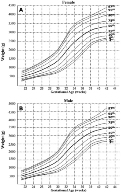
RESULTS
In Table I one can find the 3rd, 5th, 10th, 25th, 50th,
90th, 95th and 97th percentiles of birth weights for
Figure 1 shows the graphical representation of these percentiles.
Fig. 1 – Graphical representation of the 3rd, 5th, 10th, 25th, 50th, 75th, 90th, 95thand 97thpercentiles for females and males.
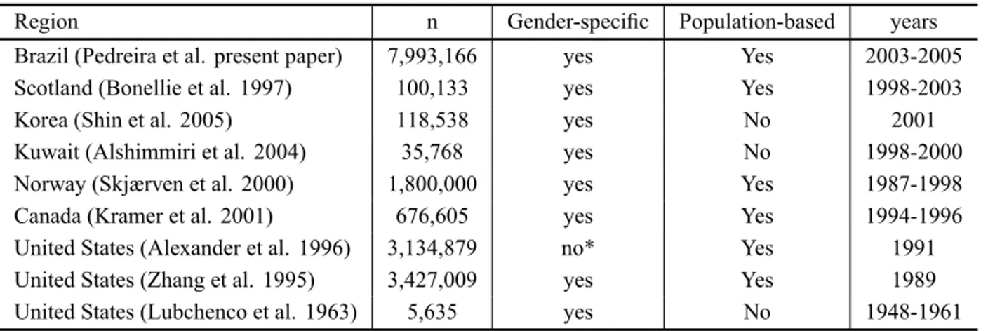
In Table II we present a comparative view concern-ing some design aspects, of the present paper and other different studies. It is worth noting that most of the pre-vious charts was based on developed countries data and that the Brazilian charts were built up with nearly 8 mil-lion newborns, more than double the sample size of the largest early studies.
In Table III one may find a comparative tabula-tion of the relative percentual differences for the 10th,
50th and 90th percentiles, for males, between four
pre-vious published charts (Kramer et al. 2001, Alshimmiri et al. 2004, Bonellie et al. 2008, Alexander et al. 1996) and the present paper, used as reference. Negative and positive quantities in Table III reflect that the
Brazil-ian percentiles were, respectively, smaller or larger, in comparison with the other four references. Negative numbers indicate that the percentiles are overestimated in comparison to the Brazilian ones, while positive values means underestimation in relation to present study percentiles. Note that all values are percentual. For instance, the male percentile 90 from Table III for 40 weeks would be overestimated by 4.1% if (Bonellie et al. 2008) reference was used.
Following the way the references are published by different authors, for Bonellie we used the average be-tween nulliparous and multiparous for comparison pur-pose. Although (Alexander et al. 1996) provide the 10th
percentile values for both males and females, the data is not gender-specific for most percentiles, and so the same values were used in Table III.
DISCUSSION
Birth weight for gestational age charts is an essential tool providing relevant information to clinicians regard-ing which newborns may be at higher risk of neonatal morbidity and subsequent mortality or developmental delay. In this paper we present the first comprehensive charts with Brazilian data.
Table II allows a comparative view of the Brazil-ian study presented in this paper and some of the pub-lished charts. It can be noticed that the only pubpub-lished population-based studies with considerably large sam-ple size refer to data collected more than 15 years ago.
From the ethnic standards point of view, the Brazil-ian population is a truly melting pot. Accordingly, the present study constitutes a portrait of the birth weight for gestational age of a mixed population based on nearly 8 million observations.
Table III should be viewed with caution since the studies over ethnic and socioeconomic diversities embrace substantial methodological differences among each other. Nevertheless, it is interesting to note that the Brazilian chart produced lower percentiles (negative values in Table III) for the majority of the gestational weeks if compared to (Alexander et al. 1996). Further-more, the 50th percentile is generally higher for the
Brazilian data at very low gestational ages (up to 26th
TABLE II
Different published studies for assessing birth weight for gestational age.
Region n Gender-specific Population-based years Brazil (Pedreira et al. present paper) 7,993,166 yes Yes 2003-2005 Scotland (Bonellie et al. 1997) 100,133 yes Yes 1998-2003 Korea (Shin et al. 2005) 118,538 yes No 2001 Kuwait (Alshimmiri et al. 2004) 35,768 yes No 1998-2000 Norway (Skjærven et al. 2000) 1,800,000 yes Yes 1987-1998 Canada (Kramer et al. 2001) 676,605 yes Yes 1994-1996 United States (Alexander et al. 1996) 3,134,879 no* Yes 1991 United States (Zhang et al. 1995) 3,427,009 yes Yes 1989 United States (Lubchenco et al. 1963) 5,635 yes No 1948-1961
* only the 10thpercentile values are provided for both males and females.
Overall, despite the methodological discrepancies and the expected consequent diversion in percentile values, the Brazilian chart is quite consistent with previous studies. The greatest divergences appeared in relation to (Alshimmiri et al. 2004) charts, which are maybe explained by its quite small sample size. It should be noticed that the differences among these studies may be in part attributed to differences in the statistical pro-cedures applied in different papers.
Several approaches are concerned with misestim-ation of the gestmisestim-ational age for a proportion of new-borns (Kramer et al. 2001, Bonellie et al. 2008, Platt et al. 2001, Oja et al. 1991, Hutcheon and Platt 2008). This misclassification may produce curves that are not smooth or biologically plausible. Undesirable ef-fects like bumps in extreme percentiles, specially around weeks 28 to 30, have been reported (Kramer et al. 2001, Bonellie et al. 2008). We did not observe these distor-tions in our curves maybe because of the huge size of the sample.
We hope the presented charts will be useful to clin-icians in classifying fetal growth. They may also con-tribute as a reference for investigations of predictors and to show the consequences of small and large patterns for gestational age delivery. At last, some limitations should be acknowledged. Our study is cross-sectional, as all population-based gestational age references. Pos-sible bias due to missing data caused by the absence of the weights of the fetuses still in utero (Hutcheon and Platt 2008) is also common to all charts, including ours. Finally, the accurate determination of the gestational age
in population-based studies is an open challenge for all charts and the practical adequacy of different measures is a stirring investigation problem (Wingate et al. 2007).
ACKNOWLEDGMENTS
This work has been partially supported by grants from Conselho Nacional de Desenvolvimento Científico e Tecnológico (CNPq), and Fundação Carlos Chagas Fi-lho de Amparo à Pesquisa do Estado do Rio de Janeiro (FAPERJ).
RESUMO
Background e objetivos.Apresentamos um retrato atualizado de peso-por-idade-gestacional, baseado em quase 8 milhões de observações em uma população etnicamente misturada. Estas constituem as primeiras tabelas com dados brasileiros. Esta contribuição pretende dar assistência aos clínicos na classi-ficação do crescimento fetal, e prover uma referência para pesquisas de prognósticos e consequências em partos com pa-drões pequenos e grandes para a idade gestacional. A maior parte dos dados de referência para estimar peso-por-idade-ges-tacional sofre de tamanho de amostra insuficiente, especial-mente em baixa idade gestacional. Os estudos populacionais com uma amostra de tamanho considerável se referem a dados coletados há mais de 15 anos.
Métodos.Nós realizamos um estudo populacional baseado em
Resultados. As tabelas e representações gráficas resultantes proveem uma referência gênero-específica para acessar a dis-tribuição de peso ao nascimento de acordo com a idade gesta-cional na população brasileira.
Conclusões. Esta é a primeira referência populacional
cons-truída com dados de um país em desenvolvimento. Estas ta-belas podem prover uma importante ferramenta para melhorar a avaliação clínica do crescimento em recém-natos.
Palavras-chave: peso ao nascimento, recém-nascido, idade
gestacional, crescimento fetal, pré-termo, pós-termo.
REFERENCES
ALEXANDERGR, HIMESJH, KAUFMANRB, MORJAND
KOGANM. 1996. A United States national reference for
fetal growth. Obstetrics & Gynecology 87: 163–168. ALSHIMMIRI MM, AL-SALEH EA, ALSAEID K, HAM
-MOUD MS AND AL-HARMI JA. 2004. Birth weight percentiles by gestational age in Kuwait. Arch Gynecol Obstet 269: 111–116.
BONELLIES, CHALMERSJ, GRAYR, GREERI, JARVISS
ANDWILLIAMSC. 2008. Centile charts for birth weight for gestational age for Scottish singleton births. BMC Pregnancy Childbirth 25: 8–15.
BONELLIESRANDRAABGM. 1997. Why are babies
get-ting heavier? Comparison of Scottish births from 1980 to 1992. BMJ 315: 1205.
CHIKE-OBI U, DAVID RJ, COUTINHO R AND WU SY. 1996. Birth weight Has Increased Over a Generation. Am J Epidemiol 164: 563–569.
FRITSCHFNANDCARLSONRE. 1980. Monotone Piecewise Cubic Interpolation, SIAM. J Num Anal 17: 238–246. HUTCHEON JA AND PLATT RW. 2008. The missing data
problem in birth weight percentiles and thresholds for small-for-gestational-age. Am J Epidemiol 167: 793–796. KAHANER D, MOLER CANDNASH S. 1989. Numerical
Methods and Software. London: Prentice Hall, 495 p.
KIERANSWJ, JOSEPHKS, LUOZC, PLATTR, WILKINS
RANDKRAMERMS. 2008. Does one size fit all? The case for ethnic-specific standards of fetal growth. BMC Pregnancy Childbirth 8: 1.
KRAMERM, PLATTRW, WEN SW, JOSEPHKS, ALLEN
A, ABRAHAMOWICZM, BLONDELB, BRÉARTGAND
FETAL/INFANTHEALTHSTUDYGROUP OF THECANA -DIAN PERINATAL SURVEILLANCE SYSTEM. 2001. A
new and improved population-based reference for birth-weight for gestational age. Pediatrics 108: E35.
LUBCHENCO L, HANSMANC, DRESSLER MAND BOYD
E. 1963. Intrauterine growth as estimated from liveborn birth weight data at 24 to 42 weeks of gestation. Pedi-atrics 32: 793–800.
OJAH, KORAINENMANDRANTAKALLIOP. 1991. Fitting
mixture models to birth weight data: a case study. Bio-metrics 47: 883–897.
PLATTRW, ABRAHAMOWICZM, KRAMER MS, JOSEPH
KS, MERYL, BLONDELB, BRÉARTGANDWENSW. 2001. Detecting and eliminating erroneous gestational ages: a normal mixture model. Stat Med 20: 3491–3503. SHIN SM, CHANG YP, LEE ES, LEE YA, SON DW, KIM MH AND CHOI YR. 2005. Birth Weight, Very Low Birthweight Rates and Gestational Age-Specific Birth weight Distribution of Korean Newborn Infants. J Korean Med Sci 20: 182–187.
SKJÆRVENR, GJESSINGHKANDBAKKETEIGLS. 2000.
Birth weight by gestational age in Norway. Acta Obst et Gynec Scand 796: 440–449.
WINGATE MS, ALEXANDERGR ANDBUEKENSP. 2007. Comparison of Gestational Age Classifications: Date of Last Menstrual Period vs. Clinical Estimate. Ann
Epi-demiol 17: 425–430.
ZHANGJANDBOWESW JR. 1995.