Incidence and multiplex PCR based detection of trichothecene chemotypes of
Fusarium culmorum
isolates collected from freshly harvested Maize kernels in
Southern India
M. Venkataramana, P. Shilpa, K. Balakrishna, H.S. Murali, H.V. Batra
Defence Food Research Laboratory, Microbiology Devision, Sidartha Nagar, Mysore, Karnataka, India.
Submitted: October 29, 2011; Approved: July 23, 2012.
Abstract
Hundred Fusarium culmorum strains, isolated from freshly harvested maize grain samples from Southern parts of India, were incubated in czapek-dox medium and analyzed for trichothecene (DON/NIV) production. The mPCR assay was standardized targeting trichothecene metabolic path-way genes viz.,Tri6,Tri7,Tri13for detection of trichothecene (DON/NIV) chemotypes andrDNA gene for specific detection ofF. culmorumspecies. Primers for targeted genes were designed and used to predict whether these isolates could produce deoxynivalenol/nivalenol, 94 isolates were able to produce DON/NIV by mPCR assay. Chemical analysis of DON/NIV was carried out for mPCR positive isolates by high performance-thin layer chromatography (HPTLC). To check the practical usefulness of developed mPCR assay, 150 field samples of maize were evaluated and results were compared with conventional HPTLC method. Out of 150 samples, 34% samples stayed as a positive for NIV contamination whereas 44% were found to have deoxynivalenol contamination. Moreover, mPCR results are equivocally matched with the HPTLC chemical analysis for field samples. Chemotyping ofF. culmorumisolates were reported for the first time from India, and highlights the important potential ofF. culmorumto contaminate maize with DON/NIV.
Key words:deoxynivalenol, nivalenol, multiplex PCR assay, HPTLC.
Introduction
The genusFusariumis a common fungal contaminant of many economically important field crops and food prod-ucts, which causes a major contamination in human and an-imal nutrition.Fusarium sp. infects many important food grains, such as maize, wheat, barley, rice, millet, oat and rye, and produce highly toxic secondary metabolites known as mycotoxins. The major classes ofFusariummycotoxins are trichothecenes and fumonisins. According to the type of trichothecene production, someFusariumspecies, likeF. culmorumand other species have been divided into two chemotypes: (i) the nivalenol chemotype, which includes isolates producing nivalenol and fusarenone X, and (ii) the deoxynivalenol chemotype, which includes isolates pro-ducing deoxynivalenol and acetyldeoxynivalenol (Langseth et al., 1999; Muthomi et al., 2000). Tricho-thecenes can cause wide range of acute and chronic effects
in humans and animals through ingestion of food and feed prepared from cereal crops contaminated with the toxins (Melloet al., 1999). The effects include skin inflammation, digestive disorders, trachycardia, oedema and haemor-rhages in several internal organs, haemolytic disorders, im-pairment of immune disorders and nervous disorders (IARC, 1993).
Contamination of maize grains with trichothecenes is a common problem throughout the World, especially in temperate regions like India (Janardhanaet al., 1999; Ven-kataramanaet al., 2011). In Southern India, maize is grown under different ecological conditions and is harvested with fairly high trichothecene contaminations (Janardhanaet al., 1999). Recent reports indicate that maize is prone to fungal infection during the pre and post harvest period (Bilgrami et al., 1980; Sinha, 1990). Vasanthkumar (Vasanthkumar, 1986) studied the infection of maize by field and storage
Send correspondence to M. Venkataramana. Defence Food Research Laboratory, Microbiology Devision, Sidartha Nagar, Mysore, 570011Karnataka. E-mail: ramana.micro@gmail.com.
moulds during pre and post harvest practices in relation to seed-borne fungal diseases of maize. Thimmappaiahet al. (1987) reported the natural occurrence ofFusariumtoxins such as T-2 toxin, deacetoxyscirpenol and zearalenone in peanut, sorghum and maize from Mysore districts of South-ern India. Pre-harvest succession of fungi in ripening maize grains has been reported by Banerjeeet al.(Beyeret al., 2005).
The usual methods for chemotyping ofFusarium iso-lates are high performance liquid chromatography (HPLC) or gas chromatography/ mass spectroscopy (GS/MS) anal-ysis of extracts from substrates such as maize artificially in-oculated withFusarium(Sugiuraet al., 1990; Muthomiet al., 2000). These methods are commonly applied due to their high sensitivity and specificity. However, these meth-ods are rather labour-intensive and require sophisticated in-strumentation and skilled operators. Recent studies are fo-cusing genotypic identification of toxigenic fungi through molecular methods. DNA based methods that rely on the amplification of the genes involved in the biosynthesis of trichothecenes also are available. Specific PCR primers have been developed to the specific genes (Tri5, Tri6, Tri7 andTri13) which are involved in trichothecene metabolism (Venkataramanaet al., 2011; Leeet al., 2001). Some of the genes have been sequenced and found to be functional in NIV-producing isolates and nonfunctional in DON-pro-ducing isolates (Lee et al., 2002, 2001; Chandler et al., 2003).
The distribution of each chemotype/genotype varies by geographic region. Thus, strains ofF. culmorumwith DON and NIV chemotype/ genotype are known from sev-eral countries, including UK (Jenningset al., 2004), Ger-many (Muthomiet al., 2000), the Netherlands and Norway (Langsethet al., 1999), Italy (Gang et al., 1998), France (Bakanet al., 2001), USA (Mirochaet al., 1994), Canada (Abramsonet al., 2001). However in India, we are first time reporting incidence of trichothecene chemotypes of F. culmorumon maize kernels by multiplex PCR method.
In the present study a novel mPCR method was devel-oped for detection of trichothecene chemotypes of F. culmorum. Developed method was successfully evaluated in terms of sensitivity, specificity and reliability on to artifi-cially contaminated samples as well as contaminated field samples of maize.
Materials and Methods
Sampling area
A total of 150 freshly harvested maize kernel samples were randomly collected from Karnataka and Andhra Pra-desh, two districts from each state viz., Kusalnagara and Mysore districts of Karnataka; Srikakulam and Viziana-garam districts of Andhra Pradesh, India. Collected sam-ples were stored at 4 °C for further analysis of mycoflora and toxin detection.
Isolation and identification of moulds from infected maize grains
Isolation of mycoflora
F. culmorumand other fungal genera were isolated from the infected maize grains using Potato Dextrose Agar (PDA) media. The morphological identification of Fusariumspecies were followed by the previous reports of Nelsonet al. (1983). The pure cultures of Fusariumwas maintained on SNA plates and stored at 4 °C for analysis of toxins.
DNA extraction
DNA was extracted from pure cultures ofFusarium sp., using commercially available DNA extraction kits (QUIAZEN, Gambh).
Isolation of DNA from contaminated food grains
Twenty grams of contaminated food sample was ground in coffee grinder for 90 s and then 0.2 g ground grain was mixed in 1 mL lysis buffer (100 mMTris-HCl, 50 mM EDTA, 150 mM NaCl and 1% SDS) and homoge-nized by gentle mixing and kept it for water bath at 60 °C for 10 min. Samples were centrifuged at 12000 x g for 5 min and 500mL of supernatant was mixed with 150mL of
5 M potassium acetate and incubated on ice for 10 min. Af-ter centrifugation a 400mL of supernatant was mixed with
300mL of ice cold isopropanol to precipitate the DNA, the
resulting pellet was washed with 70% ethanol, dried under air and dissolved in 50mL ofTris-EDTA (pH 8.0).
Development of primers and PCR analysis
Designing of primers
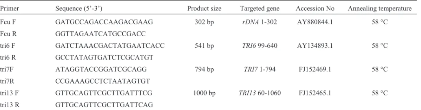
DNA sequences were analysed and aligned by Clustal method. Primers were designed using the aligned gene bank database sequences viz.,Tri6, Tri7, Tri13andrDNA genes for the specific detection of nivalenol and deoxy-nivalenol producing F. culmorum. Total of four primer pairs were designed using Gene runner software (http://www.generunner.com). Primer sequences are listed in Table 1. Before standardizing mPCR protocol, all de-signed primers were evaluated on to array of fungal species to check the specificity and sensitivity.
Multiplex PCR assay
Multiplex PCR was carried out for DON and NIV producingF. culmorumin an Eppendorf master cycler gra-dient (Hamburg, Germany) with a reaction volume of 30 mL. The amplification mixture consisted of template
DNA (1.0mL), MgCl2(2.0 mM), 1X PCR buffer (Sigma),
dNTP mix (200mM, MBI, Fermentas), Taq polymerase (1
de-naturation at 94 °C for 4 min, followed by 30 cycles of 94 °C for 1 min, 58 °C for 1 min and 72 °C for 1.5 min, with a final extension of 72 °C for 8min.
Specificity and sensitivity of mPCR on artificially contaminated maize grains
The specificity of the mPCR primers was determined against different organisms shown in (Table 2). Sterile maize grains (5 g) were experimentally spiked with F. culmorum spore suspensions at different concentrations (1x10⁵, 1x10⁴, 1x10³and 1x10²cfu g-1). Negative controls were kept without inoculation of spores. All the samples were enriched for two days, further DNA was isolated and analysed by mPCR assay.
Application of mPCR on field samples
Analysis of field samples
Maize samples were collected from various fields of Andhra Pradesh and Karnataka, India and processed as de-scribed earlier and all of these samples were subjected to mPCR assay and toxins are analyzed by HPTLC.
Chemical analysis of DON and NIV
Extraction and cleanup
Briefly, 50 g of the well ground sample was extracted with 250 mL of acetonitrile-water (60:40, v/v) using high speed blending for 2 min. The extract was filtered through Whatman No. 4 filter paper and an equal volume of ethyl acetate was added to the filtrate and separated the lower chloroform fraction and evaporated to dryness. Dried com-pound was diluted with PBS and passed through immu-noaffinity columns (VICAM, USA) for clean-up. After washing with water, DON /NIV were eluted with methanol. These extracts further used for HPTLC analysis.
HPTLC detection
High performance thin layer chromatography (HPTLC) technique was used to detect the specific che-motypes of (DON /NIV) F. culmorum. Chromatography was carried out on 10 x 10 cm precoated silica gel HPTLC plates (Merck). Test samples and standards were applied
with automatic TLC sampler (ATS III) from CAMAG (Muttenz, Switzerland), with a 50 mm run length using: chloroform + methanol + water (9+1+0.2, v/v/v.) as mobile phase. Fluorescence detection was carried out by post-chromatographic derivatization with 10% aluminium chlo-ride (Sigma) in methanol-water mixture. The plate was im-mersed in the derivatization solution using a dipping device III (CAMAG) and heated for 20 min at 110 °C. The plate was then scanned at 366/ > 400 nm with a densitometer TLC Scanner 3 (CAMAG) using a slit dimension of 5.0 x 0.5 mm and scanning speed of 40 mm s-1(18). DON and NIV standards (Sigma) at a concentration of 0.1 mg/mL in methanol: water (1:1) was used as positive controls for HPTLC detection.
Results
Incidence of mycotoxigenicFusarium culmorumand
other fungi
A total of 150Fusariumisolates were identified from the maize samples of the present study. Out of 150 fungal isolates, 100 isolates were identified with F. culmorum morphologically (45 isolates from Karnataka and 55
iso-Table 1- Primers used in this study.
Primer Sequence (5’-3’) Product size Targeted gene Accession No Annealing temperature
Fcu F GATGCCAGACCAAGACGAAG 302 bp rDNA1-302 AY880844.1 58 °C
Fcu R GGTTAGAATCATGCCGACC
tri6 F GATCTAAACGACTATGAATCACC 541 bp TRI699-640 AY134893.1 58 °C
tri6 R GCCTATAGTGATCTCGCATGT
tri7F ATAGGTACCGGATCGCAGG 794 bp TRI71-794 FJ152469.1 58 °C
tri7R CCGAAAGCCTCTAATAGTGT
tri13 F GTTGCAGTTCGCTTGATTTCG 1000 bp TRI1360-1060 FJ152465.1 58 °C
tri13 R GTTGCAGTTCGCTTGATTCAG
Table 2- Standard cultures used in this study.
SNO Name Source
1 Fusarium culmorumITCC146 IMTECH 2 Fusarium culmorumITCC149 ITCC 3 Fusarium culmorumITCC148 ITCC 4 Fusarium graminearumMTCC 1893 MTCC 5 Fusarium graminearumITCC 1805 ITCC 6 Fusarium moniliformeITCC 3362 ITCC 7 Fusarium moniliformeATCC 14164 IMTECH 8 Fusarium oxysporumNCIM 1072 NCIM 9 Fusarium oxysporumIMTECH 2480 IMTECH 10 Fusarium solaniITCC3359 IMTECH
lates from Andhra Pradesh) and remaining 50 isolates from other species including F. sporotrichioides, F. verticillioidesandF. proliferatum(Table 3).
Multiplex PCR assay application and chemical analysis of pure cultures of fungi
Primer concentrations (100 nM of Tri6, 150 nM of Tri7, 200 nM of Tri13 and 50 nM of rDNA) and annealing temperature (58 °C) were standardized to get a uniform am-plification of all the genes targeted for mPCR assay (Figu-re 1). Out of 150 Fusarium isolates, 100 were showed positive signal forrDNAgene specific toF. culmorum(94 toxigenic to DON/NIV and remaining were non toxigenic by mPCR). However, 54 and 34 strains of them were posi-tive for DON and NIV respecposi-tively, and rest of the 7 strains were stayed as negative for the chemical analysis by HPTLC.
Contamination studies
The detection limit for deoxynivalinol and nivalenol producing F. culmorum strains from spiked samples by mPCR was 1x10³cfu g-1maize grains. Samples tested with initial fungal load of 1x10³cfu g-1and above concentrations following 48 h enrichment at 30 °C were positive for both toxins.
mPCR and chemical analysis of field samples
Out of 150 maize samples collected for mPCR stud-ies, 78% samples were showed positive for toxigenic Fusariumspecies. However, only 34% of the total analysed samples were positive for NIV and 44% positive for DON chemotypes. Toxin analysis by HPTLC providing equivo-cal results with mPCR for both the groups of toxins.
Discussion
It would be more meaningful if analytical systems are made available for low cost, simple to use qualitative and
for quantitative assessments of the mycotoxigenic fungi and mycotoxins present in the different food matrices. Con-ventional methods for the detection ofFusariumbased on sporodochia with abundant macroconidia on the chaff sur-face is time consuming and laborious, however, PCR as-says have proven to be very useful and sensitive where sporodochia are absent or in poorly developed state (Ni-cholsonet al., 1998). Multiplex PCR assays have been ap-plied for the detection of air samples and for diseased plant, animal and human tissues for the presence of bacteria, para-sites, viruses and fungi (Yennyet al., 2009; Mohdet al., 2010). In this study, we attempted a mPCR assay with an objective to obtain simultaneous detection of trichothecene chemotypes ofF. culmorumspp. that are commonly associ-ated withFusariumdisease.
A total of 150 Fusarium isolates originated from Andhra Pradesh and Karnataka, India were assayed by mPCR/HPTLC for their mycotoxin chemotypes. Results were showed that 100 strains have therDNAgene specific toF. culmorum. Additionally mPCR amplification ofTri6, Tri7 and Tri13 alleles suggests that 94% of the F. culmorumisolates are able to produce DON/NIV. Some other researchers observed that, the same NIV and DON ac-cumulation byF. culmorumin wheat grains (Kammounet al., 2009; Lobnaet al., 2010). However, to our knowledge in India, we are reporting first time DON/NIV chemotypes in maize kernels. In the present study, DON chemotypes were more aggressive when compared to NIV chemotypes ofF. culmorum. The higher incidence of DON chemotypes may be due to intra specific variation of gene clusters ofF. culmorum strains (Bakan et al., 2001). The findings of Jennings et al. (2004) were confirmed our results, that DON chemotypes dominance in F. culmorum strains. Whereas, Laurenet al. (1992) and Lee et al. (2002) re-ported contradictory results that NIV dominants in F. culmorum.
The four DNA amplicons scored (Figure 1) were serve as a diagnostic tool for the early detection of tricho-thecene chemotypes ofF. culmorum. The 300 bp amplicon is signifies therDNAgene specific toF. culmorum, 546 bp region ofTri6gene is specific to all trichothecene produc-ingFusariumspecies (both type A and type B) whereas, 900 bpTri7gene region specific to NIV chemotype and 1000 Bp region ofTri13gene is specific to DON chemo-types ofF. culmorumwas used in this study.
Results of the molecular assays (mPCR) were firmed by chemical analysis by HPTLC. Thus HPTLC con-ducted on 94 mPCR positiveF. culmorumisolates revealed that 54 isolates were positive for DON and 33 were positive for NIV toxin, whereas, 7 strains were negative for chemi-cal analysis. The variation in mPCR and HPTLC analysis is not unexpected since Quartaet al.(2005) and Ramana et al.,(2011) compared the molecular analysis with chemical analysis for toxigenicFusariumspecies and made the simi-lar kind of findings. Thus while a positive trichothecene
Figure 1 - Multiplex PCR photograph for DON and NIV producing
genotype indicates the potential for trichothecene biosyn-thesis, only a test for the toxin itself can be used to deter-mine if and how much toxin a strain has produced. In vitro, DON/NIV may be produced by utilizing very different cul-ture conditions such as whole grain, solid substrate fermen-tation, or liquid cultures using a defined minimal medium. Our results corroborate that growth conditions greatly in-fluence the amount of mycotoxin produced. trichothecene biosynthesis may be regulated by temperature (Ramirezet al., 2006), relative humidity (Beyeret al., 2005), and sub-strate composition (ONeillet al., 1993). Results lead to the conclusion that in vitro assays could not appropriate to pre-dict production of DON in the field as suggested by Ganget al.(1998). The physiology of plant-hosts and pathogenesis of the strain itself may further influence mycotoxin accu-mulation under field conditions. However, in the case of field samples mPCR results are equivocally matched with the chemical analysis. So, the newly developed mPCR as-say is an alternative for the time consuming and laborious conventional culture methods for early assessment of tri-chothecene chemotypes ofF. culmorum from field sam-ples. The present research has demonstrated that, the occur-rence ofF. culmorumin Southern India (Andhra Pradesh and Karnataka) is dominant; it may due to cool climatic conditions and high moisture and rainfalls. Global variation in DON/NIV production by isolates ofF. culmorum and distribution of these isolates geographically and by host are important in Plant pathology and food safety and security.
Conclusion
High levels of toxigenicF. culmorumincidence in maize samples demonstrates the need for better surveil-lance and monitoring by policy makers or food toxicolo-gists to reduce the exposure of human and animal life to toxic compounds produced by fungi.
Acknowledgments
Authors are thankful to the director DFRL, Mysore, for his providing necessary facilities to carry out the work.
References
Abramson D, Clear RM, Gaba D, Smith D, Patrick SK, Saydak D (2001) Trichothecene and moniliformin production by Fusarium isolates from western Canadian wheat. J Food Prote 64:1220-1225.
Bakan B, Pinson L, Cahagnier B, Melcion D, Sémon E, Richard-Molard D (2001) Toxigenic potential of Fusarium culmorumstrains isolated from French wheat. Food Addi Contami 18:998-1003.
Banerjee A, Shetty HS, Majumdar SK (1988) Succession of mycoflora in ripening maize grain. Indian Phytopathology 41:562-566.
Beyer M, Verreet JA, Ragab WSM (2005) Effect of relative hu-midity on germination of ascospores and macro conidia of
Gibberella zeaeand deoxynivalenol production. Int J Food Microbiol 98:233-240.
Bilgrami KS, Prasad T, Misra RS, Sinha KK (1980) Survey and study of mycotoxin producing fungi associated with the grains in standing maize crops. Final Technical Report. ICAR project, Bhagalpur University, India.
Chandler EA, Simpson DR, Thomsett MA, Nicholson P (2003) Development of PCR assays toTri7andTri13trichothecene biosynthetic genes, and characterisation of chemotypes of Fusarium graminearum, Fusarium culmorumandFusarium cerealis. Phys Mol Plant Pathol 62:355-367.
Chu FS (1996) Recent studies on immunoassays for mycotoxins. In: Beier RC, Stanker LH (eds) Immunoassays for Residue Analysis: Food Safety. ACS Symposium Series 621. Ameri-can Chemical Society, Washington DC, pp 294-313. Gang G, Miedaner T, Schuhmacher U, Schollenberger M, Geiger
HH (1998) Deoxynivalenol and nivalenol production by Fusarium culmorumisolates differing in aggressiveness to-ward winter rye. Phytopathology 88:879-884.
IARC (1993) Some Naturally Occurring Substances: Food Items and Constituents, Heterocyclic Aromatic Amines and Mycotoxins, Volume 56. WHO, Lyon, 599 pp.
Janardhana GR, Raveesha KA, ShekarShetty H (1999) Myco-toxin contamination of maize grains grown in Karnataka (India). Food Chem Toxicol 37:863-868.
Jennings P, Coates ME, Turner JA, Chandler EA, Nicholson P (2004) Determination of deoxynivalenol and nivalenol chemotypes ofFusarium culmorumisolates from England and Wales by PCR assay. Plant Pathol 53:182-190. Kammoun GL, Gargouri S, Hajlaoui MR, Marrakchi M (2009)
Occurrence and disTribution of Microdochium and Fusariumspecies isolated from durum wheat in northern Tunisia and detection of mycotoxins in naturally infested grain. J Phytopathol 157:546-551.
Langseth W, Bernhoft A, Rundberget T, Kosiak B, Gareis M (1999) Mycotoxin production and cytotoxicity ofFusarium strains isolated from Norwegian cereals. Mycopathologia 144:103-113.
Lauren DR, Sayer ST, Dimenna ME (1992) Trichothecene pro-duction byFusariumspecies isolated from grain and pasture throughout New Zealand. Mycopathologica 120:167-176. Lee T, Han YK, Kim KH, Yun SH, Lee YW (2002)Tri13andTri7
determine deoxynivalenol and nivalenol producing chemo-types ofGibberella zeae. App Environ Microbiol 68:2148-2154.
Lee T, Oh DW, Kim HS, Lee J, Kim YH, Yun SH, Lee YW (2001) Identification of deoxynivalenol and nivalenol producing chemotypes ofGibberella zeaeby using PCR. App Environ Microbiol 67:2966-2972.
Lobna GK, Samia G, Christian B, Florence R, Mohamed RH (2010) Trichothecene chemotypes ofFusarium culmorum infecting wheat in Tunisia. Int J Food Microbiol 140:84-89. MarioV, Daniela C. (2006) Determination of Deoxynivalenol in
wheat by validated GC/ECD method: Comparison with HPTLC/FLD. Electronic. J Food Plant Chem 1:16-20. Mello JPFD, Placinta CM, MacDonald AMC (1999)Fusarium
mycotoxins: A review of global implications for animal health, welfare and productivity. Anim Feed Sci Technol 80:183-205.
my-cotoxins by Fusarium graminearum and Fusarium culmorumor barley and wheat. Mycopathologia 128:19-23. Mohd E, Elhassan N, Kwai LT (2010) Differentiation of
Salmo-nella enterica based on PCR detection of selected somatic and flagellar antigens. Afr J Microb Res 4:871-876. Muthomi JW, Schutze A, Dehne HW, Mutitu EW, Oerke EC
(2000) Characterization ofFusarium culmorumisolates by mycotoxin production and aggressiveness to winter wheat. Z Pflanzenk Pflanzen 107:113-123.
Nelson PE, Toussoun TA, Marasa WFO (1983)Fusariumspecies. An Illustrated Manual for identification. Pennsylvania State University Press, University Park.
Nicholson P, Simpson DR, Weston G, Rezanoor HN, Lees AK, Parry DW, Joyce D (1998) Detection and quantification of Fusarium culmorumandFusarium graminearumin cereals using PCR assays. Phy Mole P Pathol 53:17-37.
ONeill K, Damaglou AP, Patterson MF (1993) Toxin production by Fusarium culmorumIMI 309344 andF. graminearum NRRL 5883 on grain substrates. J App Bacteriol 74:625-628.
Quarta A, Mita G, Haidukowski M, Santino A, Mule G, Visconti A (2005) Assessment of trichothecene chemotypes of Fusarium culmorum occurring in Europe. Food Addi Contam 22:309-315.
Ramirez ML, Chulze S, Magan N (2006) Temperature and water activity effects on growth and temporal deoxynivalenol
pro-duction by two Argentinean strains of Fusarium graminearum on irradiated wheat grain. Int J Food Microbiol 106:291-296.
Sinha K (1990) Incidence of mycotoxins in maize grains in Bihar State (India). Food Addi Contam 7:55-61.
Sugiura Y, Watanabe Y, Tanaka T, Yamamoto S, Ueno Y (1990) Occurrence of Gibberella zeaestrains that produce both nivalenol and deoxynivalenol. App Environ Microbiol 56:3047-3051.
Thimmappaiah N, Bhavanishankar P, Shantha T (1987) Natural occurrence of Fusarium toxins in peanut, sorghum and maize from Mysore (India). J Sci Food Agric 40:127-132. Vasanthkumar. (1986) Studies on some seed-borne fungal
dis-eases of maize in Karnataka. PhD Thesis, University of My-sore, India.
Venkata Ramana M, Balakrishna K, Murali HS, Batra HV (2011) Multiplex PCR-based strategy to detect contamination with mycotoxigenic Fusariumspecies in rice and fingermillet collected from southern India. J Sci Food Agric 91:1666-1673.
Yenny S, Maribeth A, Cousin B, Charles P, Woloshuk (2009) Multiplex real-time PCR for detection and quantification of mycotoxigenic Aspergillus, Penicillium and Fusarium. J Stor Pro Res 45:139-145.