online | memorias.ioc.fiocruz.br
Dipyridamole potentiated the trypanocidal effect
of nifurtimox and improved the cardiac function
in NMRI mice with acute chagasic myocarditis
Sonia Santeliz1, Peter Caicedo2, Elidiosmar Giraldo2, Carmen Alvarez1, María-Daniela Yustiz2,
Claudina Rodríguez-Bonfante3, Romina Bonfante-Rodríguez2, Rafael Bonfante-Cabarcas2/+
1Decanato de Ciencias Veterinarias, Unidad de Biomedicina, Departamento de Medicina y Cirugía, Barquisimeto, Estado Lara, Venezuela 2Decanato de Ciencias de la Salud, Unidad de Bioquímica, Barquisimeto, Estado Lara, Venezuela
3Universidad Centroccidental Lisandro Alvarado, Unidad de Parasitología Médica, Barquisimeto, Lara, Venezuela
BACKGROUND As chronic Chagas disease does not have a definitive treatment, the development of alternative therapeutic protocols is a priority. Dipyridamole (DPY) is an alternative to counteract the pathophysiological phenomena involved in Chagas cardiomyopathy.
OBJECTIVE To evaluate the therapeutic efficacy of DPY associated with nifurtimox (Nfx) in epimastigote axenic cultures and in mice with acute Chagas disease.
METHODS NMRI adult male mice were divided into nine groups: three healthy and six Trypanosoma cruzi-infected groups. Mice received vehicle, Nfx or DPY, alone or combined. The doses assayed were Nfx 10 and 40 mg/kg and DPY 30 mg/kg. The treatment efficacy was evaluated by clinical, electrocardiographic, parasitological, biochemical and histopathological methods.
FINDINGS In vitro, DPY and Nfx had a trypanocidal effect with IC50 values of 372 ± 52 and 21.53 ± 2.13 µM, respectively; DPY potentiated the Nfx effect. In vivo, Nfx (40 mg/kg) with or without DPY had a therapeutic effect, which was reflected in the 84-92% survival rate and elimination of parasitaemia and heart tissue amastigotes. Nfx (10 mg/kg) had a subtherapeutic effect with no survival and persistence of amastigotes, inflammation and fibrosis in heart tissue; adding DPY increased the survival rate to 85%, and all tested parameters were significantly improved.
MAIN CONCLUSION DPY has a trypanocidal effect in vitro and enhances the Nfx therapeutic effect in an in vivo murine model.
Key words: Chagas disease - nifurtimox - dipyridamole - Trypanosoma cruzi - treatment
doi: 10.1590/0074-02760160499
Financial support: National Fund for Science and Technology (FONACIT) under the Ministry of Popular Power for Science and Technology (Venezuela) (Project No 2007001425) and by CDCHT at Universidad Centroccidental Lisandro Alvarado (Venezuela) (Doctoral project number 001-DVE-2014). + Corresponding author: [email protected]
Received 15 November 2016 Accepted 22 April 2017
In the last two decades, sufficient data have been generated to suggest that Chagas disease is a global pub-lic health problem. Approximately 6 million to 7 million people worldwide, mostly in Latin America, are estimated to be infected with Trypanosoma cruzi, the parasite that causes Chagas disease, which is found mainly in endemic areas of 21 Latin American countries, where it is estimated that there is a population of 70,199,360 individuals at risk of acquiring the infection; 5,742,167 infected people; and 29,925 new cases per year with a mortality of 12,000 indi-viduals per year (Dias et al. 2016, WHO 2017). Active vec-tor transmission remains, which is reflected in the onset of acute Chagas disease cases in regions infested by vectors (Añez et al. 2004), and there is re-emergence of the disease manifested by the appearance of circumscribed outbreaks by oral transmission (Alarcón de Noya et al. 2015). The migration of infected people from underdeveloped South American countries to developed countries has enabled
disease globalization, reporting a prevalence of 4.2% in these immigrant Latin-American populations in those countries (Requena-Méndez et al. 2015).
DPY enhanced Nfx effect • Sonia Santeliz et al. 597
is no guarantee that a parasitological “cure” in chronic Chagas disease patients translates into a change in the clinical prognosis of the patient, especially when sero-logical titres remain positive. Side effects depend on the generation of free radicals, and reactive oxygen species described for both drugs are an important issue, which causes demotivation in doctors who lack expertise in the use of both drugs. However, side effects are no longer a reason not to treat (Murcia et al. 2012).
An alternative view to developing new therapeutic drugs is the use of protocols in which the classical drugs Nfx and BZL are combined with potentially useful drugs, from the parasitological and pathophysiological point of view. The drugs to be tested should ideally have a trypano-cidal effect or enhance the trypanotrypano-cidal effect of Nfx or BZL, counteract the side effects of both drugs, stimulate the immune system, have an anti-inflammatory effect, and improve cardiac function; also, there must be sufficient ex-perience in the therapeutic use of the drug in humans.
Dipyridamole (DPY) is an old drug whose mechanism of action seems to indicate that it meets the aforementioned characteristics. It has been used primarily as a platelet aggregation inhibitor in preventing cerebral thrombo-embolic diseases. Its mechanism of action is based on in-ducing elevated levels of extracellular adenosine and by inhibiting the phosphodiesterase five (PDE5) enzyme. Adenosine has cardioprotective effects by decreasing the metabolic rate due to its chronotropic, inotropic and dromotropic negative effects, and it causes coronary va-sodilation, improving the coronary flow and preventing platelet aggregation, which reduces the chances of platelet thrombi development (Layland et al. 2014). Additionally, adenosine is an autacoid anti-inflammatory hormone with immunoregulatory properties that can limit the damage induced by inflammation and hypoxia (Palmer & Trev-ethick 2008). Furthermore, the molecular structure of DPY allows it to accept electrons, working as a free radi-cal scavenger and antioxidant agent (Kim & Liao 2008).
Parasite persistence and immune-mediated cardiac damage are considered the main mechanism for Chagas disease progression. However, an interesting point in the pathophysiology of Chagas cardiomyopathy involves mi-crovasculature disorders with increased platelet aggre-gation, resulting in vasospasm and thrombus formation and causing ischaemia and necrosis, which progressively leads to cardiac remodelling and chronic cardiomyopa-thy (Rossi et al. 2010). In addition, intense inflammation and free radical generation are key factors in the residual damage caused by T. cruzi (Vyatkina et al. 2004).
There is a need for new drugs because of a lack of efficacy of the current trypanocidal drugs to change the natural history of chronic Chagas disease cases. There-fore, given the pharmacological and therapeutic charac-teristics of DPY, it could help treat Chagas disease as an adjuvant drug by acting as a cardioprotective drug that should counteract some pathophysiological phenomena induced by infection and side effects induced by Nfx, decreasing the disease sequelae.
In the present paper, we study the therapeutic effect of DPY (30 mg/kg) combined with Nfx (10 or 40 mg/ kg) in NMRI male mice with acute chagasic myocarditis that were treated from the third week post-infection.
MATERIALS AND METHODS
T. cruzi axenic cultures - The strain of T. cruzi used in this work was M/HOM/VE/92/2-92-YBM. The strain has been maintained through successive passages from the vector to a susceptible host. The vectors were Rhod-nius prolixus stage III nymphs, and NMRI mice were used as a susceptible host. Drug inhibitory effects were tested at concentrations between 1 µM and 1 mM in a fi-nal volume of 10 mL of liver infusion tryptose (LIT) me-dium in 50-mL Falcon tubes in triplicate. Parasitic pro-liferation began with an OD of 0.2 (4 x 10⁶ parasites/mL)
when parasites were in a logarithmic phase of growth. Epimastigotes were cultured under continuous stirring in a rotary incubator at 28ºC for 48 to 72 h.
In vivo model - Animals used in this study were NMRI albino mice, adult males, 30-40 g weight, from the animal facilities of Universidad Centroccidental Lisandro Alvarado, Venezuela. Mice were kept in 40 x 25 x 15 cm stainless steel cages, with 5-10 animals per cage, at a temperature between 25-30ºC, 12-h light/dark-ness cycles and relative humidity of 65%. Feeding was based on concentrated feed (Perrarina®, Protinal, Ven-ezuela), and sterilised water was provided ad libitum.
The tested drugs were as follows. (i) Nifurtimox, Lampit® (Bayer Corporation Bonima SA, El Salva-dor), which was packaged in 120 mg tablets. For in vi-tro experiments, tablets were pulverised in a mortar and suspended in dimethyl sulphoxide (DMSO) in a final volume (VF) of 10 mL; they were extracted under con-tinuous stirring for 24 h and then centrifuged at 2000 rpm for 30 min. The supernatant with drug dissolved at a concentration of 41.77 mM was obtained, and the drug was tested at final concentrations between 0.1-100 µM. For in vivo experiments, one or four tablets were crushed and suspended in 12 mL of 1% carboxymethylcellulose, resulting in concentrations of 10 or 40 mg/mL, respec-tively. The dose was 10 or 40 mg/kg weight and orally administered using a micropipette. (ii) Dipyridamole (Sigma-Aldrich) stock solution of 1x10⁻² M in absolute
ethanol was prepared for in vitro experiments, and the drug was tested at concentrations between 0.01-1 mM. For in vivo experiments, suspensions of 30 mg/mL were prepared in 1% carboxymethylcellulose, and the admin-istered dose was 30 mg/kg weight, which was orally ad-ministered with a micropipette.
The therapeutic protocol consisted of nine groups of 20 NMRI adult male mice each, including three groups of healthy mice receiving vehicle, Nfx 40 mg/kg or DPY 30 mg/kg and six groups of T. cruzi-infected mice treated with vehicle, Nfx 10 or 40 mg/kg, DPY 30 mg/kg, Nfx 10 mg/kg plus DPY 30 mg/kg and Nfx 40 mg/kg plus DPY 30 mg/kg.
Electrocardiographic (ECG) studies - These studies were conducted at the end of the treatment protocol. Un-der general anaesthesia with pentobarbital 50 mg/kg and xylazine 1 mg/kg ip, mice were placed in the supine po-sition and electrodes were placed into the subcutaneous tissue. We worked with four ECG leads: DI, DII, DIII and AVF. Recording was performed in a bipolar configura-tion, and analogue signal was amplified with a bioampli-fier (BIO Amp - ADInstruments) and transformed into a digital signal by an interface Power Lab/8sp (ADInstru-ments, USA). The signal generated was filtered at 60 Hz and captured at 10 kHz. The electrocardiographic signal was displayed during the experiment, stored and anal-ysed using the LabChart program.
Behavioural assessment - Since it has been report-ed that the most serious side effects resulting from the administration of Nfx are peripheral neuropathies and central nervous system disorders of the higher functions, we decided to evaluate nociception by the hot plate and formalin tests, while motor function was evaluated by assessing motility on an open field.
The hot plate test consisted of individually placing each mouse in a box with dimensions of 32x32x26 cm. The walls were constructed of transparent acrylic and thermally con-ductive granite floor. On the pre-trial day, the animal was placed in the behavioural box for 10 min with the floor at room temperature. On the trial day, the animal was placed in the behavioural box with the conductive floor at 40ºC and the latency time it takes the mouse to lick his hind legs is measured up to a maximum of 5 min.
The formalin test consisted of injecting 50 µL of 5% formalin into the subcutaneous tissue in the dorsal surface of the paw. Then, each mouse was individually placed in a glass cylinder with a diameter of 30 cm and height of 30 cm at room temperature; the number of licking events in the injected paw was quantified in 5-min periods for a to-tal of 30 min. The experiment requires a prior condition-ing period a day before the measurement, placcondition-ing the ani-mal in the glass cylinder for 10 min at room temperature.
Biochemical tests - Creatine kinase MB (CKMB) and glutamic oxaloacetic transaminase (SGOT) plasma levels were determined using commercial kits according to the manufacturer’s instructions. Blood samples were taken by cardiac puncture using heparinised syringes. Samples were centrifuged at 2500 rpm for 5 min, and plasma was aspi-rated and stored in small aliquots at -70ºC until their use.
Histopathological studies - Mice were euthanised by exsanguination with cardiac puncture under general anaesthesia. Autopsy was performed, and heart tissue samples were fixed in 10% formalin in PBS, embedded in paraffin, cut into 200-micron slices and stained with haematoxylin-eosin or Masson trichrome.
Data analysis - The obtained data were expressed in absolute, percentage or average values ± standard deviation (SD; for tables) or standard error (SEM; for figures). Nor-mality was determined with the D’Agostino Pearson omni-buss normality test. The analysis of the observed difference between two groups was performed using Student’s t test and that between more than three groups was performed
using ANOVA followed by Bonferroni’s post-test. In all cases, a value of p < 0.05 was accepted as significant.
To get an IC50, the results obtained in the inhibition curves were analysed by nonlinear regression using the inhibition dose-response curves type based on a sigmoid equation. All analyses were performed using GraphPad Prism (San Diego, CA).
For ECG analysis, 54 healthy mice and 111 mice with acute Chagas ECG traces were included. These mice be-long to the database of our laboratory raised from other studies; these mice were handled similarly as mice from the experimental protocol. ECG analysis was systema-tised to detect rhythm, conduction and repolarisation disorders. For quantitative disorders, we based the val-ues of the 10th and 90th percentiles (p10 and p90) of the ECG parameters calculated in the healthy mice, ac-cepting abnormal values as those below and above these percentiles. Also, the ECG traces were tracked for mor-phologic qualitative disturbances.
For rhythm disorders, we defined bradycardia and tachycardia based on lower than p10 and higher than p90 values of the heart rate, respectively. Extrasystole was defined as a premature QRS complex followed by a compensatory pause. The origins of the quantitative dis-orders and extrasystoles were classified as follows: sinus when the QRS complex was preceded by a P wave, nodal when there was not a P wave and the QRS complex mor-phology was similar to the basal traces, and ventricular when there was no P wave and the QRS morphology was different from the basal traces. Atrial fibrillation was defined based on the absence of a P wave or atrioven-tricular dissociation, and both had variable RR intervals. Conduction disorders were defined as lengthening of the PR segment or QRS complex, and the period’s du-rations were corrected by the Bazett method. Sinoatrial conduction disorders were classified in I, II and III de-gree blockages. Those of I dede-gree involved PR length-ening with values greater than p90 of healthy animals. Degree II was defined when PR intervals remain con-stant; however, sometimes there was no QRS complex following P waves. Degree III blockage (atrioventricular dissociation) was defined as P waves not synchronised with QRS complexes with different PP and RR interval values, while intraventricular conduction disorders were defined based on the p10 and p90 QRS complex duration.
DPY enhanced Nfx effect • Sonia Santeliz et al. 599
Ethics - The project was approved on January 24, 2014, by the Ethics Committee of the Health Sciences School, Universidad Centroccidental Lisandro Alvara-do, Barquisimeto, Venezuela. Murine manipulation was performed according to the ethical criteria for the use of experimental animals of the National Endowment for Science Technology and Innovation (http://www.cdc. fonacit.gob.ve/boletin/libro2_311003.html).
RESULTS
Nfx and DPY potency in vitro - Inhibition curves performed on epimastigotes in the logarithmic phase of growth gave IC50 values of 21.53 ± 2.13 and 372 ± 52 µM for Nfx and DPY, respectively (Fig. 1A). To study the NFX/DPY interaction, we determined the effect of 20 µM DPY on the inhibition induced by 5, 10 and 20 µM NFX. Fig. 1B clearly shows that DPY enhances the inhibitory effect of Nfx on parasite proliferation.
Parasitaemia - Parasitaemia values obtained at the end of the protocol in the Nfx-treated groups compared with the control Chagas group are shown in Fig. 2, panel A. All treated groups, including the DPY group, showed significantly lower parasitaemia compared to the Chagas control group. In panel B, the evolution of parasitaemia is displayed in relation to the time course of treatment with Nfx to 40 mg/kg, alone and combined with DPY, obtain-ing t50 values (days needed to decrease parasitaemia by 50%) of 6.9 and 9.3 days, respectively. In panel C, the percentages of survival are shown, demonstrating that the control Chagas, DPY and Nfx 10 mg/kg groups showed 100% mortality, while mice treated with Nfx 40 mg/kg, Nfx 10 mg/kg or 40 mg/kg associated with DPY showed survival percentages of 92, 84 and 85%, respectively.
ECG - To visualise clearly ECG changes induced by treatment, we first analysed ECG traces in 74 healthy mice and 131 infected mice in the acute phase of Cha-gas disease and then compared them with the traces ob-tained in mice belonging to the experimental groups.
Fig. 1: effect of nifurtimox (Nfx) and dipyridamole (DPY) in epimastigote axenic culture. After epimastigote growth in liver infusion tryptose (LIT) medium at 28ºC under continuous rotational shaking until they achieved logarithmic growth with a density of 4 x 10⁶ parasites/mL, moment Nfx (1 µM to 1 mM) and/or DPY (10 µM to 1 mM) was added and then incubat-ed for 48 h. (A) Normalisincubat-ed dose response curves for Nfx (left) and DPY (right); observe that Nfx had 10 times more potency than DPY on the inhi-bition of epimastigote proliferation. (B) DPY (20 µM) was assayed at 5, 10 and 15 µM doses of Nfx; observe that DPY potentiated the effect of Nfx.
In chagasic mice, compared with healthy mice, ECG disorders in virtually all parameters were observed, ex-cept for the S wave amplitude, P wave axis, second com-ponent of the T wave decay (t2) and in the QT interval length. Healthy mice displayed very few ECG qualita-tive disturbances; we only observed sinus extrasystole in 6 (8.1%) individuals. By contrast, 65.4% of chagasic mice displayed some type of disturbance, which was most frequently a post-depolarisation (U) wave (65.4%), sinus extrasystole (23.5%), ventricular extrasystole (21.3%), two independent P waves (19.8%) and a bifid P wave (17.6%). Also, we observed severe rhythm disor-ders as non-sustained supraventricular (10.2%) or ven-tricular tachycardia (1.4%), atrial fibrillation (4.6%) and atrioventricular dissociation (6.2%) (Tables I, II, III).
In Nfx-treated healthy mice, an increase in the cor-rected QRS length and decrease in the value of the sec-ond component of the T wave decay (t2) were observed. In healthy mice treated with DPY, a decrease in the heart rate and R wave amplitude and an increase in the QRS length and in QRS and T axes values was obtained. No ECG qualitative disturbances were induced with both drugs.
In mice with acute Chagas disease treated with DPY, the same ECG disorders as observed in the Chagas control group were seen; however, DPY significantly shortened the QRS length compared with chagasic mice (Table II). Notwithstanding, ECG traces in these mice were improved and they were close to ECG traces from chagasic mice treated with Nfx 10 mg/kg plus DPY (Fig. 4). Moreover, the ECG qualitative disturbances were lower compared with the Nfx 10 mg/kg treated group (Fig. 4, Table III).
Chagas disease Nfx-treated mice had improved atrial depolarisation and ventricular repolarisation disorders; however, a significant increase in the T amplitude decay values at 5 and 20 ms persisted compared with control mice. Although the QRS length value was significantly reduced in the treated group compared to chagasic mice, the corrected QRS length value was similar to that ob-served in chagasic mice and significantly higher than in healthy mice, indicating that intraventricular conduction disorders persisted in treated mice (Table II). Noticeably, in these mice, there were increases in ECG qualitative
dis-TABLE I
Electrocardiographic (ECG) parameters found in untreated and dipyridamole (DPY) or nifurtimox (Nfx) treated healthy NMRI mice
ECG/Healthy
Untreated DPY Nfx
Mean ± SD
Percentile
Mean ± SD p value Mean ± SD p value
10% 90%
HR 333.8 ± 103.1 216.6 453.5 252.8 ± 21.9 ↓ 0.04 344.7 ± 112.6 0.80
PR 39.7 ± 7 34.9 52.9 43.4 ± 4.7 0.67 44.1 ± 14.6 0.59
PR Bazett 96.2 ± 14.1 80 108 89.1 ± 9.7 0.19 103.3 ± 23.5 0.24
QRS 11.2 ± 1.8 9 14.1 13.2 ± 1.1 ↑ 0.00 12.3 ± 2.15 0.18
QRS Bazett 25.7 ± 3.6 21.5 30.5 27.2 ± 2.5 0.26 29.3 ± 4.5 ↑ 0.02
QT 79.7 ± 42.5 25.9 141.4 75.5 ± 29.4 0.79 80.4 ± 70.2 0.97
QTc 176.8 ± 79.2 68.4 291.9 155.6 ± 63.7 0.49 182.5 ±136.7 0.86
Pamp 89.3 ± 42.9 43.7 143.3 74.7 ± 39.4 0.38 86.9 ± 70 0.89
Ramp 1140 ± 312.2 740.5 1548 855.4 ±153.5 ↓ 0.01 1069 ± 282.1 0.58
Samp 214 ± 189.1 18.4 411.9 267 ± 161.4 0.47 290.3 ±241.2 0.34
Tamp 409.7 ± 125.6 255.4 589.1 395.2 ± 74.1 0.76 387.4 ± 75.8 0.66
P axis 47.7 ± 26.5 8 80.5 40.2 ± 38.1 0.44 30.6 ± 18.6 0.13
QRS axis 83.1 ± 9.3 75 89 91.5 ± 5.1 ↑ 0.02 77.8 ± 14.5 0.19
T axis 85.9 ± 3.4 81 89 89.4 ± 4.8 ↑ 0.01 85.5 ± 7.1 0.77
t1 4.1 ± 0.9 2.8 5 4.4 ± 0.8 0.39 3.7 ± 1.6 0.40
t1% 90.5 ± 9.7 75.8 100 89.4 ± 6.9 0.76 84.6 ± 22.8 0.19
t2 67.3 ± 43.8 38.4 113.3 90.4 ± 76.3 0.14 31.2 ± 48.3 ↓ 0.01
t2% 9.4 ± 9.7 0 24.1 10.6 ± 3.8 0.87 15.3 ± 22.8 0.26
Tad 5ms 34.2 ± 7.4 23.9 43 34.6 ± 5.5 0.94 34.7 ± 11.2 0.92
Tad 10 ms 18.3 ± 6.4 8.9 26.5 18.4 ± 4.4 0.85 18.9 ± 15.1 0.98
Tad 20ms 11.5 ± 6.2 2.6 18.1 10.8 ± 4.5 0.69 12.4 ± 16.8 0.87
Tad 40ms 8.1 ± 5.5 1 14.9 8.6 ± 4.5 0.99 13.7 ± 7.9 0.09
Tad 60ms 6 ± 5.3 0 11.8 6.8 ± 1.3 0.09 9.1 ± 4 0.40
D
PY e
nh
an
ce
d N
fx e
ffe
ct • S
on
ia S
an
te
liz e
t a
l.
6
01
TABLE II
Electrocardiographic (ECG) parameters of acute Chagas NMRI mice treated with dipyridamole (DPY), nifurtimox (Nfx) alone or combined with DPY, compared to untreated control group
ECG Parameters
Untreated DPY Nfx DPY + Nfx
Mean ± SD
Percentile
Mean ± SD
Analysis
Mean ± SD
Analysis
Mean ± SD
Analysis
10% 90% Healthy Chagas Healthy Chagas Healthy Chagas
HR ↑ 382.9 ± 86.2 279.1 503.6 405.6 ± 32.6 ↑ 0.03 0.75 419 ± 100.5 ↑ 0.00 0.36 377.3 ± 89.6 0.07 0.96
PR ↑ 54.8 ± 12.5 40.2 70.5 56.1 ± 10.9 ↑ 0.00 0.94 55.5 ± 10.7 ↑ 0.00 0.86 55.8 ± 12.6 ↑ 0.00 0.93
PR Baz ↑ 137.9 ± 27.4 106.3 177.4 144.3 ± 25.7 ↑ 0.00 0.69 146.2 ± 18.11 ↑ 0.00 0.42 136.9 ± 25.5 ↑ 0.00 0.99
QRS ↑ 13.9 ± 2.9 10.3 17.4 11.4 ± 1.2 0.98 ↓ 0.03 12.2 ± 2.4 0.99 ↓ 0.00 11.4 ± 1.5 0.96 ↓ 0.01
QRS Baz ↑ 32.4 ± 9.8 15.1 45.2 32.5 ± 8.4 0.06 0.99 32 ± 10.6 ↑ 0.03 0.96 28.2 ± 3.6 0.64 0.08
QT 90.1 ± 40.4 37.2 141 75.9 ± 23.4 0.96 0.60 74.5 ± 32.2 0.91 0.41 81.1 ± 34.6 0.99 0.68
QTc ↑ 215.6 ± 84.5 85.3 319.6 198 ± 55.4 0.76 0.82 187.1 ± 68.5 0.91 0.48 194.9 ± 68 0.70 0.60
P amp ↓ 47.3 ± 30.5 10.2 82.9 55.1 ± 19.8 ↓ 0.01 0.76 75 ± 27.6 0.24 ↑ 0.01 78.6 ± 27.5 0.52 ↑ 0.00
R amp ↓ 664.3 ± 311.5 270.6 1033 861.2 ± 453.9 ↓ 0.04 0.15 783.5 ± 333.7 ↓ 0.01 0.34 956.1 ± 161.4 0.08 ↑ 0.00
S amp 265.8 ± 190.5 34.9 564.7 276.6 ± 230.8 0.51 0.98 206.7 ± 162.8 0.99 0.52 286.2 ± 227.6 0.21 0.90
T amp ↓ 129.6 ± 191 95.2 398 184.9 ± 131.1 ↓ 0.01 0.81 326.1 ± 234.8 0.23 ↑ 0.00 441.3 ± 124.4 0.73 ↑ 0.00
P axis 54.3 ± 44.3 15.1 94.6 51.4 ± 28.3 0.97 0.98 79.7 ± 6.5 ↑ 0.01 0.06 83.6 ± 8.13 ↑ 0.00 ↑ 0.01
QRS axis ↓ 71.3 ± 43.2 20.8 95.6 76.1 ± 7.9 0.86 0.88 77.6 ± 15 0.87 0.75 81.8 ± 8.78 0.99 0.39
T axis ↓ 58.7 ± 58.2 77.1 93.9 76.5 ± 9.3 0.85 0.57 81.5 ± 11.2 0.94 0.24 85.6 ± 5.6 0.99 0.05
t1 ↑ 7.6 ± 3.8 3.5 14.3 7.9 ± 3 ↑ 0.00 0.74 4.7 ± 1.2 0.70 ↓ 0.01 4.6 ± 0.9 0.75 ↓ 0.01
t1% ↓ 55.2 ± 31.7 8.2 100 89 ± 16.46 0.98 ↑ 0.00 79.7 ± 24.5 0.38 ↑ 0.01 81.4 ± 21.6 0.36 ↑ 0.01
t2 60.3 ± 44.9 13.4 124.8 144.2 ± 152.9 ↑ 0.01 ↑ 0.00 72.6 ± 32.6 0.95 0.76 61.1 ± 23.6 0.92 0.99
t2% ↑ 48.1 ± 28.5 8.5 91.1 29.3 ± 11.8 0.30 0.34 37.1 ± 21.4 ↑ 0.01 0.48 35 ± 17.1 ↑ 0.00 0.22
Tad 5ms ↑ 71.6 ± 12.8 51.9 86.8 62.7 ± 13.4 ↑ 0.00 0.07 48.1 ± 18.9 ↑ 0.00 ↓ 0.00 47.9 ± 12.6 ↑ 0.00 ↓ 0.00
Tad10 ms ↑ 48.5 ± 15.7 24.6 70.3 35.4 ± 13.6 ↑ 0.01 0.05 27.3 ± 16.7 0.12 ↓ 0.00 28.5 ± 13.4 ↑ 0.01 ↓ 0.00
Tad 20ms ↑ 33.2 ± 17.5 8.2 59.7 23.4 ± 14.6 0.05 0.17 21.9 ± 14.1 ↑ 0.04 ↓ 0.04 19.7 ± 11.2 0.05 ↓ 0.00
Tad 40ms ↑ 27.6 ± 16.2 4.4 49.1 17.7 ± 10.6 0.17 0.14 13.9 ± 11.6 0.42 ↓ 0.00 15.2 ± 10.12 0.14 ↓ 0.00
Tad 60ms ↑ 16.8 ± 12 1.2 31.9 8.5 ± 6 0.69 0.11 3.8 ± 6.3 0.82 ↓ 0.00 10.3 ± 7.5 0.11 0.05
orders compared with all groups (Table III), especially se-vere rhythm disorders, such as non-sustained ventricular tachycardia and multiples ventricular ectopics, as well as ischaemic disorders such as flattened or inverted T wave and post-depolarisation wave (Fig. 4, Table III).
In mice with acute Chagas disease treated with Nfx 10 mg/kg combined with DPY, an improvement in most of the analysed parameters was observed. However, a persistence of the prolonged PR interval was observed and ventricular repolarisation improvement was partial because an increase in the T wave decay amplitudes at 5 and 10 msec persisted compared to values obtained in healthy mice (Table II). As expected, ECG traces were similar to healthy mice; however, ECG qualitative distur-bances were higher in the Chagas mice (Fig. 4, Table III).
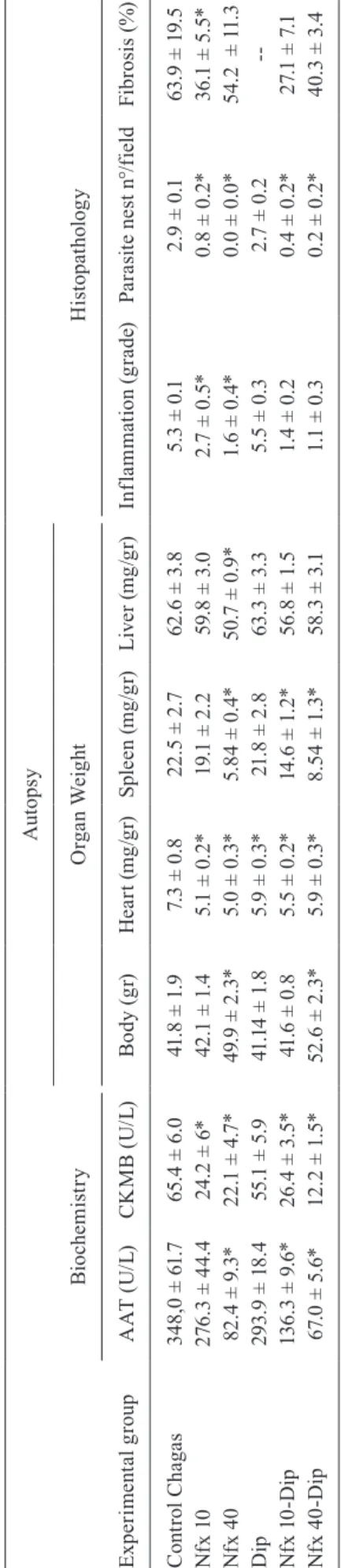
Histopathology - The hearts from the groups treat-ed with Nfx 10 or 40 mg/kg, alone or in combination with DPY, had a significant decrease in their weights compared with the Chagas control group. Spleens from groups treated with Nfx 40 mg/kg alone, Nfx 10 mg/kg plus DPY and Nfx 40 mg/kg plus DPY had a significant decrease in the weight compared with the control Chagas group. In mice treated with Nfx 40 mg/kg, a significant decrease in liver weight was also observed (Table IV).
In groups treated with Nfx 10 and 40 mg/kg alone or in combination with DPY, a significant decrease in in-flammatory cell infiltrates was observed compared to the control group. The DPY-treated group showed an inflam-matory cell infiltrate similar to the Chagas control group (Fig. 5, Table IV).
TABLE III
Electrocardiographic (ECG) rhythm, conduction and repolarisation qualitative disorders
Heart rhythm disorders
Group
Healthy Chagasic Nifurtimox (Nfx) Dipyridamole (DPY) DPY + Nfx
n (%) n (%) n (%) n (%) n (%)
Sinus extrasystole 6 8.1 32 23.5 2 25 2 25 0 0
Ventricular extrasystole 0 0 29 21.3 3 37.5 0 0 1 12.5
Nodal extrasystole 0 0 4 2.9 1 12.5 1 12.5 0 0
Supraventricular tachycardia 0 0 14 10.2 0 0 0 0 0 0
Ventricular tachycardia 0 0 2 1.4 2 25 0 0 0 0
Atrial ectopics beats 0 0 8 6.2 0 0 0 0 0 0
Atrioventricular dissociation 0 0 8 6.2 0 0 0 0 0 0
Atrial fibrillation 0 0 6 4.6 0 0 0 0 0 0
Second-degree AV block 0 0 3 2.3 0 0 0 0 0 0
Two P waves 0 0 27 19.8 7 87.5 3 37.5 5 62.5
Bifid P wave 0 0 24 17.6 0 0 0 0 1 12.5
Post-depolarisation (U) wave 0 0 89 65.4 4 50 2 25 3 37.5
Flattened T wave 0 0 12 8.8 3 37.5 0 0 0 0
Total n were 74 and 136 for healthy and chagasic mice respectively; for treated groups were eight each one. Nfx and DPY doses were 10 and 30 mg/Kg respectively.
DPY enhanced Nfx effect • Sonia Santeliz et al. 603
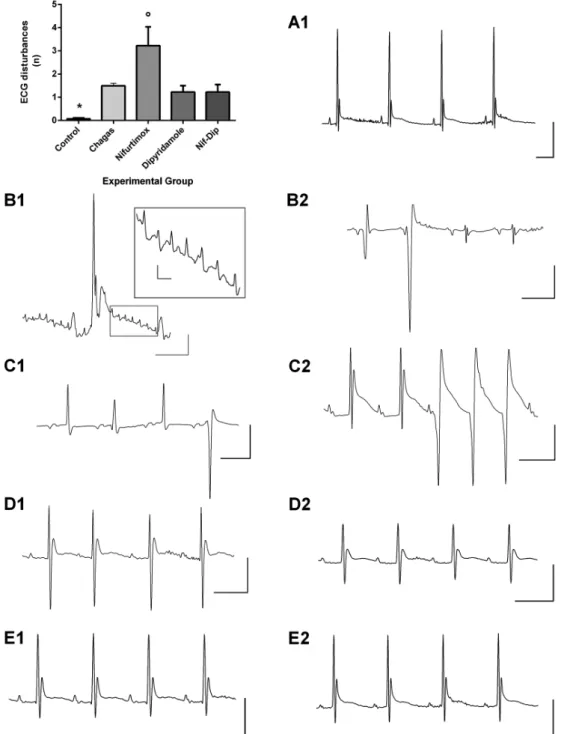
Fig. 4: typical electrocardiographic (ECG) traces and qualitative ECG disorders observed in healthy, untreated and treated chagasic mice. The mean ± standard error (SEM) of the qualitative ECG disorders (rhythm, conduction and repolarisation disorders) observed in mice of the different experimental groups are shown in the upper panel on the left. Healthy mice have practically no disorders, whereas the group of chagasic mice treated with nifurtimox (Nfx) had a significantly greater increase in these disorders compared with the remaining groups. In panel A1, we show a typical record of a healthy mouse. Traces of mice with chagasic myocarditis are shown in panels B1 and B2; in B1, we can observe low-voltage QRS complexes with a Rr’ wave suggestive of His intranodal block and multiple rhythmical atrial ectopics that are magnified in the inserted box suggestive of atrial flutter; in B2, we can perceive a complex rhythm disorder with ventricular extrasystoles, a very low voltage QRS complex, a deep S wave and a negative T wave. Traces of mice with chagasic myocarditis treated with Nfx at doses of 10 mg/kg of body weight are shown in panels C1 and C2 where there is a worsening in the qualitative disorders. In C1, there is atrioventricular dissociation, a negative-positive biphasic P wave, and diminished voltage QRS complex with variable morphology, including ventricular extrasystole and negative T wave. In C2, there is a double P wave and peaked T wave with an evident delay in its decay, which delineates a post depolarisation phenomenon (U wave) and no sustained ventricular tachycardia. In D1 and D2, traces from mice with chagasic myocarditis treated with dipyridamole are shown with clear improvement in the qualitative aspects of the electrocardiogram; however, repolarisation disorders and a decrease in the voltage of the QRS complex persist (D2). Traces of mice with chagasic myocarditis treated with Nfx 10 mg/kg plus dipyridamole are shown in E1 and E2, where there are practically no ECG disorders (compare with trace A1). *: indicates p < 0.05 when comparing the control with the other experimental groups, º: it indicates p < 0.05 when comparing the group treated with Nfx compared to the other
experimental groups. The vertical bars are equivalent to 500 μV and horizontal bars are 100 ms for all traces, by except of panel B1 where vertical and
T A B L E I V B io ch em is tr y a n d m o rp h o h is to p at h o lo g ic al p ar am et er s i n N M R I m ic e t re at ed o r n ot w it h n if u rt im o x ( N fx ) a n d /o r d y p ir id am o l ( D P Y ) E x p e ri m e n ta l g ro up Bi o ch e m is tr y Au to p sy H is to pa th o lo g y O rg a n W ei g h t A A T ( U /L) C K M B ( U /L) B o d y ( g r) H e a rt ( m g /g r) S p le en ( m g /g r) L iv e r ( m g /g r) In fl a m m at io n ( g rad e) P a ra sit e n e st n °/ fi eld F ib ros is ( % ) C o n tro l C h aga s 34 8
,0 ± 6
1
.7
6
5
.4 ± 6
.0 41 .8 ± 1 .9 7.
3 ± 0
.8
2
2
.5 ± 2
.7
6
2
.6 ± 3
.8
5
.3 ± 0
.1
2
.9 ± 0
.1
6
3
.9 ± 1
9. 5 N fx 1 0 2 7 6 .3 ± 4 4.4 2 4
.2 ± 6
*
4
2
.1 ± 1
.4
5
.1 ± 0
.2
*
1
9.
1 ± 2
.2
5
9.
8 ± 3
.0
2
.7 ± 0
.5 * 0. 8 ± 0. 2 * 3 6
.1 ± 5
.5 * Nf x 4 0 8 2
.4 ± 9
.3
*
2
2
.1 ± 4
.7
*
4
9.
9 ± 2
.3
*
5
.0 ± 0
.3
*
5
.8
4 ± 0
.4 * 5 0. 7 ± 0. 9 * 1
.6 ± 0
.4 * 0 .0 ± 0 .0 * 5 4
.2 ± 1
1 .3 D ip 2 9 3
.9 ± 1
8. 4 5 5 .1 ± 5 .9 41 .1 4 ± 1 .8 5
.9 ± 0
.3 * 21 .8 ± 2 .8 6 3. 3 ± 3. 3 5
.5 ± 0
.3
2
.7 ± 0
.2 --N fx 1 0 -D ip 1 3 6
.3 ± 9
.6
*
2
6
.4 ± 3
.5
*
41
.6 ± 0
.8
5
.5 ± 0
.2
*
14
.6 ± 1
.2
*
5
6
.8 ± 1
.5
1
.4 ± 0
.2 0. 4 ± 0. 2 * 2 7. 1 ± 7. 1 N fx 4 0 -D ip 6 7. 0 ± 5 .6 * 1 2
.2 ± 1
.5
*
5
2
.6 ± 2
.3
*
5
.9 ± 0
.3
*
8.
5
4 ± 1
.3
*
5
8.
3 ± 3
.1
1
.1 ± 0
.3 0. 2 ± 0. 2 * 4 0
.3 ± 3
.4 * : m e a n
s p < 0
.0 5 a s c o m p a re d w it h c o n tr o l C h ag a s.
In groups treated with Nfx 10 and 40 mg/kg alone or in combination with DPY, a significant decrease in the amastigote nest number was observed, while the group treated with DPY alone showed a similar number as in the Chagas control group (Fig. 5, Table IV).
Likewise, in the groups treated with Nfx 10 or 40 mg/kg associated with DPY, a significant decrease in the fibrosis was observed compared to the control Chagas group (Fig. 6, Table IV).
Plasma enzymes - In the Chagas control and DPY-treated groups, a significant increase in the serum CKMB activity was observed, while mice treated with Nfx alone at both doses or combined with DPY had de-creased CKMB levels, achieving values similar to those in healthy mice. The SGOT serum activities were also significantly increased in the Chagas control, DPY and Nfx 10 mg/kg groups, while in mice treated with Nfx 40 mg/kg alone or in combination with DPY at both doses, the serum SGOT levels were significantly decreased and similar to healthy mice (Table IV).
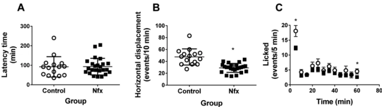
Behavioural effect of Nfx in healthy mice - Mice treat-ed with Nfx exhibit similar behaviour in the nociceptive hot plate test and formalin test late licking behaviour; however, a significant decrease in early licking behav-iour during the formalin test was observed. Moreover, in the group treated with Nfx 40 mg/kg, a significant decrease in horizontal displacement movements was ob-served compared with healthy mice (Fig. 3).
DISCUSSION
In the present paper, we confirm that Nfx is an ef-ficient trypanocidal drug, although its effect depended on the dose. In our animal model, the dose of 40 mg/kg was therapeutic; parasitaemia was suppressed on day 20 post-treatment and remained negative until the end of the experiment. Also, 92% of the treated mice survived and showed no cardiomegaly, hepatomegaly or spleno-megaly, and heart histopathological studies did not re-veal free or nest amastigotes, while the inflammatory infiltrate was mild. By contrast, Nfx at 10 mg/kg was subtherapeutic, as revealed by the 80% mortality, con-tinuous parasitaemia increase in the first three weeks of treatment, persistence of amastigotes in heart tissue, splenomegaly, abnormal EKG and elevated blood SGOT activity. The 40 mg/kg Nfx therapeutic dose is above the usually recommended dose of 10 mg/kg for treating Chagas disease in humans.
DPY enhanced Nfx effect • Sonia Santeliz et al. 605
depend on the assayed strain of T. cruzi, which is appar-ent from the work of Andrade et al. (1985), who found that type I strains (high and early parasitaemia with macrophage tropism) had high susceptibility (56 ± 16% cure), type II strains (high and late parasitaemia with heart tropism) had medium to high susceptibility (52 ± 11% cure), and type III strains (low parasitaemia with skeletal muscle tropism) had low susceptibility (0.45 ± 0.45% cure) to therapeutic schemes based on Nfx 200 mg/kg for four days followed by 50 mg/kg for five days/ week for 90 days. Furthermore, Faúndez et al. (2008) found decreased parasitaemia at doses of 2.5 and 10 mg/ kg with 25 and 100% survivals, respectively.
When comparing the effectiveness obtained in this work in cultured epimastigotes (IC50 = 21.53 µM) with Nfx
doses used in in vivo experiments, where 10 and 40 mg/kg are equivalent to 34.8 and 139.23 µmol/kg, respectively, these data confirm that 10 mg/kg is a subtherapeutic dose, while 40 mg/kg is almost a therapeutic dose based on the IC50. However, when we analysed the drug bioavailability, which is very low (5.25%) due to rapid and extensive me-tabolism in the liver, where it is subjected to nitroreduction by cytochrome P-450 reductase, the serum concentration of Nfx achieved after an oral dose of 15 mg/kg (52 µmol/ kg) would be 2.73 µM serum (Paulos et al. 1989), which is far below the therapeutic concentrations. Therefore, the drug potency in vivo is increased. Increased potency could be explained through drug metabolism during which host cells generate free radicals that facilitate parasite lysis (Letelier et al. 2004, Bartel et al. 2007).
Fig. 5: histopathological findings in haematoxylin-eosin stained heart tissue. (A) Shows a heart tissue sample from healthy mice; (B) from Trypano-soma cruzi-infected mice where amastigote nests (arrows), mononuclear infiltrate and myocitolisis can be observed; (C) 40 mg/kg Nfx-treated mice lacked amastigotes present, but fibrosis areas were present; and (D) 40 mg/kg Nfx plus 30 mg/kg-treated mice lacked evident lesions or parasite forms.
Two points have been crucial to the reluctance to Nfx routine use: the reasoned but controversial lack of efficacy and frequent side effects (Murcia et al. 2012). In this paper, we evaluated the side effects of Nfx at a dose of 40 mg/kg in healthy mice and found slight but significant disturbances in the red blood cell count (even when diminished, it remained within normal limits for the species), decreased motility and decreased early no-ciceptive response to formalin; we did not observe disor-ders in animal weight, in the counting and distribution of white blood cells and in nociception to thermal stimuli. Therefore, our results indicate that psychic excitability and polyneuropathies with paraesthesia observed in hu-mans are not observed in mice treated with Nfx.
In a phase I study performed in paediatric patients on the therapeutic effect of Nfx alone or in combination with cyclophosphamide and topotecan for treating refractory neuroblastoma with multiple relapses, it was determined that the maximum tolerated dose in these compromised patients was 30 mg/kg (Sholler et al. 2011), which is very close to the therapeutic dose raised in the present paper. In addition, we observed that chagasic mice treated with Nfx 40 mg/kg had an increased weight and maintained a healthy trophism in terms of the coat appearance and in-stinctive behaviours. By contrast, in chagasic mice treat-ed with Nfx 10 mg/kg, alopecia, piloerection, crusttreat-ed lesions and defensive muscle tone were observed (results not shown). To reconcile data reported in the literature with data reported in the present study, we propose that the side effects are a greater reflection of concurrent Chagas disease pathophysiological phenomena than the drug itself. In fact, the toxic effect of Nfx may depend on its metabolism by host cells and parasites.
Within reductions catalysed by the oxidative cyto-chrome P450 system in the host, the best known is the single electron reduction of nitro groups, leading to the formation of -NO2-, which can undergo redox recycling
and generate radical superoxide anion (O2-) and hydroxyl
radicals (HO-) in the presence of O
2 and can generate
oxi-dative stress (Letelier et al. 2004, Bartel et al. 2007). The cytochrome P450 oxidative system is strongly expressed in leukocytes; therefore, in an inflammatory process such as Chagas disease, treatment with Nfx in the presence of inflammatory cells could amplify oxidative stress and may explain the increase in ECG disturbances observed in chagasic mice treated with subtherapeutic doses. Using this dose, the inflammatory cell infiltrate continues given the persistence of the parasite in tissues and the side ef-fects are therefore dependent on oxidative stress generat-ed by the metabolism of Nfx in inflammatory cells. With therapeutic doses, the parasite is removed; inflammation ceases, and oxidative stress induced by Nfx metabolism in leukocytes is prevented, decreasing collateral side effects.
In this paper, we demonstrate for the first time that DPY has a trypanocidal effect in vitro on T. cruzi epimas-tigote proliferation and decreases parasitaemia in infected NMRI mice. However, the amastigote density in cardiac tissue was not affected by the drug. The effect of DPY on epimastigote proliferation has previously been reported in T. brucei, where an IC50 of 30 µM was obtained (de Koning et al. 2012). The dose of DPY used in the in vivo
model was subtripanocidal because 30 mg/kg equals 60 µM. Moreover, assuming a bioavailability between 37 and 66%, we would reach serum concentrations between 22.2 and 39.6 µM, respectively, which is 9.39 to 16.75 times lower than the IC50in vitro. At these doses, between 5 and 15% of the maximum trypanocidal effect would be obtained, which is in perfect agreement with the results obtained in vivo with the use of DPY alone. The lack of effect on tissue amastigotes may reflect that these para-sitic forms do not depend on the extracellular adenosine as a source of endogenous purines; therefore, they are not sensitive to DPY (Guimarães & Gutteridge 1987).
Notwithstanding the trypanocidal action of DPY, its functional effect should be considered; like other au-thors, we think that the cardiovascular benefits of this drug have been undervalued, and the beneficial effect observed in this study could be related to its mechanism of action. First, DPY induces coronary vasodilation mediated by nitric oxide and inhibits platelet aggrega-tion mediated by PGI2. This effect results from PDE5 enzyme inhibition, causing elevation of the intracellular cAMP and cGMP levels (Kim & Liao 2008). Moreover, by blocking nucleotide transporters, DPY elevates extra-cellular adenosine levels, which act on A2 receptors and potentiate intracellular elevation of cAMP levels (Lay-land et al. 2014). The consequence of this mode of action is improved coronary irrigation and prevention against new platelet and hematic thrombi formation, counter-acting pathophysiological phenomena related to diffuse ischaemia in the coronary microvasculature. Moreover, it has been reported that DPY improves cardiac contractil-ity of hypokinetic areas and ejection fraction in patients with Chagas cardiomyopathy (Kuschnir et al. 1983).
Second, DPY is cardioprotective, an effect medi-ated by adenosine acting on A1 and A3 receptors, which would optimize cardiac work according to the availabil-ity of energy sources, inhibit early apoptotic phenomena and prevent the occurrence of lethal arrhythmias (Lay-land et al. 2014). In this sense, it has been reported that endogenous adenosine acting on A1 receptors generates negative chronotropic and dromotropic effects, reducing the incidence of ventricular arrhythmias induced by isch-aemia-reperfusion in isolated rat hearts (Lee et al. 1994). Third, DPY is an immunomodulator drug, which is mainly mediated by A2A receptors. Adenosine acting on A1 and A3 receptors promotes the recruitment of imma-ture dendritic cells to sites of inflammation, while it pro-motes the differentiation of T cells to an anti-inflammatory TH2 profile via A2A receptors. This scenario promotes decreased levels of IL6 and TNFa, while the IL10 levels are increased. This balance promotes an anti-inflammatory state in heart tissue that counteracts necrotic phenomena, fibrosis and cardiac remodelling associated with severe
in-flammation (Haskó et al. 2008). Elevated levels of TNF-α
DPY enhanced Nfx effect • Sonia Santeliz et al. 607
Finally, the molecular structure of DPY allows it to accept electrons, acting as a free radical scavenger,
whose capacity is even greater than α-tocopherol and
vitamin C (Kim & Liao 2008). During Chagas cardio-myopathy development, the myocardium is continuously exposed to injury caused by the release of free radi-cal products of mitochondrial damage (Vyatkina et al. 2004); therefore, the beneficial effect of DPY could be associated with its antioxidant capacity.
In conclusion, in the present study, we show that 40 mg/kg is the therapeutic trypanocidal dose of Nfx in NMRI mice with acute Chagas disease, while the 10 mg/ kg dose is subtherapeutic and has little utility in prevent-ing or reversprevent-ing Chagas cardiomyopathy development in an NMRI murine model. The side effects observed in healthy mice are mild; therefore, in NMRI mice, severe side effects may be related to drug metabolism by persis-tent inflammatory cells. DPY enhanced the trypanocidal effect of Nfx, transforming subtherapeutic doses to thera-peutic levels, and this effect could be related to their try-panocidal capacity and its mechanism of action related to the prevention or reversal of pathophysiological phe-nomena related to microvascular theory. Because of their anti-inflammatory, immunomodulatory and antioxidant effects, these medications may improve cardiac function.
The results obtained here in NMRI mice should be interpreted with caution when considering clinical im-plications in humans with Chagas disease. For example, the 10 mg/kg Nfx dose is subtherapeutic in mice, but this is not applicable for other species, including humans, be-cause the same drug dose will produce different blood levels and tissue concentrations in different species be-cause the bioavailability and biodistribution of the drug differ between different species.
Notwithstanding, given the extensive experience that medical doctors have regarding the clinical use of DPY, its wide therapeutic window, safety and mechanism of action make it an interesting drug that should be tested as an adjunct drug in the treatment of Chagas disease in chronic animal models and then its value assessed in moving on to clinical trials.
AUTHORS’ CONTRIBUTION
SS - Conception and drafting of the general project, execu-tion, analysis and interpretation of in vitro and in vivo para-sitological experiments, histopathological experiments, elec-trocardiographic and biochemical experiments, participated in writing and gave final manuscript approval; PC - execution and analysis of behavioural experiments, writing and interpretation of behavioural experiments and final approval of the manu-script; EG - execution and analysis of in vivo parasitological and electrocardiographic experiments, wrote and interpreted electrocardiographic and in vivo parasitological experiments and gave final approval of the manuscript; CA - execution of in vitro and in vivo parasitological experiments, writing and interpretation of in vitro parasitological experiments and fi-nal approval of the manuscript; DY - execution and afi-nalysis of electrocardiographic experiments, writing and interpreta-tion of experiments and electrocardiographic data, and writ-ing, critical review and final approval of the manuscript; RB - execution and analysis of biochemical experiments, writing
and interpretation of biochemical experiments, and writing, critical review and final approval of the manuscript; CR - co-general director of the project, participated in the conception and analysis of the project for its approval, participated in the design, execution, monitoring, analysis and interpretation of histopathological and in vitro and in vivo parasitological exper-iments, participated in consolidating and enriching the writing as well as in the critical review and final approval of the manu-script; RB-C - director general of the project, participated in the design and drafting of the project for approval, participated in the design, execution, monitoring, analysis and interpreta-tion of in vitro and in vivo parasitological experiments, histo-pathological, electrocardiographic, behavioural and biochemi-cal experiments, participated in the configuration of the final manuscript, in consolidating and enriching his writing, and in the critical review and final approval of the manuscript.
REFERENCES
Alarcón de Noya B, Díaz-Bello Z, Colmenares C, Ruiz-Guevara R, Mauriello L, Muñoz-Calderón A, et al. Update on oral Chagas dis-ease outbreaks in Venezuela: epidemiological, clinical and diag-nostic approaches. Mem Inst Oswaldo Cruz. 2015; 110(3): 377-86.
Andrade SG, Magalhães JB, Pontes A. Evaluation of chemotherapy with benznidazole and nifurtimox in mice infected with Try-panosoma cruzi strains of different types. Bull World Health Or-gan. 1985; 63(4): 721-6.
Añez N, Crisante G, Rojas A. Update on Chagas disease in Venezuela - A Review. Mem Inst Oswaldo Cruz. 2004; 99(8): 781-7.
Bartel LC, de Mecca MM, Fanelli SL, de Castro CR, Diaz EG, Cas-tro JA. Early nifurtimox-induced biochemical and ultrastructural alterations in rat heart. Hum Exp Toxicol. 2007; 26(10): 781-8.
Bustamante JM, Craft JM, Crowe BD, Ketchie SA, Tarleton RL. New, combined, and reduced dosing treatment protocols cure Trypano-soma cruzi infection in mice. J Infect Dis. 2014; 209(1): 150-162.
Coura JR, Borges-Pereira J. Chronic phase of Chagas disease: why should it be treated? A comprehensive review. Mem Inst Oswaldo Cruz. 2011; 106(6): 641-5.
de Koning HP, Gould MK, Sterk GJ, Tenor H, Kunz S, Luginbuehl E, et al. Pharmacological validation of Trypanosoma brucei phospho-diesterases as novel drug targets. J Infect Dis. 2012; 206(2): 229-37.
Dias JCP, Ramos Jr AN, Gontijo ED, Luquetti A, Shikanai-Yasuda MA, Coura JR, et al. II Consenso Brasileiro em doença de Cha-gas, 2015. Epidemiol Serv Saude. 2016; 49(núm esp.): 7-86.
Faúndez M, López-Muñoz R, Torres G, Morello A, Ferreira J, Kem-merling U, et al. Buthionine sulfoximine has anti-Trypanosoma cruzi activity in a murine model of acute Chagas’ disease and enhances the efficacy of nifurtimox. Antimicrob Agents Che-mother. 2008; 52(5): 1837-9.
Guimarães RC, Gutteridge WE. Purine base uptake in Trypanosoma cruzi: adaptations and effects of inhibitors. Braz J Med Biol Res. 1987; 20(1): 1-10.
Haskó G, Linden J, Cronstein B, Pacher P. Adenosine receptors: ther-apeutic aspects for inflammatory and immune diseases. Nat Rev Drug Discov. 2008; 7(9): 759-70.
Kim HH, Liao JK. Translational therapeutics of dipyridamole. Arte-rioscler Thromb Vasc Biol. 2008; 28(3): s39-42.
Layland J, Carrick D, Lee M, Oldroyd K, Berry C. Adenosine: physi-ology, pharmacphysi-ology, and clinical applications. JACC Cardiovasc Interv. 2014; 7(6): 581-91.
Lee Y, Chern J, Yen M. Antiarrhythmic effects of BN063, a newly
synthesized adenosine A1 agonist, on myocardial ischaemia in rats. Br J Pharmacol. 1994; 112(4): 1031-6.
Letelier ME, Izquierdo P, Godoy L, Lepe AM, Faúndez M. Liver micro-somal biotransformation of nitro-aryl drugs: mechanism for poten-tial oxidative stress induction. J Appl Toxicol. 2004; 24(6): 519-25.
López L, Arai K, Giménez E, Jiménez M, Pascuzo C, Rodríguez-Bonfante C, et al. Las concentraciones séricas de interleucina-6 y proteína C reactiva se incrementan a medida que la enfermedad de Chagas evoluciona hacia el deterioro de la función cardíaca. Rev Española Cardiol. 2006; 59(1): 50-6.
Medrano-Mercado N, Ugarte-Fernández R, Butrón V, Uber-Busek S, Guerra HL, Araújo-Jorge TC, et al. Urban transmission of Cha-gas disease in Cochabamba, Bolivia. Mem Inst Oswaldo Cruz. 2008; 103(5): 423-30.
Mendoza EA, Bonfante CR, Camacho I, Martínez J, Perdomo T, Ca-brera A, et al. Pacientes con cardiomiopatía dilatada chagásica y cardiopatía no chagásica presentan niveles elevados del factor de necrosis tumoral a. Invest Clin. 2005; 46(3): 229-40.
Murcia L, Carrilero B, Segovia M. Limitations of currently available Chagas disease chemotherapy. Rev Esp Quimioter. 2012; 25(1): 1-3.
Palmer TM, Trevethick MA. Suppression of inflammatory and im-mune responses by the A(2A) adenosine receptor: an introduc-tion. Br J Pharmacol. 2008; 153(Suppl. 1): S27-34.
Paulos C, Paredes J, Vasquez I, Thambo S, Arancibia A, González-Martin G. Pharmacokinetics of a nitrofuran compound, nifurti-mox, in healthy volunteers. Int J Clin Pharmacol Ther Toxicol. 1989; 27(9): 454-7.
Requena-Méndez A, Aldasoro E, de Lazzari E, Sicuri E, Brown M, Moore DA, et al. Prevalence of Chagas disease in Latin-Amer-ican migrants living in Europe: a systematic review and meta-analysis. PLoS Negl Trop Dis. 2015; 9(2): e0003540.
Rossi MA, Tanowitz HB, Malvestio LM, Celes MR, Campos EC, Blefari V, et al. Coronary microvascular disease in chronic Chagas cardio-myopathy including an overview on history, pathology, and other pro-posed pathogenic mechanisms. PLoS Negl Trop Dis. 2010; 4(8): e674.
Sguassero Y, Cuesta CB, Roberts KN, Hicks E, Comandé D, Ciap-poni A, et al. Course of chronic Trypanosoma cruzi Infection after treatment based on parasitological and serological tests: a systematic review of follow-up studies. PLoS ONE. 2015; 10(10): e0139363.
Sholler GLS, Bergendahl GM, Brard L, Singh AP, Heath BW, Bingham PM, et al. A phase 1 study of nifurtimox in patients with relapsed/re-fractory neuroblastoma. J Pediatr Hematol Oncol. 2011; 33(1): 25-30.
Sousa G, Gomes J, Fares R, Damásio MS, Chaves A, Ferreira K, et al. Plasma cytokine expression is associated with cardiac morbidity in Chagas disease. PLoS ONE. 2014; 9(3): e87082.
Vyatkina G, Bhatia V, Gerstner A, Papaconstantinou J, Garg N. Im-paired mitochondrial respiratory chain and bioenergetics during chagasic cardiomyopathy development. Biochim Biophys Acta. 2004; 1689(2): 162-73.
WHO - World Health Organization. Chagas disease (American try-panosomiasis). Fact sheet 2017. 2017. Available from: http://www. who.int/mediacentre/factsheets/fs340/en/.