Volume 2013, Article ID 754580,9pages http://dx.doi.org/10.1155/2013/754580
Research Article
Leishmania major
Self-Limited Infection Increases Blood
Cholesterol and Promotes Atherosclerosis Development
Luciana R. Fernandes,
1Ana Cecília C. Ribeiro,
1Marcela Segatto,
1Luís Felipe F. F. Santos,
1Joana Amaral,
2Luciane R. Portugal,
1and Jacqueline I. A. Leite
11Departamento de Bioqu´ımica e Imunologia, Universidade Federal Minas Gerais, 31270-901 Belo Horizonte, MG, Brazil 2Centro Universit´ario, Av. Prof. M´ario Werneck, 1685-Estoril, 30455-610 Belo Horizonte, MG, Brazil
Correspondence should be addressed to Jacqueline I. A. Leite; jalvarezleite@gmail.com
Received 6 February 2013; Revised 30 March 2013; Accepted 31 March 2013
Academic Editor: Gloria L. Vega
Copyright © 2013 Luciana R. Fernandes et al. This is an open access article distributed under the Creative Commons Attribution License, which permits unrestricted use, distribution, and reproduction in any medium, provided the original work is properly cited.
Leishmania major infection of resistant mice causes a self-limited lesion characterized by macrophage activation and a Th1 proinflammatory response. Atherosclerosis is an inflammatory disease involving hypercholesterolemia and macrophage activation. In this study, we evaluated the influence of L. major infection on the development of atherosclerosis using atherosclerosis-susceptible apolipoprotein E-deficient (apoE KO) mice. After 6 weeks of infection, apoE KO mice exhibited reduced footpad swelling and parasitemia similar to C57BL/6 controls, confirming that both strains are resistant to infection withL. major.L. major -infected mice had increased plasma cholesterol levels and reduced triacylglycerols. With regard to atherosclerosis, non-infected mice developed only fatty streak lesions, while the infected mice presented with advanced lesions containing a necrotic core and an abundant inflammatory infiltrate. CD36 expression was increased in the aortic valve of the infected mice, indicating increased macrophage activation. In conclusion,L. majorinfection, although localized and self-limited in resistant apoE KO mice, has a detrimental effect on the blood lipid profile, increases the inflammatory cell migration to atherosclerotic lesions, and promotes atherogenesis. These effects are consequences of the stimulation of the immune system byL. major, which promotes the inflammatory components of atherosclerosis, which are primarily the parasite-activated macrophages.
1. Introduction
Leishmania major is a protozoan parasite transmitted by sandflies of the genusLutzomyiathat inject the promastigote form into the dermis of the host. Once injected, the parasite is rapidly enclosed by phagocytic cells and transforms into the replicative intracellular amastigote form [1]. In immuno-competent hosts, such as C57BL/6 mice,L. majorinfection is a self-contained cutaneous lesion that elicits a Th1 immune response. In infected mice, the immune cells (macrophages, dendritic cells, natural killer cells, and T cells) produce cytokines and bioactive molecules, such as IFN-𝛾, IL-12, and nitric oxide (NO), which act against the protozoan [1].
Atherosclerosis is a chronic inflammatory disease associ-ated with a high level of total cholesterol and proatherogenic lipoproteins (VLDL, IDL, and LDL), a prothrombotic status,
an L. major infection. However, even localized infections, such as odontologic ones, may be associated with the devel-opment of atherosclerosis [12].
Apolipoprotein (apo) E is a component of chylomicron and very low-density lipoproteins (VLDL) and mediates the uptake of these lipoproteins and possesses anti-inflammatory and antioxidant effects [13]. ApoE KO mice have impaired clearance of VLDL and chylomicrons from the blood, which results in hypercholesterolemia and favors the develop-ment of atherosclerotic lesions [11]. ApoE also has anti-inflammatory and antioxidative properties, and its absence is partially responsible for the higher inflammatory profile of apoE-deficient animals compared to the wild-type control, C57BL/6 mice [14]. ApoE KO mice spontaneously develop atherosclerosis even when fed a normolipidemic diet [15]. The atherosclerotic lesions of apoE KO mice exhibit a similar distribution, microscopic appearance, and cellular composi-tion to those found in humans. The atherosclerotic lesions are characterized by endothelial dysfunction, macrophage, and T cell infiltration and production of proinflammatory cytokines, such as TNF-𝛼, INF-𝛾, and IL-6, which indicates a typical Th1 polarized immune response [2].
This work aimed to determine if a self-limitedL. major infection would be sufficient to modify the lipid metabolism and promote the development of atherosclerosis similar to what occurs with systemic infections.
2. Material and Methods
This protocol was approved by the Animal Care Committee of Universidade Federal de Minas Gerais (CETEA #147/05).
2.1. Mice and Diet. Twenty-eight 11-week-old female apoE KO mice were separated into control (noninfected) and Leish-mania major(L. major) infected groups. For the background control, twenty wild-type C57BL/6 mice were divided into control (noninfected) orL. majorinfected groups. Animals of both strains were equally distributed according to weight and blood cholesterol and were fed on AIN-93G standard diet [15].
2.2. Leishmania major Infection. The mice were infected with L. major (clone WHO MHOM/IL/80/Friedlin). The inocu-lum was prepared under sterile conditions from five-dayL. majorcultures, which corresponds to the stationary growth phase rich in the promastigote metacyclic infectious forms. The medium containingL. majorwas centrifuged, and the pellet was resuspended in 1 mL of PBS. Approximately 1×10⁶ parasites were inoculated into the left footpad, and the right footpad was used as a control after inoculation with PBS as previously described [16]. The footpad was measured weekly with a caliper, and the amount of swelling was calculated as the difference between the measures of the infected (left) and control (right) footpads.
The animals’ body weight and food intake were measured weekly for 6 weeks after infection. All groups received the same amount of food to avoid confounding the experiment with different amounts of nutrients. After 6 weeks and an
overnight fast, the mice were euthanized under anesthesia. Plasma, footpads, and tissues were collected for subsequent analyses. The infected footpad was weighed without the skin and used for parasite quantification as previously described [16].
2.3. Lipids Assay. The total cholesterol, cholesterol in high density lipoprotein (HDLc) form, and triacylglycerols in the plasma were measured using commercial kits (Doles, Brazil) as previously described [17]. The amount of non-HDLc (that represents the atherogenic fractions VLDLc and IDLc) in the plasma was determined by calculating the difference between the total cholesterol and the HDLc.
The liver was washed in PBS and dried on filter paper. The contents of the cecum were separated from the lumen. The hepatic and cecal lipid extractions were performed as previously described by Folch et al. [18].
2.4. Histological Analysis. For the analysis of atherosclerosis, the aortic valves were washed with PBS, gently perfused with 10% neutral buffered formalin, and embedded in paraffin. The analysis was performed on 10 samples per group. The specimens were fixed in 3.7% paraformaldehyde and pro-cessed in paraffin. Briefly, every consecutive section (10𝜇m thick) through the aortic valve (300𝜇m) was taken and stained with hematoxylin and eosin [19]. The sections used for immunofluorescence were deparaffinized by 2 xylene washes (30 min) and then transferred to 3 washes (15 minutes each) of ethanol at concentrations of 100%, 85%, and 70%. The atherosclerotic lesion area was quantified using the Image Pro Plus software (Media Cybernetics, USA) from the sum of the atherosclerotic lesion areas obtained from the selected 10 sections, spaced 20𝜇m apart, as previously described [11]. The inflammatory cells per field were counted automatically by the Image Pro Plus software (𝑛 = 5mice, 3 nonconsecutive sections per animal) [11].
2.5. L. major Parasitism. The DNA in liver and spleen was extracted with proteinase K (Qiagen USA) according to the manufacturer’s instructions. A PCR analysis was conducted [20]. Briefly, 2𝜇L of DNA sample was added to 1.2𝜇L of Taq polymerase buffer, 1.0𝜇L of dNTPs (2.5 mM), 1.0𝜇L of primers, 0.05𝜇L of Taq polymerase, and 5.75𝜇L of Milli-Q sterile H2O. The samples were placed in a thermocy-cler and the amplification conditions were as follows (35 cycles): denaturation at 94∘C for 1 min, annealing at 54∘C for 1 min, elongation at 72∘C for 1.5 min, and extension at 72∘C for 10 min. The PCR products were then sub-jected to gel electrophoresis polyacrylamide and visualized by silver staining. The sequences of the primers used, which correspond to the minicircle kDNA [20], are Uni 21-5GGGGTTGGTGTAAAATAAGGCC 3and LmJ4-5CTA.
anti-goat antibody served as negative controls. Sections were mounted with Vectashield medium and were visualized with an Axioscope 2 Plus fluorescence microscope (Carl Zeiss).
2.7. RT-PCR. The popliteal lymph node was homogenized using the TH-01 (OMNI-INC). The total RNA from the lymph node and aortic valve was extracted using the TRIzol reagent according to the manufacturer’s protocol. The reverse transcription was performed using 2𝜇g of the total RNA, 200 U of the reverse transcriptase, 2.5𝜇L of the 5X RT buffer, 1.8𝜇L of the 10 mM dNTPs, 0.2𝜇L of the 10000 U/mL RNasin, and 1.0𝜇L of the 50𝜇M oligo dT. The temperature settings for this reaction were 70∘C for 5 min, on ice for 2 min, 42∘C for 60 min, 70∘C for 15 min, and 4∘C for the final step. The resulting cDNA was used for real-time PCR as described later. The specific primers were designed using the Primer Express software and synthesized by IDT. Real-time PCR was carried out on a StepOne sequence detection system (Applied Biosystems) using the Power SYBR Green PCR Master Mix (Applied Biosystems). The dissociation curve indicated that only one product was obtained in each reaction. The relative levels of gene expression were determined using theΔΔcycle threshold method as described by the manufacturer, in which data for each sample is normalized to the𝛽-actin expression and the data are shown as the relative expression (the fold increase over the control). The PCR results were analyzed with the SDS 2.1 software (Applied Biosystems), and the amount of mRNA of each gene of interest was normalized to the amount of the murine𝛽-actin gene. mRNA expression levels were calculated as the fold difference relative to the housekeeping gene by the formula: relative expression =
2−(CT[target gene]−CT[𝛽-actin-1])
.
The sequences of the primers used are as follows:
VCAM-1: 5-CCCCAAGGATCCAGAGATTCA-3 and 5-ACTTGACCGTGACCGGCTT-3;
CD36: 5GTACAGATTTGTTCTTCCAGCCAAT-3 and 5-TCAGTGCAGAAACAATGGTTGTC-3; MCP1/CCL2: 5 -AGGAAGATCTCAGTGCAGAG-3and 5-AGTCTTCGGAGTTTGCCTTTG-3; 𝛽-actin: 5-CTGCCTGACGGCCAAGTC-3 and 5 -CAAGAAGGAA GGCTGGAAAAGA-3;
IL-6: 5-ACAACCACGGCCTTCCCTACTT3 and 5-CACGATTTCCCAGAGAACATGTG-3;
IFN-𝛾: 5-TGGCTCTGCAGGATTTTCATG-3 and 5-TCAAGTGGCATAGATGTGGAAGAA-3; IL-10: 5-GGTTGCCAAGCCTTATCGGA-3and 5 -ACCTGCTCCACTGCCTTGCT-3;
IL-4: 5-ACAGGAGAAGGGACGCCAT-3 and 5 -GAAGCCCTACAGACGAGCTCA-3;
LmjF36.5520: 5 -GGCCATCACCACAAACAGAG-3and 5-GCTCAGGTCATACAAGGGGA-3.
2.8. Statistical Analyses. The data were initially analyzed by the Kolmogorov-Smirnov test to check their symmetry and by the Mann-Whitney or Student’s t-test using GraphPad
Table 1: Final body weight, food intake, and cecal and hepatic lipids of non-infected (control) andL. major-infected (L. major) apoE KO mice 6 weeks after the inoculation of 1×10⁶parasites (clone WHO MHOM/IL/80/Friedlin) into footpad.
Parameter Control ApoE KO L. majorApoE KO
Final body weight (g) 22.18±0.77 20.67±0.7 Food intake (g/week/mouse) 25.10±0.95 24.72±0.85 Liver lipids
Total lipids (mg/g) 252.0±12.0 172.0±21.0∗ Total cholesterol (mg/g) 14.1±1.3 13.2±0.9 Triacylglycerols (mg/g) 49.7±4.4 30.2±4.5∗ Cecum lipids
Total lipids (mg/g) 51.1±8.9 70.0±11 Total cholesterol (mg/g) 2.8±0.3 2.4±0.3 Triacylglycerols (mg/g) 0.97±0.2 0.86±0.21 Control group,𝑛= 8–11, andL. majorgroup,𝑛= 10–13. Results are expressed as the means±SE.∗𝑃 ≤ 0.05.
Prism, version 5.00, for Windows, GraphPad Software, San Diego, CA, USA. A𝑃value of<0.05 was considered signifi-cant.
3. Results
3.1. L. major Infection in ApoE KO and Wild-Type Control C57BL/6 Mice. Our first aim was to confirm thatL. major causes a self-limited infection in apoE KO mice similar to that observed in the wild-type control C57BL/6 mice. The results showed that after 6 weeks of infection, although apoE KO mice presented higher levels of the cytokines TNF alpha and IL-10 (Figures1(a)to1(d)), these mice controlled parasite load and footpad swelling similarly to C57BL/6 mice (Figures1(e) and 1(f)). Therefore, only apoE KO animals were used for further experiments.
There were no differences in the food intake between apoE experimental and control groups. As expected,L. major infection did not cause a significant weight loss in these animals (Table 1).
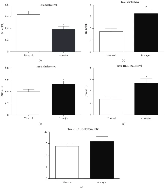
3.2. Lipid Profile. L. major infection was associated with increased serum cholesterol due to an increase in both HDL and non-HDL fractions (Figure 2). These alterations in the lipid profile were not due to differences in the hepatic cholesterol content or cecal excretion because the levels of these were similar between groups (Table 1).
0 1 2 3 4 5 6
C57BL/6 L. major
ApoE KO L. major ApoE KO
Control
F
o
ld incr
ea
se
o
ver co
n
tr
o
l
INF-𝛾 ∗
(a)
IL-6
0 1 2 3 4 5 6
C57BL/6 L. major
ApoE KO L. major ApoE KO
Control
F
o
ld incr
ea
se
o
ver co
n
tr
o
l
(b)
IL-4
0 1 2 3 4 5 6
C57BL/6
L. major
ApoE KO
L. major
ApoE KO Control
F
o
ld incr
ea
se
o
ver co
n
tr
o
l
(c)
C57BL/6
L. major
ApoE KO
L. major
ApoE KO Control IL-10
0 1 2 3 4 5 6
F
o
ld incr
ea
se
o
ver co
n
tr
o
l ∗
∗
(d)
1 2 3 4 5 6
0 0.2 0.4 0.6 0.8 1
C57BL/6 L. major ApoE KO L. major
Footpad swelling
Weeks of infection
(mm)
(e)
0 2 4 6 8
C57BL/6 ApoE KO
Parasite quantification (footpad)
P
arasi
te
s (log)
(f)
Triacylglycerol
Control L. major
0 0.2 0.4 0.6 0.8
(mmo
l/L) ∗
(a)
Total cholesterol
Control L. major
4 5 6 7 8
(mmo
l/L)
∗
(b) HDL cholesterol
Control L. major
0 0.2 0.4 0.6 0.8
(mmo
l/L)
∗
(c)
(mmo
l/L)
Non-HDL cholesterol
Control L. major
4 5 6 7 8
∗
(d) Total/HDL cholesterol ratio
Control L. major
0 5 10 15 20
(e)
Figure 2: Blood lipid profile of noninfected (𝑛 = 9) andL. major-infected (𝑛 = 12) apoE KO mice 6 weeks after the inoculation of 1×10⁶ parasites (clone WHO MHOM/IL/80/Friedlin) into left footpad. (a) Total triacylglycerols; (b) total cholesterol; (c) high density lipoprotein (HDL) cholesterol; and (d) non-HDL cholesterol (sum of cholesterol in low density (LDL), intermediate density (IDL) lipoproteins, and remnant chylomicron), calculated as the difference between cholesterol and HDL cholesterol. Bars = average and vertical lines = standard error.∗𝑃 < 0.05.
3.3. Atherosclerotic Lesions. Animals infected withL. major presented larger and more developed atherosclerotic lesions when compared to the controls (Figures3(a),3(c), and3(e)). In the control group, atherosclerotic lesions were formed by fatty streaks and several layers of foam cells. On the other
Control
(a)
L. major
(b)
×106 Aortic valve
Control L. major
0 0.5 1 1.5 2
L
esio
n a
re
a (
𝜇
m
2)
∗
(c)
Control
(d)
L. major
(e)
Inflammatory infiltrate
Control L. major
5000 6000 7000 8000 9000
10000 ∗
C
ells/mm
2
(f)
Aortic root
Control
CD11b+
(g)
Aortic root
L. major
CD11b+
(h)
Aortic root
L. major
CD36+
(i)
Figure 3: Atherosclerotic lesions and inflammatory infiltrate in apoE KO mice infected or noninfected (control) withL. majorfor 6 weeks.
(a, b) Histology of the aorta from noninfected (a) andL. major-infected (b) apoE KO mice, 6 weeks after infection.(c) Atherosclerosis lesion area in aortic valve of noninfected (Control) andL. major-infected apoE KO mice. The results represent the average of the lesion area (𝜇m2) in the aortic valve of animal from the control (𝑛 = 7) and the infected groups (𝑛 = 10), 6 weeks after infection. The major lines represent the means and the minor lines represent the standard error.∗𝑃 = 0.05. (d, e) Inflammatory infiltrate around the atherosclerotic lesion in aortic valve of non-infected (d) andL. major-infected (e) apoE KO mice. (f) Average number of cells/mm2of aortic valve of non-infected (𝑛 = 5) and infected (𝑛 = 5) apoE KO mice 6 weeks after infection. The bars represent the average and vertical lines represent standard error, ∗𝑃 < 0.05. (g, h) Immunofluorescence: CD11b FITC conjugated positive cells in aortic root of control (g) andL. major-infected (h) mice. (i) Anti-CD36 antibody Alexa488 conjugated in aortic valve ofL. major-infected group. In (a), (b), (d), and (e), the sections were stained with H&E. Image magnification: 20x in (a), (b), (g), and (h) and 100x in (d), (e), and (i).
system, including monocytes and macrophages as suggested by the higher number of CD11b positive cells around the atherosclerotic lesion of infected mice (Figures3(g)and3(h)).
3.4. Expression of VCAM-1, MCP1/CCL2, and CD36 in the Aortic Valve. The expression of the adhesion molecule VCAM-1, the monocyte chemotactic protein MCP-1/CCL2, and the scavenger receptor CD36 was also assessed in athe-rosclerotic lesions.
Confirming the histological data, the results showed that CD36 expression was significantly increased in theL. major group (𝑃 = 0.02). There was a tendency for increased (𝑃 = 0.11) MCP1/CCL2 expression, and there were no changes in VCAM 1 (𝑃 = 0.67) expression (Figure 4).
3.5. L. major DNA in Peripheral Tissues. Spleen and liver were tested for the presence ofL. majorDNA. The parasite was not detected in the spleen or liver of mice from both groups, suggesting the absence of viable parasites in these organs (Figure 5).
4. Discussion
VCAM-1 mRNA (aortic valve)
Control L. major
0 1 2 3 4 5
F
o
ld incr
ea
se
o
ver co
n
tr
o
l
𝑃 = 0.670
(a)
MCP-1/CCL2 (aortic valve)
Control L. major
0 1 2 3 4 5
F
o
ld incr
ea
se
d o
ver co
n
tr
o
l
𝑃 = 0.112
(b) CD36 mRNA (aortic valve)
Control L. major
0 1 2 3 4 5
F
o
ld incr
ea
se
o
ver co
n
tr
o
l
∗ 𝑃 = 0.020
(c)
Figure 4: Relative increase of (a) VCAM-1, (b) MCP1/CCL2, and (c) CD36RNA expression in the aortic valve of non-infected and infected apoE KO mice, 6 weeks after infection. Bars = average and vertical lines = standard error,𝑛 = 4/group.∗𝑃 < 0.05. The values of apoE KO control andL. majorgroups were, respectively, VCAM = 1.01±0.07 and 0.89±0.21; CD36 = 1.05±0.19 and 2.16±0.20; MCP-1 = 1.22±0.22 and 2.75±0.83.
Ct Leis Leis Leis Leis Ct Ct Leis Leis Leis Leis Ct +
650 bp
Spleen Liver Footpad
Figure 5: PCR amplification ofL. majorDNA in the spleen and liver of non-infected andL. major-infected apoE KO mice.First line: molecular weight marker; last line: positive control DNA from anL. major-infected footpad. Ct = apoE KO control mice; Leis- apoE KOL. major-infected mice.
model used in the present study, the authors did not analyze atherosclerosis development and lipid profile. Moreover, cytokine determinations were done mainly in vitro, after parasite antigen restimulation. Finally, the resolution of L. major infection in C57BL/6 mice (measured by footpad swelling) started only after 10 weeks of infection while in ours and other studies [22–24] it occurred after 5 weeks of infection.
Our previous studies showed thatT. gondii, which induces a strong systemic inflammation, promotes atherogenesis in apoE KO mice [11]. BecauseL. majorinfection is self-limited in mice, we expected thatL. majorwould have little influence on atherosclerosis development compared toT. gondii infec-tion. However, our results suggest an important role forL. majorinfection in macrophage activation and atherosclerosis development.
Several mouse strains, including C57BL/6, effectively controlL. majorinfection, while others, such as the BALB/c strain, develop progressive damage to the site of infection and systemic disease [25]. Our results indicate that apoE KO ani-mals present the same pattern of resistance toL. major infec-tion as that observed in the C57BL/6 animals, as determined by similar degrees of parasite load and footpad swelling. However, the pattern of cytokine expression after 6 weeks of infection shows a strong tendency to be higher in ApoE KO infected mice compared to the ApoE control and the C57BL/6 infected groups. This higher inflammatory status of ApoE KO mice has been previously presented as a consequence of the absence of anti-inflammatory effects of apoE as well as the proinflammatory stimulus of hypercholesterolemia and oxidized lipoproteins [26].
L. majorinfection leads to hypercholesterolemia associ-ated with a reduction in blood and hepatic triacylglycerols without changes in cholesterol excretion. This pattern of lipid alteration was not found during aT. gondii infection [11], which reduces cholesterolemia with no alterations in the level of triacylglycerols in the blood.These different findings reflect the specific metabolic characteristics of each proto-zoan with regard to their use of host lipids.T. gondiiis unable to produce cholesterol on its own. Thus, it is dependent on host cholesterol taken from the blood. Additionally, T. gondiicauses a more severe infection that induces a reduction in food intake and weight loss in the mice. One of the consequences of this is the preferential use of acetyl CoA to supply energy, reducing its availability to the cholesterol synthesis pathway [27]. Both factors contribute (host choles-terol uptake and acetyl-coA redirection) to the reduction of cholesterolemia during aT. gondii infection.Leishmania, on the other hand, is able to synthesize its own cholesterol [28] but is dependent on host fatty acids, an important source of energy for amastigotes [29]. It is possible that L. major acquires fatty acids from triacylglycerol-rich lipoproteins, particularly IDL, which is the primary lipoprotein in apoE KO mice, resulting in the decrease of circulating triacylglyc-erol from lipoproteins in infected mice. On the other hand, the reduction of triacylglycerol could be related to the weight loss and a catabolic state. Independently of the cause, the reduction of the blood triacylglycerols stimulates the rapid conversion of the IDL into the more atherogenic, small, and dense LDL [30], which could explain the higher cholesterol and atherogenic fractions in theL. majorgroup [31,32].
L. major infection also stimulated the migration of inf-lammatory cells to the atherosclerotic lesion site. Aside from the hypercholesterolemia, these results could be explained by the oxidative stress and endothelial dysfunction resulting from inflammation [33], which leads to rapid migration of
immune cells to the atherosclerotic lesion, where they accel-erate atherogenesis. Moreover,L. majorinfected leukocytes could egress from the footpad tissue or draining lymph node and, following chemotactic stimulus, and migrate to other inflammatory regions such as the atherosclerotic site. Macrophages are connected to the progression of atheroscle-rosis through the production of inflammatory cytokines, which activate endothelial mediators and prothrombotic factors that are important in the atherosclerosis development. The increased movement of L. major-activated leukocytes to the site of atherosclerosis is suggested by the higher inflammatory infiltration and the higher expression of CD36, the major macrophage scavenger receptor for abnormal (oxidized) LDL. The introduction of minimally oxidized LDL into the intimae of arteries causes endothelial activation and release of leukocyte chemotactic factors that attract macrophages and other immune cells, which aggravates the inflammation and enhances plaque formation. In our study, parasite DNA was not found in the spleen or liver, refuting the hypothesis of visceralization.
In conclusion,L. majorinfection, although localized and self-limited in resistant apoE KO mice, has a detrimental effect on the blood lipid profile, increasing the inflammatory cell migration to atherosclerotic lesions and accelerating atherogenesis. The latter is the consequence of the increase of the inflammatory component of atherosclerosis that could be trigged by the parasite-activated macrophages.
Acknowledgments
This work was supported by PRPq/UFMG, Pr´o-reitoria de Pesquisa of Universidade Federal de Minas Gerais, CNPq (Conselho Nacional de Desenvolvimento Cient´ıfico e Tec-nol´ogico), and CAPES (CAPES—Coordenac¸˜ao de Aper-feic¸oamento de Pessoal de N´ıvel. Superior). The authors are grateful to Maria Helena Alves de Oliveira, who was respon-sible for the animal facility. This work was conducted at the Universidade Federal de Minas Gerais, Brazil.
References
[1] J. Liese, U. Schleicher, and C. Bogdan, “The innate immune response against Leishmania parasites,” Immunobiology, vol. 213, no. 3-4, pp. 377–387, 2008.
[2] G. K. Hansson, “Inflammatory mechanisms in atherosclerosis,”
Journal of Thrombosis and Haemostasis, vol. 7, no. 1, pp. 328–331, 2009.
[3] R. Ross, “Atherosclerosis—an inflammatory disease,”The New England Journal of Medicine, vol. 340, no. 2, pp. 115–126, 1999. [4] G. K. Hansson and P. Libby, “The immune response in
athero-sclerosis: a double-edged sword,”Nature Reviews Immunology, vol. 6, no. 7, pp. 508–519, 2006.
[5] S. Kiechl, G. Egger, M. Mayr et al., “Chronic infections and the risk of carotid atherosclerosis: prospective results from a large population study,”Circulation, vol. 103, no. 8, pp. 1064–1070, 2001.
[7] F. F. Mussa, H. Chai, X. Wang, Q. Yao, A. B. Lumsden, and C. Chen, “Chlamydia pneumoniae and vascular disease: an update,”Journal of Vascular Surgery, vol. 43, no. 6, pp. 1301–1307, 2006.
[8] L. J. Murray, K. B. Bamford, D. P. J. O’Reilly, E. E. McCrum, and A. E. Evans, “Helicobacter pylori infection: relation with cardio-vascular risk factors, ischaemic heart disease, and social class,”
British Heart Journal, vol. 74, no. 5, pp. 497–501, 1995.
[9] M. S. Burnett, S. Durrani, E. Stabile et al., “Murine cytomega-lovirus infection increases aortic expression of proatheroscle-rotic genes,”Circulation, vol. 109, no. 7, pp. 893–897, 2004. [10] M. J. Doenhoff, R. G. Stanley, K. Griffiths, and C. L. Jackson,
“An anti-atherogenic effect of Schistosoma mansoni infections in mice associated with a parasite-induced lowering of blood total cholesterol,”Parasitology, vol. 125, no. 5, pp. 415–421, 2002. [11] L. R. Portugal, L. R. Fernandes, G. C. Cesar et al., “Infection with Toxoplasma gondii increases atherosclerotic lesion in ApoE-deficient mice,”Infection and Immunity, vol. 72, no. 6, pp. 3571– 3576, 2004.
[12] J. H. Meurman, M. Sanz, and S. J. Janket, “Oral health, athero-sclerosis, and cardiovascular disease,”Critical Reviews in Oral Biology and Medicine, vol. 15, no. 6, pp. 403–413, 2004. [13] H. Zhang, L. M. Wu, and J. Wu, “Cross-talk between
apolipo-protein E and cytokines,”Mediators of Inflammation, vol. 2011, Article ID 949072, 10 pages, 2011.
[14] L. S. Capettini, S. F. Cortes, J. F. Silva, J. I. Alvarez-Leite, and V. S. Lemos, “Decreased production of nNOS-derived hydrogen peroxide contributes to endothelial dysfunction in atheroscle-rosis,”British Journal of Pharmacology, vol. 164, no. 6, pp. 1738– 1748, 2011.
[15] P. G. Reeves, F. H. Nielsen, and G. C. Fahey, “AIN-93 purified diets for laboratory rodents: final report of the american insti-tute of nutrition ad hoc writing committee on the reformulation of the AIN-76A rodent diet,”The Journal of Nutrition, vol. 123, no. 11, pp. 1939–1951, 1993.
[16] L. Q. Vieira, M. Goldschmidt, M. Nashleanas, K. Pfeffer, T. Mak, and P. Scott, “Mice lacking the TNF receptor p55 fail to resolve lesions caused by infection withLeishmania major, but control parasite replication,”The Journal of Immunology, vol. 157, no. 2, pp. 827–835, 1996.
[17] S. Fazio, V. R. Babaev, A. B. Murray et al., “Increased athero-sclerosis in mice reconstituted with apolipoprotein E null macrophages,”Proceedings of the National Academy of Sciences of the United States of America, vol. 94, no. 9, pp. 4647–4652, 1997.
[18] J. Folch, M. Lees, and S. G. H. Sloane, “A simple method for the isolation and purification of total lipides from animal tissues,”
The Journal of biological chemistry, vol. 226, no. 1, pp. 497–509, 1957.
[19] B. Paigen, A. Morrow, P. A. Holmes, D. Mitchell, and R. A. Williams, “Quantitative assessment of atherosclerotic lesions in mice,”Atherosclerosis, vol. 68, no. 3, pp. 231–240, 1987. [20] G. Anders, C. L. Eisenberger, F. Jonas, and C. L. Greenblatt,
“DistinguishingLeishmania tropicaandLeishmania majorin the Middle East using the polymerase chain reaction with kinetoplast DNA-specific primers,”Transactions of the Royal Society of Tropical Medicine and Hygiene, vol. 96, supplement 1, pp. S87–S92, 2002.
[21] A. T. Shamshiev, F. Ampenberger, B. Ernst, L. Rohrer, B. J. Mar-sland, and M. Kopf, “Dyslipidemia inhibits Toll-like recep-tor-induced activation of CD8𝛼-negative dendritic cells and
protective Th1 type immunity,” Journal of Experimental Medicine, vol. 204, no. 2, pp. 441–452, 2007.
[22] H. C. Santiago, C. F. Oliveira, L. Santiago et al., “Involvement of the chemokine RANTES (CCL5) in resistance to experimental infection withLeishmania major,”Infection and Immunity, vol. 72, no. 8, pp. 4918–4923, 2004.
[23] C. F. Oliveira, D. Manzoni-de-Almeida, P. S. Mello et al. , “Char-acterization of chronic cutaneous lesions from TNF-receptor-1-deficient mice infected byLeishmania major,” Clinical and Developmental Immunology, vol. 2012, Article ID 865708, 12 pages, 2012.
[24] F. Benhnini, M. Chenik, D. Laouini, H. Louzir, P. A. Cazenave, and K. Dellagi, “Comparative evaluation of two vaccine can-didates against experimental leishmaniasis due toLeishmania major infection in four inbred mouse strains,” Clinical and Vaccine Immunology, vol. 16, no. 11, pp. 1529–1537, 2009. [25] D. Sacks and N. Noben-Trauth, “The immunology of
suscep-tibility and resistance to Leishmania major in mice,” Nature Reviews Immunology, vol. 2, no. 11, pp. 845–858, 2002. [26] X. Zhou, G. Paulsson, S. Stemme, and G. K. Hansson,
“Hyper-cholesterolemia is associated with a T helper (Th) 1/Th2 switch of the autoimmune response in atherosclerotic apo E-knockout mice,”Journal of Clinical Investigation, vol. 101, no. 8, pp. 1717– 1725, 1998.
[27] G. R. Jansen, M. E. Zanetti, and C. F. Hutchison, “Stdies on lipogenesis in vivo. Effects of starvation andre-feeding, and studies on cholesterol synthesis,”Biochemical Journal, vol. 99, no. 2, pp. 333–340, 1966.
[28] M. L. Ginger, M. C. Prescott, D. G. Reynolds, M. L. Chance, and J. L. Goad, “Utilization of leucine and acetate as carbon sources for sterol and fatty acid biosynthesis by old and new worldLeishmaniaspecies,Endotrypanum monterogeiiand
Trypanosoma cruzi,”European Journal of Biochemistry, vol. 267, no. 9, pp. 2555–2566, 2000.
[29] D. T. Hart and G. H. Coombs, “Leishmania mexicana: energy metabolism of amastigotes and promastigotes,”Experimental Parasitology, vol. 54, no. 3, pp. 397–409, 1982.
[30] M. Rizzo and K. Berneis, “Low-density lipoprotein size and cardiovascular risk assessment,”QJM, vol. 99, no. 1, pp. 1–14, 2006.
[31] E. Esteve, W. Ricart, and J. M. Fern´andez-Real, “Dyslipidemia and inflammation: an evolutionary conserved mechanism,”
Clinical Nutrition, vol. 24, no. 1, pp. 16–31, 2005.
[32] C. G. Nieto, R. Barrera, M. A. Habela et al., “Changes in the plasma concentrations of lipids and lipoprotein fractions in dogs infected withLeishmania infantum,”Veterinary Parasitol-ogy, vol. 44, no. 3-4, pp. 175–182, 1992.
Submit your manuscripts at
http://www.hindawi.com
Stem Cells
International
Hindawi Publishing Corporation
http://www.hindawi.com Volume 2014
Hindawi Publishing Corporation
http://www.hindawi.com Volume 2014
INFLAMMATION
Hindawi Publishing Corporation
http://www.hindawi.com Volume 2014
Behavioural
Neurology
Endocrinology
International Journal ofHindawi Publishing Corporation
http://www.hindawi.com Volume 2014 Hindawi Publishing Corporation
http://www.hindawi.com Volume 2014
Disease Markers
Hindawi Publishing Corporation
http://www.hindawi.com Volume 2014 BioMed
Research International
Oncology
Journal ofHindawi Publishing Corporation
http://www.hindawi.com Volume 2014
Hindawi Publishing Corporation
http://www.hindawi.com Volume 2014 Oxidative Medicine and Cellular Longevity Hindawi Publishing Corporation
http://www.hindawi.com Volume 2014
PPAR Research
The Scientific
World Journal
Hindawi Publishing Corporation
http://www.hindawi.com Volume 2014
Immunology Research Hindawi Publishing Corporation
http://www.hindawi.com Volume 2014 Journal of
Obesity
Journal ofHindawi Publishing Corporation
http://www.hindawi.com Volume 2014
Hindawi Publishing Corporation
http://www.hindawi.com Volume 2014
Computational and Mathematical Methods in Medicine
Ophthalmology
Journal ofHindawi Publishing Corporation
http://www.hindawi.com Volume 2014
Diabetes Research
Journal ofHindawi Publishing Corporation
http://www.hindawi.com Volume 2014
Hindawi Publishing Corporation
http://www.hindawi.com Volume 2014
Research and Treatment
AIDS
Hindawi Publishing Corporation
http://www.hindawi.com Volume 2014
Gastroenterology Research and Practice
Hindawi Publishing Corporation
http://www.hindawi.com Volume 2014
Parkinson’s
Disease
Evidence-Based Complementary and Alternative Medicine
Volume 2014