Trypanosoma cruzi
: Desferrioxamine decreases mortality and parasitemia
in infected mice through a trypanostatic effect
Jerusa Marilda Arantes
a,f, Amanda Fortes Francisco
b, Paula Melo de Abreu Vieira
b, Maisa Silva
c,
Márcio Sobreira Silva Araújo
a, Andréa Teixeira de Carvalho
a, Maria Lúcia Pedrosa
c,
Cláudia Martins Carneiro
b,d, Washington Luiz Tafuri
b, Olindo Assis Martins-Filho
a,
Silvana Maria Elói-Santos
a,e,f,⇑aLaboratório de Biomarcadores de Diagnóstico e Monitoração, Centro de Pesquisas René Rachou, Fundação Osvaldo Cruz, Belo Horizonte, MG, Brazil bLaboratorio de Imunopatologia, Núcleo de Pesquisas em Ciências Biológicas (NUPEB), Instituto de Ciências Exatas e Biológicas (ICEBII),
Universidade Federal de Ouro Preto (UFOP), MG, Brazil
cLaboratório de Bioquímica e Biologia Molecular, Núcleo de Pesquisas em Ciências Biológicas (NUPEB), Instituto de Ciências Exatas e Biológicas (ICEBII),
Universidade Federal de Ouro Preto (UFOP), MG, Brazil
dDepartamento de Análises Clínicas, Escola de Farmácia, UFOP, MG, Brazil
eDepartamento de Propedêutica Complementar, Faculdade de Medicina, Universidade Federal de Minas Gerais, Belo Horizonte, MG, Brazil fPós-Graduação em Patologia, Faculdade de Medicina, Universidade Federal de Minas Gerais, Belo Horizonte, MG, Brazil
a r t i c l e
i n f o
Article history:
Received 5 October 2010
Received in revised form 7 April 2011 Accepted 12 May 2011
Available online 18 May 2011
Keywords:
Trypanosoma cruzi(T. cruzi) Desferrioxamine (DFO) Etiological treatment Experimental infection Trypanostatic effect
a b s t r a c t
Desferrioxamine (DFO) is a potent iron chelator that is also known to modulate inflammation and act as an efficient antioxidant under normal conditions and under oxidative stress. Manyin vitroandin vivo studies have shown the efficacy of DFO in the treatment of viral, bacterial and protozoan infections. DFO is known to reduce the intensity ofTrypanosoma cruziinfections in mice even during a course of therapy that is not effective in maintaining anaemia or low iron levels. To further clarify these findings, we investigated the action of DFO on mouseT. cruziinfection outcomes and the direct impact of DFO on parasites.
Infected animals treated with DFO (5 mg/animal/day) for 35 days, beginning 14 days prior to infection, presented lower parasitemia and lower cumulative mortality rate. No significant effect was observed on iron metabolism markers, erythrograms, leukograms or lymphocyte subsets.
In the rapid method for testingin vivo T. cruzisusceptibility, DFO also induced lower parasitemia. In regard to its direct impact on parasites, DFO slightly inhibited the growth of amastigotes and try-pomastigotes in fibroblast culture. Trypan blue staining showed no effects of DFO on parasite viability, and only minor apoptosis in trypomastigotes was observed. Nevertheless, a clear decrease in parasite mobility was detected.
In conclusion, the beneficial actions of DFO on miceT. cruziinfection seem to be independent of host iron metabolism and free of significant haematological side effects. Through direct action on the parasite, DFO has more effective trypanostatic than trypanocidal properties.
Ó2011 Elsevier Inc. All rights reserved.
1. Introduction
Desferrioxamine (DFO) is a hexadentate iron chelator that com-plexes with iron in a 1:1 M ratio to yield the stable complex fer-rioxamine (stability constant 1031). It is also known to modulate inflammation and to be an efficient antioxidant under normal con-ditions and under oxidative stress, functioning via free radical scavenging and lipid chain breaking (Minotti and Aust, 1987; Rachidi et al., 1994).
The utilisation of chelators has been proposed as a strategy to disrupt the progression of a multitude of diseases, including
0014-4894/$ - see front matterÓ2011 Elsevier Inc. All rights reserved. doi:10.1016/j.exppara.2011.05.011
Abbreviations:d.p.i., day post-infection; i.p., intraperitoneally; PI, propidium iodide; I/T, infected/treated with DFO; I/NT, infected/non-treated with DFO; NI/T, non-infected/treated with DFO; NI/NT, non-infected/non-treated with DFO; TIBC, serum iron binding capacity; BZ, benznidazole.
⇑ Corresponding author at: Departamento de Propedêutica Complementar, Faculdade de Medicina/Universidade Federal de Minas Gerais, Avenida Professor Alfredo Balena, 190, Bairro Santa Efigênia, Belo Horizonte 30130-100, Brazil. Fax: +55 31 3409 9782.
E-mail address:eloisil@medicina.ufmg.br(S.M. Elói-Santos).
Contents lists available atScienceDirect
Experimental Parasitology
atherosclerosis and cancer (Yu et al., 2006) as well as neurologic (Richardson, 2004) and infectious diseases (Spellberg et al., 2005). Manyin vitroandin vivostudies have demonstrated that DFO is effective in the treatment of protozoan infections. DFO leads to the inhibition of growth inPlasmodium falciparum(Hershko and Peto, 1988) and bloodstream forms of Trypanosoma brucei(Breidbach et al., 2002). It is also considered a promising drug for the treat-ment of Toxoplasma gondii in mouse models of toxoplasmosis (Mahmoud, 1999). Studies of patients with malaria showed that treatment with DFO alone or in combination with standard therapy enhanced the parasite clearance in both asymptomatic and severe malaria (Traore et al., 1991; Gordeuk et al., 1992; Mabeza et al., 1996).
In mice infected with theTrypanosoma cruzi Y strain,Arantes et al. (2007)showed that DFO treatment starting 14 days before infection and continuing until 21 days after infection led to lower parasitemia and reduced rates of mortality when compared to un-treated animals, although there were no changes in host serum iron or haemoglobin levels. More recently, combination therapy with DFO and benznidazole (BZ), the only etiological treatment commercially available for Chagas disease in Brazil, showed high efficacy and a mortality rate of 0% in mice (Francisco et al., 2008). However, other authors have shown that a short-course treatment with DFO (until the 5th d.p.i.) did not lead to a less se-vere infection in Swiss mice infected with the Y and CL strains of
T. cruzi(Pedrosa et al., 1990).
Despite clear evidence that DFO is able to reduce the intensity of murineT. cruzi infections, the mechanisms accounting for its beneficial effects are still unclear and may involve a mechanism that is independent of host iron depletion. In this paper, we try to explain some of the previous findings that were not fully under-stood, such as a lower parasitemia in the absence of anaemia or low iron levels. Here, we have investigated both parasite and host parameters to identify mechanisms underlying the impact of DFO on the course of experimentalT. cruziinfections. This is the first study to suggest that DFO exerts trypanostatic action onT. cruzi.
2. Materials and methods
Ethical issues:This research was reviewed and approved in ad-vance by the institutional Animal Care and Use Committee (CETEA 153/07 UFMG).
2.1. T. cruzi infections and DFO treatment
Experimental animals comprised 100 male Swiss mice approx-imately 20 days old. All experimental animals were submitted to the same conditions throughout the study period. The animals re-ceived a commercial diet in pellet form (Nuvilab CR1) together with sterilised water ad libitum.
2.1.1. T. cruzi mice infections
This work investigated both short- and long-term infections of mice with theT. cruziY strain. This strain was chosen to evaluate the infectionin vivobecause it is moderately resistant to BZ and it has been routinely used for bothin vitroandin vivostudies of drug activity, which may ultimately be useful for comparing the efficacy of various compounds (Filardi and Brener, 1984, 1987; Oliveira et al., 2006; Romanha et al., 2010).
2.1.2. Short-term infections
In the short-term infection model, we used the assay standard-ised byFilardi and Brener (1984). It consists of a rapid method for testing the susceptibility of the circulating blood forms to various drugs. In our experiment, mice (n= 20) were infected with a high
inoculum (50,000 blood forms) of the Y strain ofT. cruzi. At the parasitemia peak, which occurred seven days after infection, ani-mals were treated with a single dose of 25 mg/animal of DFO (Des-feral, Novartis, Basel, Switzerland) (n= 5), approximately 12.5 mg/ animal of BZ (n= 5) or both (n= 5). Drug doses were established following protocols previously established byBrener (1962). The control untreated group (n= 5) was also evaluated. Parasitemia was determined immediately prior to treatment and 3 and 6 h after drug administration.
2.1.3. Long-term infections
For longitudinal studies, 80 animals were infected via i.p. injec-tion of 500 bloodstream forms of the Y strain ofT. cruzi, as de-scribed previously (Francisco et al., 2010).
Forty mice received a daily dose (5 mg/animal) of DFO by i.p. injection for 35 days, beginning 14 days prior to infection and continuing for 21 d.p.i. A second set of 40 animals received a daily i.p. injection of 0.05 mL of sterile water. Parasitemia and mortality studies were carried out in infected and treated animals (I/T;
n= 10) and in infected but untreated animals (I/NT;n= 10). Parasi-temia was checked and quantified daily according to the method-ology described byBrener (1962), starting 4 days after inoculation. Mortalities were recorded on a daily basis and expressed as a cumulative percentage.
For the rest of thein vivoexperiments, four groups of animals were examined: infected/treated (I/T;n= 15), infected/non-treated (I/NT; n= 15), non-infected/treated (NI/T; n= 15) and non-infected/non-treated (NI/NT;n= 15).
2.2. Biochemical analysis of iron metabolism (ferritin, serum iron, and total iron-binding capacity)
Serum ferritin was determined by ELISA (Immunoperoxidase as-say for determination of ferritin in mouse seraKit #E-90F, Immunol-ogy Consultants Laboratory, Newberg, OR, USA). Serum iron was determined in nonhaemolysed serum samples by spectrophoto-metric analysis using a commercially available kit (Ferrozine #38, Labtest, Lagoa Santa, Brazil) and employing an iron standard of 89.5
l
mol/L. The iron binding capacity was determined in non-haemolysed serum samples by spectrophotometric analysis using a commercially available kit (Ferrozine #41, Labtest) employing an iron standard of 170l
mol/L. Total iron-binding capacity (TIBC) was calculated by serum iron + latent capacity of iron binding.2.3. Mouse blood cell counts
The blood cell counts were determined using an electronic hae-matology particle counter from ABC Vet (Horiba, ABX Diagnostics). Differential leukocyte counting was performed on Giemsa stained blood smears, and a total of 100 cells were counted. Blood was col-lected by orbital plexus puncture.
2.4. Flow cytometry immunophenotyping of cultured spleen cells
Suspensions of spleen cells were prepared as described by Taylor et al. (1987). The spleens were immersed in 5 mL of cold RPMI 1640 (GIBCO, Grand Island, NY, USA) in a Petri dish and placed on ice for maceration. Fragments were pressed using a blunt glass rod and then filtered through stainless steel gauze to obtain a single cell suspension. The cell suspension was washed twice in RPMI-1640 and resuspended at a concentration of 1107cells/mL.
The suspensions of spleen cells were incubated in the presence of 1 mL of RPMI-1640 in polypropylene tubes (Falcon, BD Pharmin-gen) for 12 h at 37°C in a 5% CO2humidified incubator. This
Louis, MO, USA) at a final concentration of 10
l
g/mL for an addi-tional 4 h.At the end of the incubation period, cells were treated with EDTA (Sigma) at a final concentration of 20 mM for 10 min at room temperature, washed once with FACS buffer consisting of PBS with 0.5% bovine serum albumin, pH 7.4 (Sigma), by centrifu-gation at 600gfor 7 min at room temperature, and resuspended to half of the original volume in FACS buffer. Samples of 400
l
L of cell suspension from the cultures were dispensed into 5 mL poly-styrene tubes (Falcon), each containing either 3l
L of anti-mouse CD4, anti-mouse CD8 and anti-mouse CD19 (Caltag Laboratories) or 3l
L of anti-mouse CD49 (BD Pharmingem), all labelled with FITC.After incubation for 30 min at room temperature in the dark, cell surface-stained samples were treated with 2 mL of FACS Lyse/Fix Buffer (BD Pharmingen) and then immediately vortexed and re-incubated for an additional 3 min. Subsequently, the sus-pension was centrifuged at 400gfor 10 min at room temperature, and the supernatant was removed. The cells were washed twice with 2 mL PBS (0.15 M phosphate buffered saline, pH 7.2) and then fixed with 100
l
L of FACS FIX solution (10.0 g/L paraformaldehyde, 10.2 g/L sodium cacodylate and 6.65 g/L sodium chloride).Flow cytometry acquisition was performed using a FACSCalibur flow cytometer (BD Pharmingen) examining a total of 30,000 events per tube. CELL QUEST software (Becton–Dickinson, San Jose, CA, USA) provided by the manufacturer was used for data acquisi-tion and analysis.
2.5. In vitro T. cruzi growth inhibition assay (bgalactosidase assay)
To evaluate thein vitrotrypanocidal action of DFO, we used a modified protocol established by Buckner et al. (1996)that was recommended by the Experimental Models in Drug Screening and Development for Chagas Disease workshop, held in Rio de Ja-neiro, Brazil, on the 25th and 26th of November 2008 by the Fio-cruz Program for Research and Technological Development on Chagas Disease and Drugs for Neglected Diseases Initiative (Romanha et al., 2010).
For this experiment, the TulahuenT. cruzistrain expressing the
Escherichia colib-galactosidase gene was used. Infective trypomas-tigote forms were obtained through culture in monolayers of mouse L929 fibroblasts in RPMI-1640 medium without phenol red (Gibco BRL) containing 10% foetal bovine serum and 2 mM glutamine.
For the bioassay, 4000 L929 cells in 80
l
L of supplemented medium were added to each well of a 96-well microtitre plate. After an overnight incubation, 40,000 trypomastigotes in 20l
L were added to the L929 cells and incubated for 2 h. Medium con-taining parasites that did not penetrate the cells was replaced with 200l
L of fresh medium, and the plate was incubated for an addi-tional 48 h to establish infection.After 48 h, the medium was again discarded and replaced with 180
l
L of fresh medium with 20l
L of DFO. DFO was tested against the trypanosomes in triplicate in different concentrations (100, 50, 20, 10, 5 and 1l
g/mL). After 4 days of incubation cells were lysed by octyl phenoxylpolyethoxylethanol (Nonidet P-40Ò) (0.1% final concentration), in order to release intracellular amastigotes, and chlorophenol redb-D-galactopyranoside (CPRG) (100
l
M finalcon-centration) was added to the plates and incubated overnight at 37°C.
The absorbance was measured at 570 nm in an automated microplate reader. BZ at its IC50(1
l
g/mL = 3.81l
M) was used aspositive control. Cell culture was morphologically checked rou-tinely by microscopic examination in order to guarantee the cellu-lar growth and survival as a quality control measure.
The results were expressed as the percentage ofT. cruzigrowth inhibition in the compound-tested cells as compared to the un-treated cells (Oliveira et al., 2006; Romanha et al., 2010).
2.6. Mobility, viability of the parasites by trypan blue and apoptosis assays
The effect of DFO on parasite mobility, viability and apoptosis was assayed using Y strain trypomastigotes from the culture supernatant of L929 cells. L929 cells were maintained in our labo-ratory by serial passaging and kept frozen in liquid nitrogen. For the assays, 1106L929 cells were seeded in tissue culture flasks
(75 cm2, Falcon) with 10 mL of DMEM medium (GIBCO) containing
10% FBS and incubated at 37°C in humidified air containing 5%
CO2. After 2 or 3 days, the monolayer was infected with 5106
trypomastigotes of theT. cruziY strain obtained from experimen-tally infected mice. Cultures were maintained in DMEM with 10% FBS at 33°C in 5% CO2at 95% humidity (Bertelli et al., 1977). After
5–6 days, trypomastigotes were harvested from the supernatant. Cell debris and amastigotes were removed by differential centrifu-gation at 100gfor 10 min at room temperature. Supernatants con-taining most of the parasites were centrifuged at 1000gfor 15 min at 4°C. The pellets were washed three times in PBS supplemented
with 10% FBS.
Cultures were performed under different drug concentrations depending on the specific assays.
2.6.1. Mobility assay and trypan blue staining
Trypomastigotes of theT. cruziY strain were cultured in the presence of different concentrations of DFO (1, 5, 10, 20, 50 and 100
l
g/mL), gentian violet (50l
g/mL) or BZ (1l
g/mL) for 1, 3, 6, 9 or 12 h.To determine trypomastigote mobility, 10
l
L of culture super-natant containing 1107 parasites was applied to a Neubauerchamber, and the percentage of mobile parasites was calculated under light microscopy.
To determine the viability of the trypomastigotes, 1105
par-asites were resuspended in PBS and incubated with 1
l
L of 0.4% trypan blue for 10 min at room temperature before analysis by flow cytometry. A total of 10,000 events were acquired and ana-lysed using Flow-Jo software. The viable parasites did not stain with trypan blue. The percentage of viable parasites was defined by a histogram of trypan blue content.2.6.2. Annexin and PI staining
Trypomastigotes of theT. cruziY strain were cultured in the presence of different concentrations of DFO (1, 5, 10, 20, 50 and 100
l
g/mL), gentian violet (50l
g/mL) or BZ (1 and 5l
g/mL) or a combination of DFO (1, 5 and 10l
g/mL) and BZ (1l
g/mL), for 0.5, 1 or 3 h.For this experiment, 1106 parasites were resuspended in a
Ca2+-enriched binding buffer (Apoptosis Detection Kit, Sigma,
2.7. Statistical analysis
Statistical analyses of the data were carried out using GraphPad Prism software (GraphPad Software 5.0, San Diego, CA, USA). Data were initially assessed by one-way analysis of variance (ANOVA) between days. When the interactions were significant, the Tukey test was used to determine the specific differences between mean values. The results of differences between groups were statistically evaluated using the unpaired Studentt-test. Values are expressed as means ± standard deviation. Differences in mean values were considered significant at thep< 0.05 level.
3. Results
3.1. DFO decreases parasitemia in both short- and long-term infections
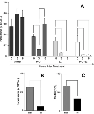
Short-term infection assays were used to evaluate the effects of DFO on highly infected animals. To test this, animals were infected with 50,000 parasites and treated 7 days after infection with DFO (25 mg/animal) and/or BZ (approximately 12,5 mg/animal) (Fig. 1A). Parasitemia was significantly reduced 3 h after treatment in DFO (65.11%), BZ (65%) and DFO + BZ (89.9%) treated mice. Nev-ertheless, 6 h after treatment, only BZ or DFO + BZ treated animals continued to exhibit lower parasitemia.
In a longitudinal study, we evaluated the capacity of DFO to re-duce parasitemia and mortality in mice infected with 500 blood-stream forms of the Y strain ofT. cruzi.
In animals treated with DFO from 14 days prior to infection to 21 d.p.i. (I/T), we observed a decrease in parasitemia and mortality
at 14 d.p.i. (p< 0.05). The average parasitemia value in I/T mice (46,444 trypomastigotes/0.1 mL of blood) was 5.4 times lower than in I/NT (250,444 trypomastigotes/0.1 mL of blood) (p< 0.05) (Fig. 1B).
Similarly, DFO was able to induce a decrease in mortality at 14 d.p.i. Mortality in the I/NT group was 67%, whereas the death rate in the I/T group was 33% (p< 0.05) (Fig 1C).
3.2. Impact of DFO on host biomarkers
To investigate the effect of DFO on host iron metabolism impairment, we analysed ferritin, serum iron and TIBC levels in in-fected and DFO treated animals. Surprisingly, we found that treat-ment with DFO did not interfere in host iron metabolism (Fig. 2), suggesting that the DFO effects on disease outcome were not dependent on the host’s iron level.
The effect of DFO on blood cells was also investigated. No difference in erythrograms (erythrocytes, haemoglobin,
Fig. 1.(A) Swiss mice (n= 20) were inoculated with 50,000 T. cruzi Y strain trypomastigotes and received no treatment (control), 25 mg/animal DFO, approx-imately 12.5 mg/animal BZ or both. Parasitemia was estimated before treatment and 3 and 6 h after treatment. Lines mean significant difference (p< 0.05). (B and C) Swiss mice (n= 20) were inoculated with 500T. cruziY strain trypomastigotes and received either no treatment (I/NT) or treatment with DFO (I/T). Parasitemia (B) and mortality (C) were observed 14 days after infection.
haematocrit), leukocytes or platelet levels was observed (data not shown), indicating that DFO treatment was not accompanied by the evident impairment of erythroid, myeloid or megakaryocytic differentiation.
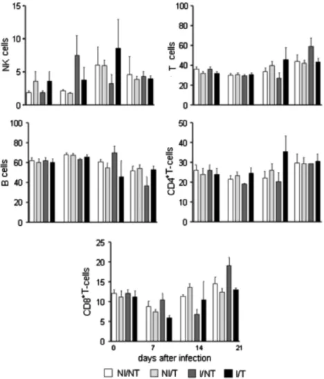
To further examine the potential unfavourable effects of DFO in lymphocyte subpopulations, we performed immunophenotypical analysis of spleen cells.
Phenotypic profiles of splenic lymphocyte populations from dif-ferent experimental groups were presented (Fig. 3). NK cells (CD49+), T lymphocytes (CD4++ CD8+), B cells (CD19+) and T cell
subpopulations (CD4+and CD8+) were analysed. We did not
ob-serve any differences between the groups.
Since these findings suggested that the sensitivity ofT. cruzito decreased iron levels could be greater than the sensitivity of the host cells, we hypothesised that DFO could act directly on the parasite.
3.3. Direct impact of DFO on T. cruzi
Using the protocol recommended by Experimental Models in Drug Screening and Development for Chagas Disease Workshop (Romanha et al., 2010), we evaluated the growth of the Tulahuen strain ofT. cruziin the presence of DFO.
This assay measures the activity ofb-galactosidase expressed by either amastigotes or trypomastigotes, since culture cells are lysed
before the measurement of enzyme activity. Using BZ as a positive control for growth inhibition, the activity of the DFO, shown by growth inhibition of amastigotes and trypomastigotes, is presented inFig. 4. DFO was shown to be active only in high concentrations.
Fig. 3.Swiss mice (n= 60) were inoculated with 500T. cruziY strain trypomastigotes and treated with DFO or left untreated. As controls, mice were left uninfected or untreated. Splenocytes were cultured for 12 h in culture medium and stained for the presence of NK cells (CD49), T cells (CD4+CD8+), B cells (CD19) and T-cell subpopulations
with anti-CD4 and anti-CD8 markers. Results are expressed as a percentage of total splenocytes.
At a concentration of 100
l
g/mL (IC50), the percentage of growthinhibition was 49%, whereas treatment with BZ (1
l
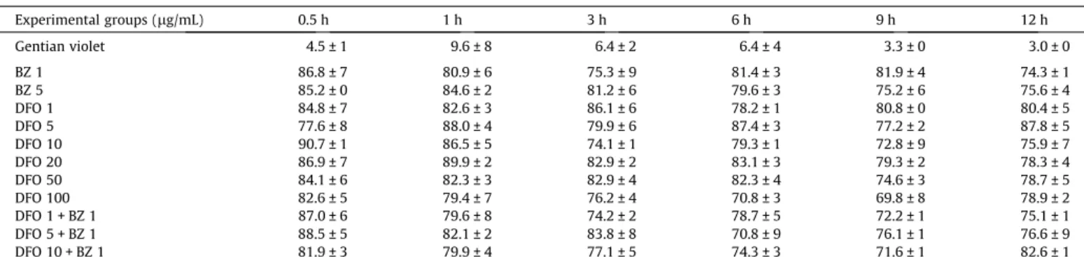
g/mL) caused a 93% growth inhibition of the parasites.We also evaluated viability by trypan blue staining. The Y strain-infected L929 cells were cultured in the presence of different concentrations of DFO (1, 5, 10, 20, 50 and 100
l
g/mL) or BZ (1 and 5l
g/mL). The parasites were incubated for 0, 0.5, 1, 3, 6, 9 and 12 h. Trypomastigotes from the supernatant were stained with try-pan blue. In all tested concentrations of DFO or BZ, 70–90% of the parasites remained viable, with no differences between the groups (Table 1), suggesting a low trypanocidal effectin vitro.Apoptosis induced by DFO was assessed by phosphatidylserine externalisation. Similar to the results of viability by flow cytome-try, the majority of the trypanosomes treated with DFO showed no staining with either PI or annexin V-FITC after up to 3 h of incu-bation (Fig. 5), suggesting parasite preservation. Because PI is a membrane-permeable nuclear stain, these results indicate mem-brane integrity.
Because DFO did not considerably decrease in vitro parasite growth, we decided to investigate whether the compound had try-panostatic action. To test the effect of DFO on parasite motility, we counted the number of mobile trypanosomes in the supernatant of L929 cells after treatment with different concentrations of DFO (1, 5, 10, 20, 50 and 100
l
g/mL) and times of incubation (1, 3, 6, 9 and 12 h).We could observe a trypanostatic effect of DFO in sublethal con-centrations. After 3 h of incubation, DFO at concentrations higher than 10
l
g/mL showed more efficiency than 1l
g/mL of BZ in reducing parasite mobility.Additionally, DFO at concentrations higher than 20
l
g/mL showed more efficiency than 1l
g/mL of DFO, which was the low-est concentration of drug used (p< 0.05) (Fig. 6).4. Discussion
Unfortunately, there is no current effective treatment for chronic Chagas disease, which is one of the world’s most neglected tropical diseases. Two drugs have been clinically used, benznidaz-ole and nifurtimox, but both were developed more than four dec-ades ago (Coura and de Castro, 2002). They are far from ideal due to multiple side effects and limited efficacy, especially in patients with the chronic form of the disease (Rocha et al., 2007; Soeiro and De Castro, 2009). Side effects vary from discrete discomfort to peripheral polyneuropathy, hypersensitivity dermatitis, and haematologic disorders that lead to treatment discontinuation. In Brazil, only benznidazole is commercially available. The use of nif-urtimox was halted owing to toxic effects.
Despite of the lack of interest from drug companies in develop-ing novel compounds for Chagas disease, a few new drugs have
undergone clinical trials, including allopurinol, itraconazole and fluconazole. The latter are last generation of anti-fungal therapies, and they act by preventing the synthesis of sterols (Coura, 2009). Nevertheless, more therapeutic options are necessary, not only as monotherapies but also for combined therapies that could be more effective and less toxic.
Table 1
Kinetic evaluation of viability (% of viable parasites unstained by trypan blue) of trypomastigotes cultured in the presence of DFO and or Bz (means ± SD).
Experimental groups (lg/mL) 0.5 h 1 h 3 h 6 h 9 h 12 h
Gentian violet 4.5 ± 1 9.6 ± 8 6.4 ± 2 6.4 ± 4 3.3 ± 0 3.0 ± 0
BZ 1 86.8 ± 7 80.9 ± 6 75.3 ± 9 81.4 ± 3 81.9 ± 4 74.3 ± 1
BZ 5 85.2 ± 0 84.6 ± 2 81.2 ± 6 79.6 ± 3 75.2 ± 6 75.6 ± 4
DFO 1 84.8 ± 7 82.6 ± 3 86.1 ± 6 78.2 ± 1 80.8 ± 0 80.4 ± 5
DFO 5 77.6 ± 8 88.0 ± 4 79.9 ± 6 87.4 ± 3 77.2 ± 2 87.8 ± 5
DFO 10 90.7 ± 1 86.5 ± 5 74.1 ± 1 79.3 ± 1 72.8 ± 9 75.9 ± 7
DFO 20 86.9 ± 7 89.9 ± 2 82.9 ± 2 83.1 ± 3 79.3 ± 2 78.3 ± 4
DFO 50 84.1 ± 6 82.3 ± 3 82.9 ± 4 82.3 ± 4 74.6 ± 3 78.7 ± 5
DFO 100 82.6 ± 5 79.4 ± 7 76.2 ± 4 70.8 ± 3 69.8 ± 8 78.9 ± 2
DFO 1 + BZ 1 87.0 ± 6 79.6 ± 8 74.2 ± 2 78.7 ± 5 72.2 ± 1 75.1 ± 1
DFO 5 + BZ 1 88.5 ± 5 82.1 ± 2 83.8 ± 8 70.8 ± 9 76.1 ± 1 76.6 ± 9
DFO 10 + BZ 1 81.9 ± 3 79.9 ± 4 77.1 ± 5 74.3 ± 3 71.6 ± 1 82.6 ± 1
Fig. 5.Trypomastigote cells were cultured in L929 cells in the presence or absence of DFO. BZ and gentian violet were used as controls. A suspension of 1106 par-asites was double stained with FITC-conjugated annexin V and PI. Parpar-asites showing no staining by either annexin or PI were considered alive (white). Parasites stained with annexin alone were considered in early apoptosis (black). Parasites stained by both PI and annexin were considered in late apoptosis (light grey). Parasites stained with PI alone were considered dead (dark grey).
DFO is known to reduce the intensity of murineT. cruzi infec-tions. Our previous data indicated beneficial effectsin vivodespite obvious interference in the host iron burden (Arantes et al., 2007; Francisco et al., 2008). Therefore we decided to further investigate the activity of DFO onT. cruziinfections.
In the present study, we used differentin vivoandin vitro ap-proaches. The use of a rapidin vivotest was supported by the fact that this methodology detects activity against circulating blood forms using a drug concentration five times higher than the recom-mended daily dose. Brener was the first to investigate the suscep-tibility ofT. cruziblood-stream forms to active compounds. Brener claimed thatT. cruzistout blood forms persist for some hours in the bloodstream without penetrating the host tissues (Brener, 1969, 1971; Filardi and Brener, 1984). In this rapid method, active drugs are supposed to induce a fast decline in the number of circulating parasites. The methodology we use here is considered efficient for characterising susceptibility to active drugs and for screening ac-tive compounds againstT. cruzi. This approach may complement data relating to the effects of drugs on differentT. cruziintracellular stages (Maria et al., 1972). This methodology has been utilised re-cently by others (Alves et al., 1999; Aguirre-Ivarado et al., 2007).
As expected, the results we obtained with this method corre-lated with those obtained by prolonged treatment schedules. Both showed lower infection intensity in the presence of DFO treatment. DFO was able to induce lower parasitemia in both short- and long-term infections and diminished mortality in long-long-term infection.
Although DFO was able to early reduce parasitemia in the rapid test, this was an unsustained effect. Six hours after treatment, par-asite levels were similar to that found in untreated animals. This finding is probably due to the short half-life in plasma and the ra-pid metabolism of DFO (Aouad et al., 2002). Nevertheless, this lim-itation was overcome by combination therapy with BZ.
Despite the clear effect of DFO on parasite growth, no interfer-ence in host iron metabolism or hematopoietic and lymphopoietic differentiation was observed. This result is supported by previous findings. In the work carried out byPedrosa et al. (1990), mice in-fected with Y, CL and YuYu strains ofT. cruziand treated with DFO exhibited no reduction of serum iron or increase in anaemia. Like-wise, Lalonde and Holbein (1984) demonstrated no significant changes in serum iron levels of either infected or uninfected mice treated with DFO. Similarly,Arantes et al. (2007)observed no de-crease in the serum iron or haemoglobin levels of mice infected withT. cruziand treated with DFO.Francisco et al. (2008)showed that serum iron concentrations were even higher in animals trea-ted with DFO or with both DFO and BZ when compared to the un-treated group.
These findings suggest that the sensitivity of T. cruzi to the unavailability of iron is greater than that of the host cell.
In the evaluation of the impact of DFO onT. cruzi, we assessed parasite growth inhibition, viability by trypan blue, apoptosis and motility assays.
Similarly to BZ, DFO did not affect the viability measured by try-pan blue staining. But DFO was able to inhibit amastigote and tryp-omastigote growth in fibroblast culture, but only when the drug was at high concentrations (100
l
g/mL). This is in contrast to BZ, which was highly effective at concentrations of 1l
g/mL.The ability of DFO in reducing parasite growth has been reported previously byLoo and Lalonde (1984). The authors have shown that DFO reduced the rate of amastigotes replication in treated cell cul-tures in a dose dependent manner by depleting host cell iron.
All of our data suggest low trypanocidalin vitroeffects of DFO, which contrasted with the beneficial effects observed in mouse infections. This led us to investigate the effects of DFO on parasite motility.
A decrease in motility was observed when trypomastigotes were cultured in the presence of DFO. This result is of great
impor-tance because it suggests a putative trypanostatic action. Little is known about DFO activity onT. cruzi, and this trypanostatic effect may be a major step in the action of DFO on parasite control since parasite motility is necessary for cell invasion and consequently parasite multiplication (Sibley, 2011). It is already described that one putative pathway for trypomastigote entry into mammalian cells involves activation of calcium signalling in the host cell and the recruitment of host cell lysosomes to the site of entry, where they fuse with the vacuole formed by the entering parasite (Rodriguez et al., 1996; Tardieux et al., 1992). The extremely active motility of the parasite likely contributes to this by causing local breaks in the plasma membrane and hence lysosomal recruitment is triggered as part of the wound healing response, akin to what happens in mammalian cells that have been damaged (McNeil and Kirchhausen, 2005).Andrade and Andrews (2004, 2005) re-ported that trypomastigotes invasion occurs either by direct recruitment and fusion of lysosomes at the plasma membrane, or through invagination of the plasma membrane followed by intra-cellular fusion with lysosomes. The lysosome-like parasitophorous vacuole is essential for preventing these highly motile parasites from exiting host cells and for allowing completion of the intracel-lular life cycle. They showed that when lysosome-mediatedT. cruzi
invasion is blocked, a significant fraction of the internalised parasites are not subsequently retained inside host cells for a productive infection.
Effects on motility have been previously observed in parasites with the use of certain drugs.Kamau et al. (2001)demonstrated that allopurinol had a leishmaniostatic effect in flow cytometry analysis. Nevertheless, the mechanism underlying this action is not fully understood.
The involvement of apoptosis as a possible mechanism of DFO action inT. cruziwas also tested. Previous work has shown that apoptosis can also be induced in unicellular parasites, including in T. cruzi(Ameisen et al., 1995; Piacenza et al., 2001). Infective forms ofT. cruziandLeishmaniaspp. display extracellular phospha-tidylserine, a marker of apoptotic cells (de Freitas Balanco et al., 2001; Damatta et al., 2007). Apoptosis of a proportion of Leish-mania parasites is required for the successful establishment of infection in the vertebrate host (van Zandbergen et al., 2006). In our data, we observed that DFO induces only minor apoptosis in trypomastigotes, indicating that this is not the major mechanism of DFO action and further studies should be conducted to better elucidate this possible mechanism.
Based on these results, we believe that DFO has more effective trypanostatic than trypanocidal properties and this effect may lead to lower infectivity by decreasing cellular invasion. It is possible thatT. cruzipresents greater sensitivity to the unavailability of iron than the host cell and therefore DFO is apparently free of major haematological side effects.
Acknowledgments
This work was supported by Coordenação de Aperfeiçoamento de Pessoal de Nível Superior (CAPES), Fundação de Amparo à Pes-quisa do Estado de Minas Gerais (FAPEMIG), Centro de PesPes-quisa René Rachou, Fundação Osvaldo Cruz – FIOCRUZ (CPq/RR), Univer-sidade Federal de Ouro Preto (UFOP) and UniverUniver-sidade Federal de Minas Gerais (UFMG), Brazil.
References
Alves, T.M., Alves, R., Romanha, A.J., Zani, C.L., dos Santos, M.H., Nagem, T.J., 1999. Biological activities of 7-epiclusianone. Journal of Natural Products 62, 369– 371.
Ameisen, J.C., Idziorek, T., Billaut-Mulot, O., Loyens, M., Tissier, J.P., Potentier, A., Ouaissi, A., 1995. Apoptosis in a unicellular eukaryote (Trypanosoma cruzi): implications for the evolutionary origin and role of programmed cell death in the control of cell proliferation, differentiation and survival. Cell Death and Differentiation 2, 285–300.
Andrade, L.O., Andrews, N.W., 2004. Lysosomal fusion is essential for the retention ofTrypanosoma cruziinside host cells. Journal of Experimental Medicine 200, 1135–1143.
Andrade, L.O., Andrews, N.W., 2005. TheTrypanosoma cruzi– host-cell interplay: location, invasion, retention. Nature Reviews Microbiology 3, 819–823. Aouad, F., Florence, A., Zhang, Y., Collins, F., Henry, C., Ward, R.J., Crichton, R.R., 2002.
Evaluation of new iron chelators and their therapeutic potential. Inorganica Chimica Acta 339, 470–480.
Arantes, J.M., Pedrosa, M.L., Martins, H.R., Veloso, V.M., de Lana, M., Bahia, M.T., Tafuri, W.L., Carneiro, C.M., 2007.Trypanosoma cruzi: treatment with the iron chelator desferrioxamine reduces parasitaemia and mortality in experimentally infected mice. Experimental Parasitology 117, 43–50.
Bertelli, M.S., Golgher, R.R., Brener, Z., 1977. Intraspecific variation inTrypanosoma cruzi: effect of temperature on the intracellular differentiation in tissue culture. Journal of Parasitology 63, 434–437.
Breidbach, T., Scory, S., Krauth-Siegel, R.L., Steverding, D., 2002. Growth inhibition of bloodstream forms of Trypanosoma brucei by iron chelator deferoxamine. International Journal of Parasitology 32, 473–479.
Brener, Z., 1962. Therapeutic activity and criterion of cure on mice experimentally infected withTrypanosoma cruzi. Revista do Instituto de Medicina Tropical de São Paulo 4, 389–396.
Brener, Z., 1969. The behavior of slender and stout forms ofTrypanosoma cruziin the blood-stream of normal and immune mice. Annals of Tropical Medicine and Parasitology 63, 215–220.
Brener, Z., 1971. Study of the action of some active drugs againstTrypanosoma cruzi
blood forms. Revista do Instituto de Medicina Tropical de São Paulo 13, 302– 306.
Buckner, F.S., Verlinde, C.L.M.J., La Flamme, A.C., Van Voorkhis, W.C., 1996. Efficient technique for screening drugs for activity against Trypanosoma cruziusing parasites expressingb-galactosidase. Antimicrobial Agents and Chemotherapy 40, 2592–2597.
Coura, J.R., 2009. Present situation and new strategies for Chagas disease chemotherapy: a proposal. Memórias Instituto Oswaldo Cruz 104, 549–554. Coura, J.R., de Castro, S.L., 2002. A critical review on Chagas disease chemotherapy.
Memórias Instituto Oswaldo Cruz 97, 3–24.
Damatta, R.A., Seabra, S.H., Deolindo, P., Arnholdt, A.C., Manhães, L., Goldenberg, S., de Souza, W., 2007. Trypanosoma cruzi exposes phosphatidylserine as an evasion mechanism. FEMS Microbiology Letters 266, 29–33.
de Freitas Balanco, J.M., Moreira, M.E., Bonomo, A., Bozza, P.T., Amarante-Mendes, G., Pirmez, C., Barcinski, M.A., 2001. Apoptotic mimicry by an obligate intracellular parasite downregulates macrophage microbicidal activity. Current Biology 11, 1870–1873.
Filardi, L.S., Brener, Z., 1984. A rapid method for testing in vivo the susceptibility of different strains of Trypanosoma cruzi to active chemotherapeutic agents. Memórias do Instituto Oswaldo Cruz 79, 221–225.
Filardi, L.S., Brener, Z., 1987. Susceptibility and natural resistance ofTrypanosoma cruzistrains to drugs used clinically in Chagas disease. Royal Society of Tropical Medicine and Hygiene 81, 755–759.
Francisco, A.F., Vieira, P.M.A., Arantes, J.M., Pedrosa, M.L., Martins, H.R., Silva, M., Veloso, V.M., de Lana, M., Bahia, M.T., Tafuri, W.L., Carneiro, C.M., 2008.
Trypanosoma cruzi: effect of benznidazole therapy combined with the iron chelator desferrioxamine in infected mice. Experimental Parasitology 120, 314– 319.
Francisco, A.F., de Abreu Vieira, P.M., Arantes, J.M., Silva, M., Pedrosa, M.L., Elói-Santos, S.M., Martins-Filho, O.A., Teixeira-Carvalho, A., Araújo, M.S., Tafuri, W.L., Carneiro, C.M., 2010. Increase of reactive oxygen species by desferrioxamine during experimental Chagas’ disease. Redox Report 15, 185–190.
Gordeuk, K., Thuma, P., Brittenham, G., McLaren, C., Parry, D., Backenstose, A., Biemba, G., Msiska, R., Holmes, L., McKinley, E., Vargas, L., Gilkeson, R., Polera, A.A., 1992. Effect of iron chelation therapy on recovery from deep coma in children with cerebral malaria. The New England Journal of Medicine 327, 1473–1477.
Hershko, C., Peto, T.E.A., 1988. Deferroxamine inhibition of malaria is independent of host iron status. Journal of Experimental Medicine 168, 375–387.
Kamau, S.W., Nunez, R., Grimm, F., 2001. Flow cytometry analysis of the effect of allopurinol and the dinitroaniline compound (Chloralin) on the viability and the proliferation ofLeishmania infantumpromastigotes. BMC Pharmacology 1, 1. Lalonde, R.G., Holbein, B.E., 1984. Role of iron inTrypanosoma cruziinfection of mice.
Journal of Clinical Investigation 23, 470–476.
Loo, V.G., Lalonde, R.G., 1984. Role of iron in intracellular growth ofTrypanosoma cruzi. Infection and Immunity 45, 726–730.
Mabeza, G.F., Biemba, G., Gordeuk, V.R., 1996. Clinical studies of iron chelators in malaria. Acta Haematologica 95, 78–86.
Mahmoud, M.S., 1999. Effect of deferoxamine alone and combined with pyrimethamine on acute toxoplasmosis in mice. Journal of the Egyptian Society of Parasitology 29, 791–803.
Maria, T.A., Tafuri, W.L., Brenner, Z., 1972. The fine structure of different bloodstream forms of Trypanosoma cruzi. Annals of Tropical Medicine and Parasitology 66, 423–431.
McNeil, P.L., Kirchhausen, T., 2005. An emergency response team for membrane repair. Nature Reviews Molecular Cell Biology 6, 499–505.
Minotti, G., Aust, S.D., 1987. The role of iron in the initiation of lipid peroxidation. Chemistry and Physics of Lipids 44, 191–208.
Oliveira, R.B., Vaz, A.B.M., Alves, R.O., Liarte, D.B., Donnici, C.L., Romanha, A.J., Zani, C.L., 2006. Arylfurans as potentialTrypanosoma cruzitrypanothione reductase inhibitors. Memórias do Instituto Oswaldo Cruz 101, 169–173.
Pedrosa, M.L., Silva Marcelo, E., Silva Márcio, E., Silva, M.E.C., Nicoli, J.R., Vieira, E.C., 1990. The effect of iron deficiency and iron overload on the evolution of Chagas’ disease produced by three strains of Trypanosoma cruzi in CFW mice. Comparative Biochemistry and Physiology 97, 235–243.
Piacenza, L., Peluffo, G., Radi, R., 2001. L-Arginine-dependent suppression of apoptosis in Trypanosoma cruzi: contribution of the nitric oxide and polyamine pathways. National Academy Sciences of the United States of America 98, 7301–7306.
Rachidi, S., Coudray, C., Baret, P., Gelon, G., Pierre, J.L., Favier, A., 1994. Inhibition of lipid peroxidation by a new family of iron chelators. Comparison with desferrioxamine. Biological Trace Element Research 41, 77–87.
Richardson, R.D., 2004. Novel chelators for central nervous system disorders that involve alterations in the metabolism of iron and other metal ions. Annals of the New York Academy of Sciences 1012, 326–341.
Rocha, M.O., Teixeira, M.M., Ribeiro, A.L., 2007. An update on the management of Chagas cardiomyopathy. Expert Review of Anti Infective Therapy 5, 727–743. Rodriguez, A., Samoff, E., Rioult, M.G., Chung, A., Andrews, N.W., 1996. Host cell
invasion by trypanosomes requires lysosomes and microtubule/kinesin-mediated transport. Journal of Cell Biology 134, 349–362.
Romanha, A.J., de Castro, S.L., Soeiro, M.N.C., Lannes-Vieira, J., Ribeiro, I., Talvani, A., Bourdin, B., Blum, B., Olivieri, B., Zani, C., Spadafora, C., Chiari, E., Chatelain, E., Chaves, G., Calzada, J.E., Bustamante, J.M., Freitas-Junior, L.H., Romero, L.I., Bahia, M.T., Lotrowska, M., Soares, M., Andrade, S.G., Armstrong, T., Wim Degrave, W., Andrade, Z.A., 2010. In vitro and in vivo experimental models for drug screening and development for Chagas disease. Memórias do Instituto Oswaldo Cruz 105, 233–238.
Sibley, D., 2011. Invasion and intracellular survival by protozoan parasites. Immunological Reviews 240, 72–91.
Soeiro, M.N., De Castro, S.L., 2009. Trypanosoma cruzi targets for new chemotherapeutic approaches. Expert Opinion Therapeutic Targets 13, 105–121. Spellberg, B., Edwards Jr., J., Ibrahim, A., 2005. Novel perspectives on mucormycosis: pathophysiology, presentation, and management. Clinical Microbiology Reviews 18, 556–569.
Tardieux, I. et al., 1992. Lysosome recruitment and fusion are early events required for trypanosome invasion of mammalian cells. Cell 71, 1117–1130.
Taylor, M.J., Hughes, B.J., Sharma, R.P., 1987. Dose and time related effects of T-2 toxin on mitogenic response of murine splenic cells in vitro. International Journal of Immunopharmacology 9, 107–113.
Traore, O., Carnevale, P., Kaptue-Noche, L., Bede, J., Desfontaine, M., Elion, J., Labie, D., Nagel, R.L., 1991. Preliminary reports in the use of desferrioxamine in the treatment ofPlasmodium falciparummalaria. American Journal of Hematology 37, 206–208.
van Zandbergen, G., Bollinger, A., Wenzel, A., Kamhawi, S., Voll, R., Klinger, M., Müller, A., Hölscher, C., Herrmann, M., Sacks, D., Solbach, W., Laskay, T., 2006.
Leishmania disease development depends on the presence of apoptotic promastigotes in the virulent inoculum. National Academy Sciences of the United States of America 103, 13837–13842.