doi: 10.1590/S1984-46702011000400008
The dark shore crab Pachygrapsus gracilis (Saussure, 1858) has a wide distribution, occurring in the western Atlantic from the Caribbean to Texas, French Guiana, Brazil (Trinidad and from Ceará to Rio Grande do Sul) and Argentina, In the east-ern Atlantic the species occurs from Senegal to Angola (MELO 1996, POUPINet al. 2005).
Pachygrapsus Randall, 1839 megalopae was described in the past from individuals collected in the natural environment: P. marmoratus (Fabricius, 1787) by GUERAOet al. (1997), P. trans-versus (Gibbes, 1850) by FLORESet al. (1998) and P. gracilis by CHÁRAZO-OLVERA & ROCHA-RAMÍREZ (2007). These studies used specimens collected from the environment, probably due to the difficulty of obtaining the stage of megalopae under labo-ratory conditions. Only two studies on the juvenile growth and morphology of this group are available in the scientific litera-ture: GUERAOet al. (1997) for P. marmoratus and FLORESet al. (1998) for P. transversus.
GUERAOet al. (1997) described the morphology of the cara-pace, pereiopods and third maxillipeds of the juvenile instar I of P. marmoratus. Their data provide information on the size of the carapace up to the juvenile IV. FLORESet al. (1998) described in detail the megalopa, the first crab instar, and the juvenile development instars of P. transversus giving emphasis on the process of pleopod differentiation up until juvenile instar VII.
Studies on crab growth are important because some mor-phological characteristics, including secondary sexual charac-ters, which arise at the juvenile phase, are useful in the identification of species, as well as age and sex determinations. The morphological characteristics found in crustaceans have contributed significantly to the understanding of the taxo-nomic, systematic and phylogenetic nature of crustaceans. They have also generated information which has helped the under-standing of their biology and assisted population management (BARUTOTet al. 2001). However, morphological descriptions of crustaceans must be accurate, and describe the setal arrange-ment, the number of the juvenile appendages and other struc-tures correctly.
In some cases, intraspecific variation has been found in individuals of different populations. Nelice M. Batistelli (unpubl. data), for example, found discrepancies in setal num-ber and setation arrangements in re-descriptions of various fid-dler crab larvae. More studies on the morphological stages and new descriptions of larval and juvenile stages of crustaceans are necessary in order to clarify these differences.
Due to the scarcity of studies detailing the morphologi-cal development of crustaceans and the growth of these ani-mals, it is necessary to conduct further studies on the growth and morphology of juvenile instars. The present study
re-de-Redescription of megalopa and juvenile development of
Pachygrapsus gracilis
(Decapoda: Grapsidae) from the Amazon region,
reared in the laboratory
Danielle C. B. Arruda
1& Fernando A. Abrunhosa
1, 21 Laboratório de Carcinologia, Instituto de Estudos Costeiros, Universidade Federal do Pará. Campus Universitário de Bragança,
Alameda Leandro Ribeiro, Aldeia, 68600-000 Bragança, PA, Brazil, E-mail: danicbarruda@yahoo.com.br; faraujo@ufpa.br
2 Corresponding author.
ABSTRACT. This present study re-describes the megalopa stage and provides detailed morphological descriptions and main growth changes observed in stages I through VII of the juvenile instars of the dark shore crab Pachygrapsus gracilis
(Saussure, 1858), from the Amazon region. The specimens in this study were reared in the laboratory and the megalopae were collected at Ajuruteua beach in northeastern Pará, Brazil. Previous studies had described the megalopa of P. gracilis
scribes the megalopa stage of P. gracilis inhabiting the Amazon region, and describes important morphological changes ob-served during the growth from juvenile instars I to VII, of.
MATERIAL AND METHODS
In February 2008, 116 megalopa were obtained at Ajuruteua (00º49’20.2”S, 46º36’02.2”W) Beach, municipality of Bragança (northeastern Pará state, Brazil). The specimens were collected off of mangrove leaves, pieces of trunks and roots brought by the tide. They were identified in the laboratory when they reached the juvenile instar. Specimen identification fol-lowed RODRIGUEZ (1980), MELO (1996) and POUPINet al. (2005).
In the laboratory, specimens were placed individually in plastic recipients with sea water (salinity 30) and a small rock shelter. The temperature was maintained at 26.5 ± 1.14ºC, pho-toperiod regime (12:12 h/light:dark cycle) and pH 8.1. Every two days the water of the recipients was changed. The indi-viduals were fed live Artemia nauplii daily and were monitored twice a day. The exuviae and dead animals were preserved in glycerol+ethylic alcohol 70% (1:1) solution.
At least 10 specimens of each stage were dissected with fine needles (BD Ultra-Fine®, 12.7 X 0.33 mm) under an opti-cal Zeiss (Axioskop 40) microscope equipped with an ocular micrometric. The appendages were extracted and transferred to another slide where they were stained with a solution of methylene blue 1% (sometimes was used). The terminology used in the descriptions follows POHLE & TELFORD (1981), GUERAO et al. (1997), FLORES et al. (1998), GARM (2004), and CHÁRAZO -OLVERA & ROCHA RAMÍREZ (2007). The studied specimens were deposited at the crustacean collection of Museu Paraense Emílio Goeldi, under the catalog number MPEG-817.
The individuals were reared until juvenile instar VII. During rearing, data on survival and intermolt periods were obtained for individuals in each juvenile instar. The carapace width (CW) was considered as an independent variable or as a reference (in axis x) because of its greater amplitude and be-cause it is the best representative dimension of organism size (HARTNOLL 1982). This variable was used for calculating the per-centage of molt increment and absolute increment after each ecdise. The abdomen width of male and female specimens was obtained by measuring the 4th abdominal somite.
The percentage of molt increment (%MI) was estimated by the following formula: %MI = [(CW2 – CW1) x 100]/CW1; where CW1 and CW2 are pre-molt and post-molt sizes, respec-tively.
The absolute increment (AI) after each ecdise was ob-tained using the following equation: AI = CW2 – CW1; where CW1 is the carapace width in pre-molt and CW2 the carapace width in post-molt.
The morphometric relationship between the carapace length (CL) and carapace width (CW) throughout the juvenile development was obtained by linear regression.
RESULTS
M
EGALOPAA total of 89 megalopae molted into the juvenile instar I. The remaining individuals died during the experiment. These were preserved. The first molting was recorded on the second day, and the last on the 18th day of culture after collections in the field (Fig. 1). Large frequencies of metamorphosed megalopae were recorded on the third and fourth days of rear-ing. The mean intermolt period was 5.72 ± 4.45 days.
J
UVENILEGROWTHDuring the culture, the mortality of juveniles was quite low: only three specimens died in the juvenile instars I, II and III, respectively. The mean intermolt period gradually increased during the juvenile development and after consecutive molts (Fig. 2). In juvenile instar I, the intermolt period mean was 6 ± 3 days whereas in the juvenile instar VII it averaged 20 ± 12 days. The carapace length of the juvenile instar I (1.96 ± 0.07 mm) was similar to that found for megalopa (1.97 ± 0.04 mm). The growth (carapace width) of each subsequent juvenile in-star showed an increase during the days of culture and molts (Fig. 3). In the juvenile instar I, the mean carapace width was 2.21 mm and in the juvenile instar VII the mean carapace width was 7.42 mm. The carapace width of male and female crabs was similar during juvenile growth until the VII instar (Fig. 4). The percentage of molt increment showed a reduction at each molt event (Fig. 5). From the juvenile instar I to juve-nile instar II, the mean rate of increment was 31.13 ± 8.75%, whereas from the juvenile instar VI to VII it was 18.51 ± 4.16%. The absolute molt increment increased at each molt event; from juvenile instar I to II the mean was 0.68 mm and from the juvenile instar VI to VII the mean was 1.17 mm (Fig. 5).
The width of the abdomen in juvenile forms increased during juvenile growth (Fig. 6). However, the width of the abdo-men of males and females presented differences only in the ju-venile instars VI and VII (Fig. 6). In the juju-venile instar I, the mean abdomen width in males and females was similar, 0.70 ± 0.04 mm and 0.60 ± 0.04 mm, respectively. In juvenile instars VI and VII, these values were very different for both sexes; fe-males had 2.10 ± 0.4 mm and 3.20 ± 0.76 mm of abdomen width, while males presented 1.70 ± 0.19 mm and 2.10 ± 0.15 mm, re-spectively.
M
ORPHOLOGICALDESCRIPTIONMegalopa
Figures 1-4. Pachygrapsus gracilis: (1) intermolt period of the megalopa stage; (2)mean of intermolt period (days) of each juvenile instar; (3) carapace width and carapace length at each juvenile instar; (4) mean carapace width of males and females at each juvenile instar. 30 25 20 15 10 5 0
1 2 3 4 5 6 7 8 9 10 11 12 13 14 15 16 17 18
Culture days Number of individuals 35 30 25 20 15 10 5 0
I II III IV V VI VII
Juvenile instars Intermolt period (days) 10 8 6 4 2 0 0 1 2 3 4 5 6 7
y = 0.600x + 0.614 R = 0.857 n = 591
2
Carapace width (mm)
Carapace length (mm) 10 8 Male Female 6 4 2 0
I II III IV V VI VII
Juvenile instars Carapace width (mm) 1 2 3 4 50 40 30 20 10 0
I-II II-III III-IV IV-V V-VI VI-VII
Percentage os size increment per molt
Absolut molt increment
2.5 2.0 1.5 1.0 0.5 0 Absolut molt increment (mm) Molt increment (%) Molt events 5 4 3 Male Female 2 1 0
I II III IV V VI
Juvenile instars VII Abdomen width (mm) 5 6
Antennule (Fig. 11): peduncle 3-segmented with 3, 5, 2+1 simple setae; endopod unsegmented with 2+1 simple setae; exopod 5-segmented with 0, 0, 2, 0, 1+3 simple setae and 0, 8, 4, 4,0 aesthetascs.
Antenna (Fig. 12) – 11-segmented with 0, 1+1, 2, 0, 0, 4, 2, 4, 0, 3, 3 simple setae; no sign of statocyst cavity in the basipod.
Mandible (Fig. 13): uncalcified, symmetrical with blade-like margin, incisive and molar processes indistinguishable; mandibular palp 3-segmented with 10 setae on distal segment, some setae are evidently pappose and some appear to be plumodenticulate but such pattern of setules is not obvious.
Maxillule (Fig. 14): coxal endite with seven terminal and three subterminal plumodenticulate setae and nine scattered simple setae; basial endite with 12 terminal cuspidate setae, 10 subterminal plumodenticulate setae, but the setules are minute and almost imperceptible, and 4 proximal simple setae; endopod 2-segmented, lacking setae.
Maxilla (Fig. 15): coxal endite bilobed, proximal lobe with five terminal simple setae and nine subterminal plumose se-tae; distal lobe with two terminal and four subterminal simple setae; basial endite bilobed, proximal lobe with 8-9 terminal simple setae, one terminal plumose setae and one subterminal simple setae; distal lobe with 9-12 terminal simple setae and one subterminal simple setae; endopod unsegmented, lacking setae; scaphognathite with approximately 70-72 marginal plu-mose setae and 3-4 simple setae on the lateral surface.
First maxilliped (Fig. 16):coxal endite with four termi-nal simple setae, one termitermi-nal plumose setae and 12 subtermi-nal simple setae; basal endite with 13 termisubtermi-nal simple setae and two subterminal simple setae; endopod unsegmented with two distal simple setae; exopod 2-segmented with three simple setae on proximal and four terminal plumose setae on distal segment; well development epipod with 10 long simple setae. Second maxilliped (Fig. 17): coxa and basis fused; endopod 5-segmented with 0, 1 long, 1, 6 simple setae, 5th seg-ment with 11 terminal and four subterminal denticulate setae; exopod 2-segmented with two simple setae on proximal seg-ment and five terminal plumose setae on distal segseg-ment; epipod with five long simple setae.
Third maxilliped (Fig. 18): coxa and basis fused bearing nine simple setae; endopod 5-segmented ischium, merus and carpus with 13+1, 5+4, 5 simple setae, propodus and dactyl bearing 9 and 8-10 strong denticulate setae; exopod 2-mented with five minute lateral simple setae on proximal seg-ment and four terminal plumose setae on distal segseg-ment; well developed epipod with 34-36 long simple setae.
Pereiopods (Fig. 7): well-developed with few setae dis-tributed on the segments (as illustrated); P1 – Chelipods simi-lar in shape and size; dactyl lacking teeth; propodus showing two truncated teeth on the posteroventral margin, propodus with one strong ventral spine close to dactyl (Fig. 9); P2-P4 – Dactyl ventrally curved with acute extremity, showing strong
teeth on the ventral margin; P5 – Dactyl with three long den-ticulate setae and a strong terminal spine, ventral margin lack-ing teeth (Fig. 10).
Abdomen (Fig. 7): 6-segmented; 2nd, 3rd, 4th and 5th seg-ments with two small setae on the posterior surface; telson semi-oval-shaped with two long setae and 4-6 small setae.
Pleopods (Fig. 19): well-developed; endopods bearing four cincinulli; exopod from pl 1 to pl 4 with 28, 23, 24, 22, plu-mose natatory setae, respectively.
Uropods (Fig. 19): endopod absent; exopod 2-segmented with three plumose setae on proximal segment and 15-16 plu-mose setae on the distal segment.
Juvenile I
Carapace (Fig. 20): length: 1.96 ± 0.07 mm; width: 2.20 ± 0.13 mm; markedly striated on metabranchial and mesobranchial regions; mesogastric, metagastric, cardiac and intestinal regions smooth; orbital extern tooth and antero-lat-eral tooth presents, the latter is smallest; frontal, orbital and lateral depressions visible; gastric, cardiac and branchial regions already distinct; eyes relatively large when compared to adult. Antennule (Fig. 21): peduncle 3-segmented, proximal segment (bulb) with minutes plumose setae arranged in trans-verse rows, 2nd segment with one terminal and four subtermi-nal simple setae, 3rd segment with 4+1 simple setae; endopod 3-segmented with 1, 2, 1+2 simple setae; exopod 6-segmented with 0, 10-12, 11, 3, 0, 0 aesthetascs and 0, 2, 2, 1, 3+1 long, two simple setae.
Antenna(Fig. 22): peduncle 3-segmented with 5+3, 0, 0 plumose setae and 0, 6, 0 simple setae; flagellum 8-segmented with 0, 4, 2, 2, 4, 0, 2, 2 simple setae; no sign of statocyst cavity in the basipod.
Mandible (Fig. 23): calcified, asymmetrical, left mandible with blade-like margin, incisive and molar processes indistin-guishable, mandibular palp 3-segmented with four and 14 plu-mose setae on 2nd and 3rd segments; right mandible incise process showing two blunt teeth and blade-like molar process, mandibular palp 3-segmented with six and 16 plumose setae on 2nd and 3rd segments.
Maxillule (Fig. 24): coxal endite with five terminal plumo-denticulate setae and 28-30 simple setae arranged in all sur-face; basial endite with 15 terminal cuspidate setae, one pappose seta on lateral margin and 13 subterminal simple setae; endopod 2-segmented with two plumose setae on proximal segment and 2+2 plumose setae on distal segment.
Figures 7-18. Pachygrapsus gracilis, megalopa appendages: (7) dorsal view; (8) ventral view of carapace showing detail of rostrum; (9) quelipods; (10) dactyl of pereiopod 5; (10) antennule; (10) antenna; (11) mandible (12) maxillule; (13) maxilla; (14) 1st maxilliped; (15) 2nd maxilliped; (16) 3rd maxilliped. Scale bars: 7 = 0.02 mm; 8-11, 15-18 = 0.01 mm; 13 = 0.006 mm; 14 = 0.08 mm.
13 7
9 8
14
18 15
17 16
12 11
First maxilliped (Fig. 26): coxal and basal endites almost entirely covered by plumose and simple setae, respectively; endopod 2-segmented, proximal segment with 12 small plumose setae on the margin, distal segment divided in two surfaces, dorsal surface with 12 and ventral surface with 9-13 plumose setae; exopod 2-segmented, proximal segment with 10 plumose setae, distal segment with four terminal plumose setae; well develop-ment epipod with 28 simple setae.
Second maxilliped (Fig. 27): coxa and basis fused with 3
plumose setae; endopod 6-segmented with 4, 6, 5+5+6, 5, 3+5 plumose setae, 6th segment bearing seven plumose and 15-17 plumodenticulate setae; exopod 2-segmented, proximal segment with a row of 12-13 dorsal plumose setae and 11-13 plumose setae on the outer margin, distal segment with 4 terminal plu-mose setae; well development epipod with 30 long simple setae. Third maxilliped (Fig. 28): coxa with 14 simple setae; basis with five plumose setae, endopod 5-segmented, ischium and merus with 38 and 20 plumose setae, carpus and propodus seg-Figure 19. Pachygrapsus gracilis, megalopa. Pleopods (Pl2-Pl5) and uropod (Urp). Scale bar: 0.01 mm.
Pl2
Pl3
Pl4
Figures 20-29. Pachygrapsus gracilis, juvenile I appendages: (20) dorsal view; (21) antennule; (22) antenna; (23) mandible; (24) maxillule; (25) maxilla; (26) 1st maxilliped; (27) 2nd maxilliped; (28) 3rd maxilliped; (29) abdomen. Scale bars: 20, 26 = 0.02 mm; 21, 23, 25 = 0.006 mm; 22, 27-29 = 0.01 mm; 24 = 0.08 mm.
20
21
24
28 26
23
22
25
ments with 20 and 18 plumodenticulate setae, dactyl segment with 18 denticulate setae; exopod 2-segmented, proximal seg-ment with 18 plumose setae, distal segseg-ment with five terminal plumose setae; well development epipod with approximately 105 long simple setae.
Pereiopods (Fig. 20): chelipods morphologically similar in shape and size, carpal tooth and laminar expansion den-ticulate of the merus already present as in adult form; pereio-pods 2-5 with striated margins, showing numerous setae and spines distributed along the segments (as illustrated).
Abdomen (Fig. 29): 2nd to 5th somites with characteristic
transverse striate; telson, a triangular in shape, bordered with 25 simple setae and four dorsal simple setae.
Pleopods: were not observed in this stage. Uropods ab-sent (not observed in all juveniles stages).
Development of other juvenile instars
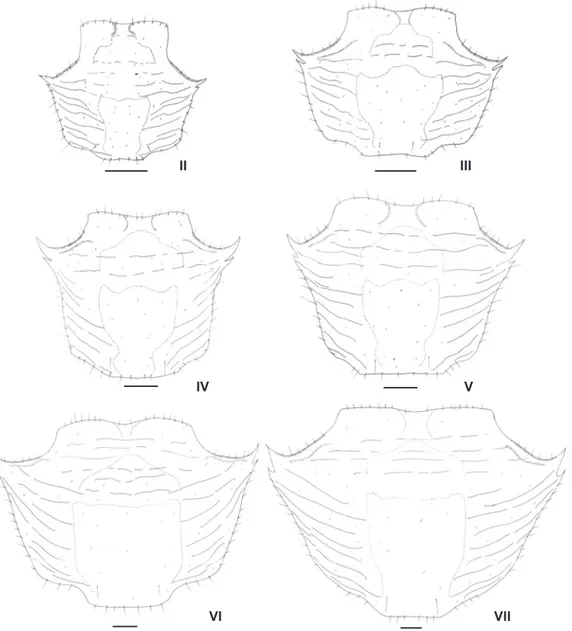
The main morphological changes were observed in the developmental sexual dimorphism in which they are described as follow. No significant morphological changes were observed in the carapace of subsequently juvenile instars (Fig. 30).
Juvenile II. Females: showing four pairs of pleopods (2nd to 5th), each one biramous and rudimentary, but due to the
Figure 30. Pachygrapsus gracilis, juvenile instars. Carapace from juvenile instar II to VII. Scale bars: Juvenile instars II, IV-V = 0.7 mm; Juvenile instars III = 0.6 mm; Juvenile instar VI-VII = 0.5 mm.
II
IV
III
V
fact that the illustrations were made from exuviae, the pleo-pods in juvenile II were damaged and they were not illustrated. Males: pleopods were not detected.
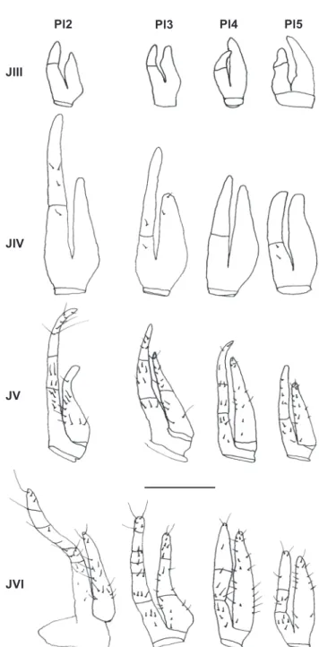
Juvenile III. Females: genital openings already present (Fig. 31); endopod of the four pairs of pleopods lacking setae; endopod 2-segmented; exopod unsegmented (Fig. 32). Males: genital opening already visible; showing only one pair of pleo-pod, biramous and rudimentary, lacking setae; in the males the second pair of pleopods is absent (Fig. 33).
ing setae. The second pair of pleopods weren’t detected in any juvenile instar (Fig. 33).
Juvenile V. Females: second, third, and fourth pairs of pleopods with endopod 3-segmented showing 7, 4, 5 simple setae on second pair, 4, 5, 3 on third pair and 3, 4, 5 on fourth pair; endopod of the fifth pair 2-segmented with 5, 4 simple Figure 31. Pachygrapsus gracilis, genital openings of males and
females in the juvenile instar III. Scale bar: 0.6 mm.
Juvenile IV. Females: pleopods larger than in juvenile III; endopods 2-segmented, endopod of the second pair of pleo-pods with 3, 1 simple setae on distal and proximal segments, respectively, endopods of the third pair with one simple setae on each segment, endopods of the fourth pair lacking setae and endopod of the fifth pair with 2, 0 simple setae on distal and proximal segments, respectively; exopods unsegmented, exopod of the thrid pair of pleopods with two simple setae (Fig. 32). Males: pleopod larger than in the previous instar,
lack-Figure 32. Pachygrapsus gracilis, juvenile instars. Female pleopods (Pl2-Pl5) from juvenile instar III to VI. Scale bar: JIII-JIV = 0.01 mm; JV-JVI = 0.02 mm.
Pl2 Pl3 Pl4 Pl5
JIV JIII
JV
setae; exopod of the second, fourth and fifth pairs unsegmented with 10, 14, 15 simple setae; exopod of the third pair 2-seg-mented with 8, 5 simple setae on proximal and distal segments, respectively (Fig. 32). Males: pleopods entirely developed. Endopod larger than exopod with a small distal groove rounded by numerous setae and with a longitudinal groove; exopod rudimentary (Fig. 33).
respectively; exopod of the second, fourth and fifth pair of pleo-pods unsegmented with 18, 22, 14 simple setae; exopod of the third pair 3-segmented with 7, 6, 8 simple setae (Fig. 32). The sexual abdominal dimorphism could also be observed (Fig. 34). Males: similar to previous instar.
DISCUSSION
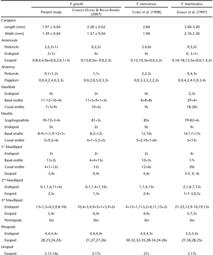
The analysis and comparisons with previous morphologi-cal studies available in the literature for thegenus Pachygrapsus (MELO 1996, POUPINet al. 2005), clearly indicates that P. Gracilis, has some unique features, including gonopod not T-shaped, lateral margins convergent posteriorly and with tooth behind exorbital angle.
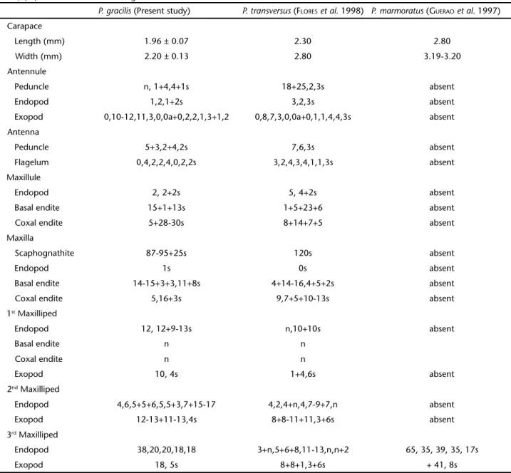
The number and type of setae in the appendages of al-ready described species (P. marmoratus, P. transversus and P. gra-cilis) are quite useful to differentiate among them, mainly the setae on the endopod of maxillule, maxilla and maxillipeds. Morphologically, all Pachygrapsus species studied have large eyes and a well-developed ocular peduncle. Beyond this, other simi-larities are found in the carapace characteristics, i.e, anterolat-eral region with lower expansion reaching orbital region and rostrum with a conspicuous medial depression. The carapace of the P. marmoratus and P. transversus are smooth and do not have a protuberance, whereas P. gracilis specimens from the Amazon and Mexico bear distinct lobes on the carapace.
Most of morphological differences are generally observed in the setal number and arrangement of: peduncle and endopod of the antennule, antenna, basal and coxal endites and endopod of the maxillule, maxilla endopod, first maxilliped exopod, fifth segment of the second maxilliped endopod, first segment of the third maxilliped exopod, pleopods and number of long setae on p5 (Tab. I).
The carapaces of P. marmoratus (GUERAOet al. 1997) is larger (CW = 2.10-2.30 mm, CL = 3.00-3.20 mm), and P. transversus (FLORESet al. 1998) (CW = 1.90 mm, CL = 2.80 mm) megalopa are larger in average than those of our species. The Mexican specimens of P. gracilis (CHÁRAZO-OLVERA & ROCHA-RAMÍREZ 2007) are larger than the Amazonian specimens. The carapace width of specimens from the Amazon was 1.39 ± 0.04 mm and the carapace length was 1.97 ± 0.04 mm, while in Mexican speci-mens the carapace length and width were 2.20 ± 0.02 mm and 1.57 ± 0.04 mm, respectively. However, many similarities were found between Mexican and Amazonian megalopae (see Tab. I). The most notable difference found in the morphology of the two populations being compared was the presence of three distinct elongated setae, or feelers, on the fifth pair of the pereiopods,and the absence of those in the Mexican popu-lation. The other two species of the genus, P. marmoratus e P. transversus also have these feelers.
Other differences found between specimens from Mexico and the Amazon region of Brazil were as follows: setal number on antennule peduncle and endopod: Amazonian P. gracilis had Figures 33. Pachygrapsus gracilis, juvenile instars. Male pleopod (Pl1)
from juvenile III to V. Scale bar: JIII-JIV = 0.01 mm; JV = 0.02 mm.
Juvenile VI. Females: pleopods fully developed; endopod of the second pair of pleopods 5-segmented with 4, 1, 1, 5, 6 simple setae; endopod of the third pair 6-segmented with 5, 1 2, 1, 6, 5 simple setae, respective; endopod of the fourth and fifth pair 3-segmented with 3, 5, 4 and 3, 4, 5 simple setae,
JV JIII
a total of 11 setae on the peduncle and three distal setae on the endopod, while Mexican P. gracilis had only four setae on the peduncle and four distal setae on the endopod. Other impor-tant distinctions were also observed between Amazonian and Mexican species (see Tab. II). The morphological variations found within P. gracilis (Tab. II) when our data is compared with previous studies may be due to mistakes or omissions in other contributions, or variations between crab populations.
Few studies are available in the literature giving detailed description of the juvenile morphology. For instance, for P. marmoratus,only some structures of the juvenile instar I were observed by GUERAOet al. (1997). On the other hand, a compre-hensive study, giving a detailed morphological description, was conducted on P. transversus from megalopa to the seven juve-nile instar (FLORESet al. 1998).
The morphologyof the juvenile instar I of P. gracilis is very similar to the morphology of the adult. The number of setae and their distribution pattern on the cephalic append-ages and maxillipeds are typical for adult crabs, as also found by FLORESet al. (1998)for P. transversus. In this juvenile, the two anterolateral teeth of the carapace are already visible in P. gra-cilis. In P. transversus, the anterolateral tooth is present only in the juvenile instar II. In P. marmoratus, by contrast, the three anterolateral teeth which can be observed in the adult, are al-ready differentiated in the juvenile instar I. This suggests that the anterolateral teeth can be used to differentiate the juvenile I of congenerics. Other similarities and differences found be-tween the species studied are shown on table II.
Similarities in the morphological characteristics and de-velopment of pleopods of juvenile males were observed in P. gracilis and Pyromaia tuberculata (Lockington, 1877) (FLORESet
al. 2002). In both species, only one pair of pleopods is present during the entire juvenile development. However, in P. trans-versus, as well as in Sesarma rectum Randall, 1840 (FRANSOZO 1987), Eriphia gonagra (J.C. Fabricius, 1781) (FRANSOZO & NEGREIROS-FRANSOZO 1987), Eurypanopeus abbreviatus (Stimpson, 1860) (FRANSOZO & NEGREIROS-FRANSOZO 1987) and Eurytium limosum (Say, 1818) (GUIMARÃES & NEGREIROS-FRANSOZO 2005), the pleopods show a varied pattern of development in which the juvenile may present one or two pairs of pleopods.
Differences are observed in the first appearance of the genital orifice of P. gracilis, P. transversus and P. tuberculata (FLORES et al. 2002). In males and females, the genital orifice arises in juvenile instars III, II and IV, respectively. Unfortunately, no comparisons could be made with other species because the authors did not mention details on the sexual dimorphism in these stages.
Studies on morphological descriptions of Pachygrapsus and other brachyuran species are very scarce, although they have been shown to be of great importance for systematic and diagnosis of species at different life stages. Thus, more studies on this topic should be made to contribute to the knowledge of the species biology and growth process.
When juvenile growths are compared, thepattern observed in P. gracilis juveniles is more similar to that of other cultured decapods. Growth patterns ascertained under culture conditions should be interpreted with caution because laboratory condi-tions and the natural environment are different. Environmen-tal factors not present in controlled conditions, may affect the juvenile growth rate. In such case, the molt increment of speci-mens reared in the laboratory may be equivalent, and probably smaller than those in the field (HARTNOLL 1982).
Table I. Morphological comparisons among megalopa stage of some Pachygrapsus species. (s) Setae, (a) aesthetascs, (es) elongated setae.
P. gracilis P. transversus P. marmoratus
Present study CHÁRAZO-OLVERA &ROCHA RAMÍREZ
(2007) FLORESet al. (1998) GUERAOet al. (1997)
Carapace
Length (mm) 1.97 ± 0.04 2.20 ± 0.02 2.80 3.00-3.20
Width (mm) 1.39 ± 0.04 1.57 ± 0.04 1.90 2.10-2.30
Antennule
Peduncle 3,5,2+1s 0,2,2s 3,4,0s 9,5,2s
Endopod 2+1s 4s 4s 0, 3+1s
Exopod 0,8,4,4,0a+0,0,2,0,1+3s 0,15,8,5a+ 0,0,2,3s 0,12,10,3a+0,0,2,2s 0,16-18,13,5a+0,0,1-2,2s
Antenna
Peduncle 0,1+1,2s 1,1s 2,2,2s 0,4,3s
Flagelum 0,0,4,2,4,0,3,3s 0,0,2,0,5,0,3,3s 0,0,3,3,3,2,2,2s 0,0,4,2,4-5,0,3,4s
Maxillule
Endopod 0s 2s 0s 2,3s
Basal endite 11-12+10+4s 11+3+9+1+2s 8+8+8s 29+4s
Coxal endite 7+3+9s 10+2s 9s 18-20s
Maxilla
Scaphognathite 70-72+3-4s 81+3s 85s 79-82+4s
Endopod 0s 2s 0s 4s
Basal endite 8-9+1+1,9-12+1s 8,2+12s 12,10s 16-17+13s
Coxal endite 5+9,2+4s 4+1+3,2+2s 3+2,10+1+6s 5+15s
1st Maxilliped
Endopod 2s 2s 2s 4s
Basal endite 13+2s 4+4+13s 10+3s 17s
Coxal endite 4+1+12s 12s 12+6s 20s
Exopod 3,4s 0,4s 4,4s 3-5, 0, 4s
2nd Maxilliped
Endopod 0,1,1,6,11+4s 0,1,1,4+1,10s 1,1,4,13s 2,1,6-7,12s
Exopod 2,5s 1,5s 2,4s 1+1-3,0,5s
3rd Maxilliped
Endopod 13+1,5+4,5,9,8-10s 10,4+3,4,9+5+1+3,9+2s 4+12+1,7+3,2+4,11,13+2s 21-23,12,9-10,19,13s
Exopod 5,4s 0,4s 4,4s 5-7,5s
Pereiopods 3es 0es 3es 3es
Pleopods
Endopod 4,4,4,4s 4,4,4,4s 4,4,4,3s 5,5,5,4s
Exopod 28,23,24,22s 21,27,27,26s 30-32,32-35,28-34,24-26s 27,30,28,25s
Uropod
Increases in the duration of the intermolt period and in carapace size at each subsequent juvenile instar accompanied a decrease in molt size increment during juvenile rearing. The mean intermolting periods of P. gracilis juveniles were: 6.2 ± 3.4, 8.2 ± 3.4, 12.5 ± 3.2, 11.5 ± 4.9, 12.6 ± 7.5, 15 ± 8, 20.4 ± 12 days in juveniles I, II, III, IV, V, VI, and VII, respectively (Fig. 2). These results could be compared with other congeneric spe-cies. However, studies on juvenile growth are scarce, with only one study on this theme reported for a Brazilian species, P. trans-versus (FLORES et al. 1998). Fortunately, the authors, as in the
Table II. Morphological comparisons among juvenile I of some Pachygrapsus species. (s) Setae, (a ) aesthetascs, (sp) spine, (n) numerous
setae, (ns) numerous setae in all segments.
P. gracilis (Present study) P. transversus (FLORESet al. 1998) P. marmoratus (GUERAOet al. 1997)
Carapace
Length (mm) 1.96 ± 0.07 2.30 2.80
Width (mm) 2.20 ± 0.13 2.80 3.19-3.20
Antennule
Peduncle n, 1+4,4+1s 18+25,2,3s absent
Endopod 1,2,1+2s 3,2,3s absent
Exopod 0,10-12,11,3,0,0a+0,2,2,1,3+1,2 0,8,7,3,0,0a+0,1,1,4,4,3s absent
Antenna
Peduncle 5+3,2+4,2s 7,6,3s absent
Flagelum 0,4,2,2,4,0,2,2s 3,2,4,3,4,1,1,3s absent
Maxillule
Endopod 2, 2+2s 5, 4+2s absent
Basal endite 15+1+13s 1+5+23+6 absent
Coxal endite 5+28-30s 8+14+7+5 absent
Maxilla
Scaphognathite 87-95+25s 120s absent
Endopod 1s 0s absent
Basal endite 14-15+3+3,11+8s 4+14-16,4+5+2s absent
Coxal endite 5,16+3s 9,7+5+10-13s absent
1st Maxilliped
Endopod 12, 12+9-13s n,10+10s absent
Basal endite n n
Coxal endite n n
Exopod 10, 4s 1+4,6s absent
2nd Maxilliped
Endopod 4,6,5+5+6,5,5+3,7+15-17 4,2,4+n,4,7-9+7,n absent
Exopod 12-13+11-13,4s 8+8-11+11,3+6s absent
3rd Maxilliped
Endopod 38,20,20,18,18 3+n,5+6+8,11-13,n,n+2 65, 35, 39, 35, 17s
Exopod 18, 5s 8+8+1,3+6s + 41, 8s
present study, described the early seven juvenile instars of P. transversus. The mean intermolt duration in each juvenile in-star of P. transversus was recorded as 17.9 ± 8, 17.8 ± 6.2, 18.6 ± 4.7, 22.5 ± 5.1, 20.5 ± 7.5, 25.8 ± 4.9, 28 days for juveniles I, II, III, IV, V; VI and VII, respectively. Such results are quite differ-ent from those found in the presdiffer-ent study, in which intermolt values were considerably lower in all instars (Fig. 2).
1998). The dimensions of carapace length of P. transversus (FLORES et al. 1998) and P. gracilis (present study) were compared. The latter species has a shorter carapace in the first six instars and a longer carapace in the juvenile VII (Fig. 3). On the other hand, P. transversus, showed carapace length of 2.3 ± 0.1 mm, 2.6 ± 0.2 mm, 3.0 ± 0.2 mm, 3.5 ± 0.3 mm, 4.1 ± 0.5 mm, 4.6 ± 0.5 mm, 5.0 ± 0.4 mm in juveniles I, II, III, IV, V, VI, and VII, respectively. In the Mediterranean species P. marmoratus (GUERAOet al. 1997), the largest carapace sizes of all studied species have been ob-served. In the 4 early instars of this crab, the carapace was 2.90, 3.20, 4.20, and 5.20 mm long for juveniles I, II, III and IV, respec-tively. Important information could be made, comparing P. gra-cilis (present study) with the Mexican P. gracilis but, unfortunately, the data was not presented by those authors. The carapace size in males and females of P. gracilis were compared in present study and no significant difference in size was observed (Fig. 4).
Pachygrapsus gracilis have also been demonstrated to be resilient, having a survival rate up75%. This result is consider-ably elevated when compared to Mexican populations which present a survival rate of approximately only 30% (CHÁRAZO -OLVERA & ROCHA-RAMÍREZ 2007). For P. transversus (FLORESet al. 1998), 17 megalopae were collected and only 1 individual sur-vived until juvenile instar VII.
The sexual dimorphism on the abdomen development has not been fully investigated (FLORESet al. 1998, 2002). Such study would be very important because abdominal develop-ment is associated with the reproductive activity of males and females. Such analysis is commonly performed in studies on the relative growth of brachyurans. The positive allometric growth of the female abdomen has generally been associated with morphological requirements for reproduction in adults. Thus, large abdomens protect and incubate a larger number of eggs, increasing the reproductive potential of females (HARTNOLL 1974). The abdominal sexual dimorphism in P. gracilis and P. transversus developed in juvenile VI. Females develop a larger abdomen with respect to males this instar (Fig. 6).
ACKNOWLEDGEMENTS
The present study was supported by a scholarship from Coordenação de Aperfeiçoamento de Pessoal de Nível Superior.
LITERATURE CITED
BARUTOT, R.A.; R.R.R. VIEIRA & P.J. RIEGER. 2001. Desenvolvimento juvenil de Callinectessapidus Rathbun, 1896 (Crustacea: Decapoda: Portunidae), em laboratório, a partir de megalopas coletadas no plâncton. Comunicações do Museu de
Ciên-cias e Tecnologia da PUCRS, Série Zoologia, 14 (1): 23-42.
CHÁRAZO-OLVERA, S. & A. ROCHA-RAMÍREZ. 2007. Morphology of the Pachygrapsus gracilis (Saussure, 1858) megalopa (Brachyura, Grapsidae) reared in the laboratory. Crustaceana 80 (1): 19-30.
FLORES, A.A.V.; M.L. NEGREIROS-FRANSOZO & A. FRANSOZO. 1998. The Megalopa and juvenile development of Pachygrapsus transversus (Gibbes, 1850) (Decapoda, Brachyura) compared with other Grapsid crabs. Crustaceana 71 (2): 197-222. FLORES, A.A.V.; MARQUES, F.P.L. & NEGREIROS-FRANSOZO, M.L. 2002. Post
larval stages and growth patterns of the spider crab Pyromaia tuberculata (Brachyura, Majidae) from laboratory-reared mate-rial. Journal of Crustacean Biology 22 (2): 314-327. FRANSOZO, A. 1987. Desenvolvimento dos estágios juvenis de
Sesarma (Holometopus) rectum Randall, 1840 (Decapoda, Grapsidae), obtidos em laboratório.Naturalia 12: 77-87. FRANSOZO, A. & M.L. NEGREIROS-FRANSOZO. 1987. Morfologia dos
primeiros estágios juvenis de Eriphiagonagra (Fabricius, 1781) e Eurypanopeus abreviatus (Stimpson, 1860) (Crustacea, Decapoda, Xanthidae), obtidos em laboratório. Papéis
Avul-sos de Zoologia 36 (22): 257-277.
GARM, A. 2004. Revising the definition of the crustacean seta and setal classification systems based on examinations of the mouthpart setae of seven species decapods. Zoological
Journal of the Linnean Society 142: 233-252.
GUERAO, G.; P. ABELLÓ & J.A. CUESTA. 1997. Morphology of the megalopa and first crab stage of the mediolittoral crab Pachygrapsus marmoratus (Brachyura, Grapsidae, Grapsinae).
Zoosystema 19 (2-3): 437-447.
GUIMARÃES, F.J. & M.L. NEGREIROS-FRANSOZO. 2005. Juvenile development and growth patterns in the mud crab Eurytium limosum (Say, 1818) (Decapoda, Brachyura, Xanthidae) under laboratory conditions. Journal of Natural History 39 (23): 2145-2161.
HARTNOLL, R.G. 1974. Variation in growth pattern between some secondary sexual characters in crabs (Decapoda, Brachyura).
Crustaceana 27: 151-156.
HARTNOLL, R.G. 1982. Growth, p. 111-196. In: L.G. ABELE (Ed).
The Biology of Crustacea: Embryology, Morphology and
Genetics. New York, Academic Press, vol. 2, 440p.
MELO, G.A.S. 1996. Manual de identificação dos brachyura
(caranguejos e siris) do litoral brasileiro. São Paulo, Museu
de Zoologia, Universidade de São Paulo, Editora Plêiade, 604p. POHLE, G. & M. TELFORD. 1981. Morphology and classification of decapod crustacean larval setae: a scanning electron microscope study of Dissodactyluscrinitichelis Moreira, 1901 (Brachyura: Pinnotheridae). Bulletin of Marine Science 31
(3): 736-752.
POUPIN, J.; P.J.F DAVIE & J.C. CEXUS. 2005. A revision of the genus Pachygrapsus Randall, 1840 (Crustacea: Decapoda: Brachyura, Grapsidae), with special reference to the Southwest Pacific species. Zootaxa 1015: 1-66.
RODRIGUEZ, G. 1980. Los crustaceos decapodos de Venezuela. Ca-racas, Instituto Venezolano de Investigaciones Científicas, 494p.