Twenty-three species of snapping shrimp in Alpheus Fab-ricius, 1798 are known from Brazilian waters (CHRISTOFFERSEN 1998). Among these species, four are restricted to estuaries: Alpheus estuariensis Christoffersen, 1984, A. chacei Carvalho, 1979,A. pontederiae Rochebrune, 1883 and A. heterochaelis Say, 1818 (CHRISTOFFERSEN 1984).
Alpheus estuariensis is often associated with hard struc-tures, such as stones, oysters or mangrove roots, or with ex-tremely soft mud, characteristic of water run-off in mangroves (CHRISTOFFERSEN 1984). This species has been recorded from Ceará to Paraná (CHRISTOFFERSEN 1984, 1998). While Alpheidae larvae often comprise a significant portion of inshore meroplankton, larval descriptions within this family are poorly known (KNOWLTON 1973, YANG 2003, YANGet al. 2003, BARTILOTTI et al. 2005). In Brazil, among those species recorded by CHRISTOFFERSEN (1998), GUTERRESet al. (2005) and MOSSOLINet al. (2006) some or all larval stages were described for A. armillatus H. Milne-Edwards, 1837 (as A. heterochaelis) by BROOKS & HERRICK (1892), A. normanni Kingsley, 1878 by BROOKS & HERRICK (1892), A. macro-cheles (Hailstone, 1835) by LEBOUR (1932) and A. heterochaelis by KNOWLTON (1973) and GROSS & KNOWLTON (1999), as described
by YANG & KIM (1996, 1999). Alpheus heterochaelis was unique in that complete development of an Alpheus in laboratory was described (YANG & KIM 1996).
Here we describe and illustrate in detail the early zoeal stages of A. estuariensis. We also compare these stages with those of other congeneric species.
MATERIAL AND METHODS
Two ovigerous females of A. estuariensis (carapace length 10.45 and 10.61 mm) were caught with a dip net in the Furo Grande tidal creek in the Bragança estuary, state of Pará, north-eastern Brazil (0°50’25.3”S, 46°38’21.7”W). In the laboratory, the females were kept in separate 1 l containers in filtered (5 µ) marine water (35‰), with constant aeration, until the eggs hatched. After hatching, the larvae were placed in small con-tainers (polyethylene, 150 ml, 10 larvae in each). Females were deposited in the Museu Paraense Emilio Goeldi (MPEG 0803) and the larvae of each larval stage in the Museu de Zoologia, Universidade de São Paulo (MUSP18452).
Temperature of the culture was maintained at 28ºC (±1.5) and pH was kept at 8.1 (±1.0). Every two days, the larvae were
ABSTRACT. Here we describe and illustrate in detail four early zoeal stages of
Alpheus estuariensis
Christoffersen,
1984 from larvae reared in the laboratory. Two ovigerous females were collected in the tidal creek of the
Bragança estuary, state of Pará, northeastern Brazil. After hatching, the larvae were placed in small containers
(with 10 larvae in each). Females were deposited in the Museu Paraense Emilio Goeldi (MPEG 0803) and the
larvae of each larval stage in the Zoological Museum of São Paulo University (MUSP18452). Ten larvae and
exuviae were dissected with fine needles under an ocular microscope. Morphological comparisons with previous
studies on larval development of the
Alpheus
species are briefly discussed.
KEY WORDS. Alpheidae; larval development; morphology.
RESUMO. Os estágios iniciais de
Os estágios iniciais de
Os estágios iniciais de
Os estágios iniciais de
Os estágios iniciais de
Alpheus
Alpheus
Alpheus
Alpheus
Alpheus estuariensis
estuariensis
estuariensis
estuariensis
estuariensis
(Cr
(Cr
(Cr
(Cr
(Crustacea:
ustacea:
ustacea:
ustacea:
ustacea: Car
Caridea) da Re
Car
Car
Car
idea) da Re
idea) da Re
idea) da Re
idea) da Região
gião
gião
gião
gião Amazônica,
Amazônica,
Amazônica,
Amazônica,
Amazônica, cultiv
cultiv
cultiv
cultiv
cultivado em
ado em
ado em
ado em
ado em
labor
labor
labor
labor
transferred to new containers. Microalgae Thalassiosira sp. was provided daily at a density of 4x10⁴/ml. Larvae were fed with rotifers (Brachionus sp., 200 ind.ml) and Artemia nauplii (8-10 ind.ml).
At least 10 larvae and exuviae from each larval stage were preserved in alcohol 70% and subsequently immersed in glycerol+ethanol 70% solution (1:1). Samples were dissected using fine needles under an ocular microscope. Carapace length was measured from the tip of the rostrum to the posterior mar-gin of the telson. Carapace length is the distance between the orbital margin to the posterior portion of the carapace.
The terminology used here follows KNOWLTON (1973), YANG & KIM (1996, 1998, 1999, 2002) and YANGet al. (2003).
RESULTS
Larvae passed through four zoeal stages in four days (at a rate of one stage per day), when they reached zoea IV. All lar-vae survived to the moult of stage zoea IV, but died on the 5th
day of culture. We describe the first zoeal stage in detail, and subsequent stages are described as they differ from zoel I.
Zoea I
Carapace length: 2.81 mm (2.7-3.0 mm). Carapace (Fig. 1): Eyes sessile; rostrum absent; pterygostomian spines present. Antennule (Fig. 5): unsegmented, with long inner plumose fla-gellum; outer flagellum with four aesthetascs and 1 short plu-mose seta. Antenna (Fig. 6): peduncle unsegmented; endopod with 1 long plumose seta and 1 small spine on apex; exopod 4-segmented distally with 9+2 plumose setae. Maxillule (Fig. 7): endopod unsegmented with 1 small terminal seta; basal endite with two stout spines; coxal endite with three distal setae. Maxilla (Fig. 8): scaphognathite with five plumose setae; basal and coxal endites similar, each endite with proximal and distal lobes fused bearing 2 and two plumose setae, respectively. Maxilliped 1 (Fig. 9): protopod lacking setae; endopod short, unsegmented, with a long terminal spine; exopod longer than endopod with 2+2 natatory setae. Maxilliped 2 (Fig. 10): Figures 1- 4. Alpheus estuariensis, zoeal stages in lateral view: (1) stage I; (2) stage II; (3) stage III; (4) stage IV. Scale bar: 0.6 mm.
1
4
3
protopod with two simple setae; endopod 4-segmented with setal formula 1-0-1-2; exopod with 2+2+2 natatory setae. Max-illiped 3 (Fig. 11): protopod lacking setae; endopod slightly longer than exopod and 4-segmented, with setal formula 0+0+0+3; exopod with 2+2+2 natatory setae. Pereiopod 1-5 (Fig. 12): Pereipods 1-5 biramous, undeveloped, lacking setae. Ab-domen (Fig. 1): 6-segmented, 6th segment fused with telson. Telson (Fig. 13): triangular, posterior margin almost straight with rounded edges bearing 14 (7+7) plumose setae.
Zoea II
Carapace length: 3.18 mm (3.15-3.20 mm). Carapace (Fig. 2): eyes stalked; rostrum short, untoothed. Antennule (Fig. 14): peduncle 2-segmented, proximal segment with two distal se-tae, distal segment with one plumose seta; outer flagellum with four aesthetascs. Antenna (Fig. 15): basal segment with 1 spine; exopod with 9+2 plumose setae; endopod with one long plu-mose seta and one spine on the apex. Maxillule (Fig. 16): endopod with one plumose seta on the apex; coxal endite with two distal plumose setae. Maxilla (Fig. 17): scaphognathite 6 with plumose setae; basal and coxal endites with proximal and distal lobes fused, coxal endite with four setae, basal endite
with three setae. Maxilliped 1 (Fig. 18): protopod with four simple setae; endopod unsegmented with setal formula 1+2 setae. Maxilliped 2 and 3 (Figs 19 and 20): similar to the previ-ous stage. Pereiopod 1-5 (Fig. 21): pereiopods 1-similar to pre-vious stage with exopods of pereiopods 1, 2 and 5 with unde-veloped natatory setae. Abdomen (Fig. 2): unchanged. Telson (Fig. 22): as previous stage with addition of two small non-plumose median spines.
Zoea III
Carapace length: 3.42 mm (3.35-3.5 mm). Carapace (Fig. 3): unchanged. Antennule (Fig. 23): peduncle 2-segmented, proximal and distal segments with two and three long plu-mose setae, respectively. Antenna (Fig. 24): exopod with 11+1 plumose setae. Maxillule (Fig. 25): coxal endite with four se-tae. Maxilla (Fig. 26): scaphognathite with five plumose sese-tae. Maxilliped 1 (Fig. 27): protopod with five simple setae. Maxil-liped II (Fig. 28): unchanged. MaxilMaxil-liped III (Fig. 29): unchanged. Pereiopod 1 (Fig. 30): well developed; endopod 4-segmented, distal segment ending with a spine; exopod developed with 2-2-2 natatory setae. Pereiopod 2 (Fig. 31): endopod undevel-oped; exopod developed with 2-2-2 natatory setae. Pereiopods Figures 5-13. Alpheus estuariensis, zoea I appendages: (5) antennule; (6) antenna; (7) maxillule; (8) maxilla; (9) first maxilliped; (10) second maxilliped; (11) third maxilliped; (12) pereiopods P1-P5; (13) telson. Scale bar: 5-6, 9-11 and 13 = 0.3 mm, 7-8 = 0.075 mm, 12 = 0.15 mm.
5
P3
6
9
8
13
12
11
10
P4
P2
P1
3-4 (Figs 32 and 33). bud, biramous. Pereiopod 5 (Fig. 34): endopod well-developed, 4-segmented with setal formula 0-0-1-1 ending with a elongate spine. Abdomen (Fig. 3): 6th seg-ment articulated with telson. Telson (Fig. 35): margin poste-rior with seven pair of setae, an additional pair of small spines on lateral margin; exopod with 6 plumose setae.
Zoea IV
Carapace length: 3.83 mm (3.75-4.0 mm). Carapace (Fig. 4): unchanged. Antennule (Fig. 36): peduncle 2-segmented, proximal and distal segments with five and four long plumose setae, respectively; outer flagellum with two aesthetascs and one small seta. Antenna (Fig. 37): endopod with two setae on apex; exopod with 11+1 plumose setae and two small spines on outer margin. Maxillule (Fig. 38): unchanged, from previ-ous stage. Maxilla (Fig. 39): scaphognathite with seven plu-mose setae; coxal endite with three setae, basal endite three and four setae on the proximal and distal lobes, respectively. Maxilliped 1 (Fig. 40): endopod 2+1 distal setae on apex. Max-illiped 2 and 3 (Figs 41 and 42): unchanged. Pereiopod 1 (Fig. 43): endopod developed, 4-segmented, with setal formula 0-0-2-2 setae; exopod with 0-0-2-2-2 natatory setae. Pereiopod 2-4 (Figs 44-46): endopod undeveloped; exopod developed with 2-2-2 natatory setae. Pereiopod 5 (Fig. 47): unchanged. Abdomen (Fig.
4): unchanged. Telson (Fig. 48): narrower than previous stage with five pairs of marginal setae; endopod developed with 9+3 setae plumose marginal setae; exopod with 11 plumose setae and one simple seta on the dorsal margin.
DISCUSSION
While larvae for species of Alpheus have been described, except for A. heterochaelis where larval development was com-plete (KNOWLTON 1973), another 21 species – A. armillatus, A. brevicristatus De Haan, 1849, A. sudara Banner and Banner, 1996, A. dentipes Guerin, 1832, A. laevis Randall, 1839, A. normanni, A. japonicus Miers, 1879, A. digitalis De Haan, 1850, A. rapacida De Man, 1908, A. rapax Fabricius, 1798, A. strenuus Dana, 1852, A. ventrosus H. Milne-Edwards, 1837, A. edwardsii (Audouin, 1827),A. euphorsyne richardsoni Yaldwyne, 1971, A. heeia Ban-ner & BanBan-ner, 1975, A. lottini Guérin-Méneville, 1829, A. macro-cheles,A. lobidens De Haan, 1850, and Alpheus albatrossaie Ban-ner, 1853 – failed to develop in culture attempts and so de-scriptions of their larval stages are incomplete (YANG & KIM 1996, 1998, 1999, 2002, 2006, YANGet al. 2003). Alpheus estuariensis was similar, in that all larvae survived for four stages, but then subsequently died (zoea IV). The intermoult interval was very short (daily) in A. estuariensis for each larval stage.
Figures 14-22. Alpheus estuariensis, zoea II appendages: (14) antennule; (15) antenna; (16) maxillule; (17) maxilla; (18) first maxilliped; (19) second maxilliped; (20) third maxilliped; (21) pereiopods P1-P5; (22) telson. Scale bar: 14-15, 18-20 and 22 = 0.3 mm, 16-17 = 0.750 mm, 21 = 0.15 mm.
14
P1
18
15
20
16
21
19
22
17
P2 P3
P4
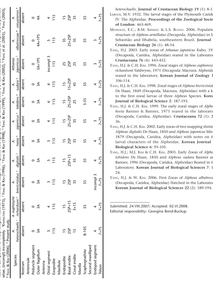
A morphological comparison shows that the first zoeal stage of A. estuariensis larvae is similar to that of other alpheids species (Tab. I). This similarity may contribute to the difficulty of specific identification. Nevertheless, differences do exist that should be useful for identification. Setation of the outer flagel-lum of the antennule is unique in bearing four aesthetascs, as compared to three in all other species.
The number of distal segments also vary, with A. estua-riensis,A. japonicus and A. euphorsyne richardsoni having 4-seg-ments while other species vary. The number of setae on the coxal endite is variable: A. estuariensis, A. euphorosyne richardsoni, A. heeia,A. digitalis,A. japonicus and A.brevicristatus all have three,
whileA. sudara,A. lobidens and A. heterochaelis vary. Also, A. estuariensis,A. brevicristatus,A. heterochaelis and A. heeia all have the same number of spines on the basal endite and all lack se-tae, while A. lobidens,A. japonicus, A. digitalis,A. euphorsyne richardsoni and A. sudara have one or two additional setae. The endopod of the second maxilliped is 4-segmented for all species exceptA. brevicristatus and A. japonicus, which are 3-segmented. Alpheus estuariensis has been confused with the very similar A. heterochaelis (CHRISTOFFERSEN 1984). However, as we show here, larvae have several differences that allow specific identification (Tab. I). The most distinct differences that separate these two spe-cies are: one seta on the maxillule coxal endite in A. heterochaelis Figures 23-35. Alpheus estuariensis, zoea III appendages: (23) antennule; (24) antenna; (25) maxillule; (26) maxilla; (27) first maxilliped; (28) second maxilliped; (29) third maxilliped; (30) first pereiopod; (31) second pereiopod; (32) third pereiopod; (33) fourth pereiopod; (34) fifth pereiopod; (35) telson. Scale bar: 23-24, 27-31, 34-35 = 0.3 mm, 25-26 = 0.75 mm, 32-33 = 0.15 mm.
23
24
27
30
31
28
26
35
32
33
29
CHRISTOFFERSEN, R.T. 1984. The Western Atlantic snapping shrimps related to Alpheus heterochaelis Say (Crustacea, Caridea), with the description of a new species. Papeis Avulsos de Zoolo-gia 53 (19): 189-208.
CHRISTOFFERSEN, R.T. 1998. Malacostraca-Eucarida-Caridea-Crangonoidea and Alpheidae, p. 353-372. In: P.S. YOUNG (Ed.).
Catalogue of Crustacea of Brazil. Rio de Janeiro, Museu Nacional.
KNOWLTON, R.E. 1973. Larval development of the snapping shrimpAlpheus heterochaelis Say, reared in the laboratory.
Journal of Natural History 7: 273-306.
GUTERRES, L.F.R.; G.A.S. MELO, & P.M.C. GUTERRES. 2005. Novos regis-tros da ocorrência de Alpheus macrocheles (Crustacea, Caridea, Alpheidae) na costa do Brasil. Biociências 13 (2): 231-233. GROSS, P.S. & KNOWLTON, R.E. 1999. Variation in larval size after
eyestalk ablation in larvae of the snapping shrimp Alpheus Figures 36-48. Alpheus estuariensis, zoea IV appendages: (36) antennule; (37) antenna; (38) maxillule; (39) maxilla; (40) first maxilliped; (41) second maxilliped; (42) third maxilliped; (43) first pereiopod; (44) second pereiopod; (45) third pereiopod; (46) fourth pereiopod; (47) fifth pereiopod; (48) telson. Scale bar: 36-37, 40-48 = 0.3 mm, 37-38 = 0.075 mm.
and three setae in A. estuariensis; 8-10 setae on the scaphognathite of the maxilla of A. heterochaelis and five setae in A. estuariensis.
ACKNOWLEDGEMENTS
Sincere thanks to Martin Christoffersen, Universidade Federal da Paraíba, and to Hoi Jeong Yang, Pusan National University. We are also grateful to Gustavo de Melo, (Museu de Zoologia, Universidade de São Paulo) for confirmation of the species identification.
LITERATURE CITED
BARTILOTTI, C.; R. CALADO & A. SANTOS. 2005. Correct diagnosis of early zoeal stages of Athanas nitescens (Leach, 1814) (Decapoda, Caridea, Alpheidae) using laboratory-raised larvae. Journal of Plankton Research 27 (11): 1189-1194.
36
37
38
39
43
44
45
41
40
42
46
Ta
b
le I. C
o m p a rison of mor p hologica l fea tur
es of fi
rs t zoea l sta ge of 1 1 sp ecies b e longing t o the Alp heida e sp ecies . ( A ) A e sthe ta sc s, (S ) se ta e , (SP setae, ( incom p l) i n com plete ly . 1K NO W LT O N (19 73) ,
2 Y
AN G & K IM ( 199 6),
3 Y
AN G & K IM (19 98) ,
4 Y
AN G &K IM (199 9), 5Y AN G &K IM (20 02) , 6Y AN G et a 8Y AN G &K IM (20 06) ,
9Present s
tudy . Sp ecies A. hetero chaelis 1 A . eupho syne ric hard so ni 2 A. br ev ic ri st a tu s 3 A. d igitalis 3 A. heeia 4 A. japo nic us 5 A. lo b idens 6 A. sud ara 6 A thanas ja p o n ic u s 7 alb Rostr u m a b sent a b sent a b sent a bsent a b sent a b se n t a b se n t a b sent a b sent Antennule Ped uncle se gme n t 0 0 0 0 0 0 0 0 0 O u ter fla ge llum 3 A 3 A 3 A 3 A 3 A 3 A 3 A 3 A 3 A + 1 P S
Antenna Distal seg
m e n t 5 4 3 5 5 4 6 6 in com p l 3 Ex op o d ite 11S 1 1 S 1 1S 11 S 1 1S 11 S 1 1 S 11S 1 1 S Ma xi llule End op od it e 1 S 1 S 1 S 1 S 1 S 1 S 1 S 1 S 2 S Ba sal e n d ite 2 S P 2 S P +1S 2S P 2 S P +1 S 2 S P 2S +2S P 2 S +2S P 1 S +2S P 2 S P Cox al e n d ite 1S 3 +1S 3S 5S 3 S 3S 2S 4S 2 S Ma xi lla S cap hog n athite 8-10 S 5 S 5 S 5 S 5 S 5 S 3 -5S 5S 5S Se c o nd m a xilli ped E n do po d segm ent 4 4 inc o m p l 3 4 4 3 4 4 3 Te ls on 7+7 S 7 +7S 7 +7S 7+ 7S 7 +7S 7+7 S 7+7 S 7 +7S 7+ 7S
Submitted: 24.VIII.2007; Accepted: 02.VI.2008. Editorial responsibility: Georgina Bond-Buckup
YANG, H.J. 2003. Early zoeas of Athanas japonicus Kubo, 1936 (Decapoda, Caridea, Alpheidae) reared in the Laboratory.
Crustaceana 76 (4): 443-452.
YANG, H.J. & C.H. KIM. 1996. Zoeal stages of Alpheus euphorosyne richardsoni Yaldwyne, 1971 (Decapoda: Macrura: Alpheidae) reared in the laboratory. Korean Journal of Zoology 39: 106-114.
YANG, H.J. & C.H. KIM. 1998. Zoeal stages of Alpheus brevicristatus De Haan, 1849 (Decapoda, Macrura, Alpheidae) with a key to the first zoeal larvae of three Alpheus Species. Korean Journal of Biological Science 2: 187-193.
YANG, H.J. & C.H. KIM. 1999. The early zoeal stages of Alpheus heeia Banner & Banner, 1975 reared in the laboratory (Decapoda, Caridea, Alpheidae). Crustaceana 72 (1): 25-36.
YANG, H.J. & C.H. KIM. 2002. Early zoeas of two snapping shrimps Alpheus digitalis De Haan, 1850 and Alpheus japonicus Miers, 1879 (Decapoda, Caridea, Alpheidae) with notes on the larval characters of the Alpheidae. Korean Journal of Biological Science 6: 95-105.
YANG, H.J.; M.J. KIM & C.H. KIM. 2003. Early Zoeas of Alpheus lobidens De Haan, 1850 and Alpheus sudara Banner and Banner, 1996 (Decapoda, Caridea, Alpheidae) Reared in the Laboratory. Korean Journal of Biological Sciences 7: 15-24.
YANG, H.J. & W. KIM. 2006. First Zoeas of Alpheus albatrossae (Decapoda, Caridea, Alpheidae) Hatched in the Laboratory.