The genus Enoplometopus A. Milne-Edwards, 1862 comprises 11 species, but only two are found in the Atlantic ocean, E. antillensis Lütken, 1865 and E. callistus Intès & Le Loeuff, 1970 (POUPIN 2003, AHYONG, & O’MEALLY 2004). The first is the most com-mon species found in the Atlantic and the only Enoplometopus lobster reported in the tropical West Atlantic (POUPIN 2003).
The nomenclature and classification of this genus have been controversial, most likely due to its rarity and morpho-logical similarities among related species which have been cap-tured (MERINO & LINDREY 2003). Thus, the Enoplometopus have been classified as an astacidean, an axiid thalassinidean or a sister to the Fractosternalia + Homarida (AHYONG & O’MEALLY 2004). However, recent revisions and studies on the phylog-eny and molecular analyses have placed it in Enoplometopidae (MARTIN & DAVIS 2001, AHYONG & O’MEALLY 2004).
In recent years, there has been an increase in the aquarium trade industry (PENHA-LOPESet al. 2006). The marine aquarists
have recognized the Enoplometopus as a very appreciated orna-mental species because of its dazzling coloration, economic value and high demand. Unfortunately, little information on larval and adult biology is available.
Because studies on the culture of ornamental decapods have improved scientific understanding, the larval development and proper culture technologies have been developed for sev-eral species (RHYNE et al. 2005). However, many attempts to culture various species of decapods have failed, one of which is the Enoplometopidae. Perhaps this is because only a few hob-byists have time and motivation to work on species resistant or difficult to culture. IWATAet al. (1991) reported the first at-tempt of Enoplometopus with the species E. occidentalis in cul-ture, in which eight zoeal stages were achieved.
A relevant aspect to be considered is the study on mor-phological development of cultured lobster. Observation of larval crustaceans has contributed to the identification of new
Universidade Federal do Ceará. Avenida Mister Hull, Pici, 60455-760 Fortaleza, Ceará, Brasil.
E-mail: [email protected]
ABSTRACT. The early stages of the tropical reef lobster
Enoplometopus antillensis
Lütken, 1865 were described and
illustrated in detail from specimens reared in the laboratory. Ovigerous females were captured in their habitat,
at a depth of about 15 meters and transported to the laboratory. The larvae were reared in a recirculation water
tank for approximately 15 days and then transferred to four 10 liters aquariums. The larvae were fed on
Artemia
sp. nauplii. Microalgae
Dunaliella viridis
was added daily to the culture. The larvae moulted seven times progressing
through the zoea VIII. Megalopa stage was not achieved. The intermoulting period of each stage averaged from
eight to 12 days. Morphological comparisons with previous reports are briefly discussed.
KEY WORDS. Crustacean; larval description; morphology; ornamental.
RESUMO. Desen
Desenv
Desen
Desen
Desen
vv
vvolvimento
olvimento
olvimento
olvimento
olvimento dos
dos
dos
dos
dos estágios
estágios
estágios iniciais
estágios
estágios
iniciais da
iniciais
iniciais
iniciais
da
da
da
da lag
lag
lag
lag
lagosta
osta
osta
osta
osta de
de rrrrrecif
de
de
de
ecif
ecif
ecif
ecife
ee
ee tr
tr
tr
tropical
tr
opical
opical
opical
opical
Enoplometopus
Enoplometopus antillensis
Enoplometopus
Enoplometopus
Enoplometopus
antillensis
antillensis
antillensis
antillensis
Lütk
Lütk
Lütk
Lütk
Lütken
en
en
en
en
(Astacidea
(Astacidea
(Astacidea
(Astacidea
water circulation similar to those used for culture of several species of spiny lobsters (KITTAKA 1994). The tanks were made with acrylic (70 cm Ø and 30 cm depth). Larvae were main-tained in this recirculation system at a density of 10 larvae/l for approximately 15 days and then transferred to four 10 l aquariums at the same density. The larvae were fed with Artemia sp. nauplii. Microalgae Dunaliella viridis was added daily to the culture at a concentration of 150 x 104cell/ml, in order to maintain the water quality by recycling inorganic nutrients, fixing carbon dioxide and supplying dissolved oxygen to the aquaculture systems by its photosynthetic activities. Tempera-ture, salinity and pH were monitored using a multi-parameter portable equipment and dissolved ammonium with a tetra ammonia kit. Salinity and temperature values in the culture were approximately 35‰ and 27-29°C, respectively.
Exuviae and death larvae were preserved in a glycerol + ethylic alcohol 70% (1:1) solution after each moulting. About 10 larvae and exuviae of each stage were dissected and illustrated.
The carapace length (CL) was measured from the ocular region to the posterior midpoint region of the carapace. The illustrations and measures of the larvae were made under a binocular Zeiss microscope equipped with a micrometer disc.
RESULTS
Larval culture
In the laboratory, hatched larvae were obtained from three females. The larvae hatch as a prezoea (Fig. 1) in which they persist for less than one hour but many prezoea failed to moult into zoea I. Moulting of the 1st to the 2nd stage occurred 7–12 days after being placed in tanks; the 2nd to the 3rd and 3rd to the 4th stages averaged seven and eight days, respectively. The intermoulting period for the subsequent stages was not possible to determine accurately since the larvae were not reared individually. High mortality occurred on the 3rd culture day in which approximately 60% of the larvae died.
Morphological malformations were frequently observed in the mouthpart of the zoeae and it appeared to be related to the individual adaptation to the laboratorial conditions. This fact needs to be researched.
proximal and medial segments with two and two distal setae respectively; basal endite with two strong cuspidate setae and two plumose setae; coxal endite with 5+2 plumose setae.
Maxilla (Fig. 13): scaphognathite with 4+1 plumose se-tae; endopod well developed and 5-segmented, with 3,2,2,1,2 long plumose setae; basal and coxal endites bilobed with long and plumose setae (as illustrated).
First maxilliped (Fig. 14): endopod short, 4-segmented with 2,2,3,4 setae; basipod bearing eight setae; coxal endite with two setae, exopod with four natatory setae.
Second maxilliped (Fig. 15): endopod 4-segmented with 3,2,2,4 setae, distal segment (one long spine and three terminal setae); basipod with four setae; exopod with four natatory setae. Third maxilliped (Fig. 16): endopod 4-segmented, (proxi-mal segment fused with basipod) with 2+2,3,2,2+2 setae (one strong and three long terminal and one subterminal setae); basipod with four setae; exopod with four natatory setae.
Abdomen (Fig. 2): with six abdominal somites lacking spines, 6th abdominal segment long and fused with telson.
Telson (Fig. 17): bifurcate showing an accentuated me-dial depression with 5+1 plumose setae and one spine on each furcal branch.
Zoea II (Fig. 3)
Carapace (Fig. 3): Length (CL) 2.24 mm (1.23-2.25 mm): rostrum with 8+8 small denticles laterally; carapace showing a pterigostomial spine elongate; eyes stalked.
Antennule (Fig. 18): with five aesthetascs and one plu-mose subdistal seta.
Antenna (Fig. 19): propod segmented with endopod with 1 small seta; exopod segmented distally showing 8-9+1 mose setae; endopod shorter than exopod with three long plu-mose setae.
Maxillule (Fig. 20): exopodite with four setae; endopod 3-segmented; basal endite with two cuspidate and three plu-mose setae; coxal endite with nine pluplu-mose setae.
Maxilla: similar to the previous stage.
Second maxilliped (Fig. 22): endopod 4-segmented with 3,2,2,4 setae, distal segment (one long spine and three termi-nal setae); basipod with four setae.
Third maxilliped (Fig. 23): endopod 4-segmented with 1,5,4,5 setae (one strong and three long terminal and one sub-terminal setae); basipod with four setae; exopod with five na-tatory setae.
First pereiopod (Fig. 24): rudimentary; endopod 2-seg-mented showing few setae; exopod small with 3-4 short distal setae.
Abdomen (Fig. 3): with six abdominal somites lacking spines, 6th abdominal segment incompletely articulated with telson.
Telson (Fig. 25): bifurcate with 6+1 plumose setae and one spine on each furcal branch.
Zoea III (Fig. 4)
Carapace (Fig. 4): length (CL) 1.96 mm (1.96-2.00 mm); carapace unchanged; eyes stalked.
Antennule (Fig. 26): peduncle 3-segmented with 4-5 long lateral setae from first segment to distal portion of last seg-ment; first segment longer than other about twice the length of the medial segment; outer flagellum unsegmented with six aesthetascs and one subdistal plumose seta; inner antennular flagellum unsegmented ending in one plumose and two simple setae.
Antenna (Fig. 27): exopod 6-segmented with 18 plumose and one simple setae and strong lateral spine; endopod 3-seg-mented with three setae.
Maxillule (Fig. 28): endopod unchanged; basal endite with four strong cuspidate and five simple setae; coxal endite with 5+5 plumose setae.
Maxilla (Fig. 29): scaphognathite with 14+1 long plumose setae; endopod unchanged; basal and coxal endites as illus-trated.
First maxilliped (Fig. 30) and second maxilliped (Fig. 31): exopod with six natatory setae.
Third maxilliped (Fig. 32): exopod with 6-8 plumose setae. First pereiopod (Fig. 33): endopod 4-segmented sub-che-late, arrangement of setation as illustrated; exopod with eight plumose setae.
Second pereiopod (Fig. 34): endopod developed and 4-segmented, arrangement of setation as illustrated; exopod with eight setae.
Third pereiopod (Fig. 35): endopod developed and 4-seg-mented, arrangement of setation as illustrated; exopod with seven setae.
Fourth pereiopod (Fig. 36): bilobed and rudimentary with two setae.
Abdomen (Fig. 4): somites 1-3 each with pair of acute posterolateral spines, somite five with pair of bifurcate acute
Figures 1-5. Enoplometopus antillensis, zoeal stages in lateral view: (1) prezoea; (2) stage I; (3) stage II; (4) stage III; (5) stage IV. Scale bar: 1-3 = 0.4 mm, 4 = 0.6 mm, 5 = 0.9 mm.
1
2
posterolateral spines; somite six articulated with telson bear-ing two small posterodorsal spines.
Telson (Fig. 37): posterior portion arcuated, with a strong and elongate median spine followed of 6+6 inner setae and two strong spines on the outer margin; exopod with 14 plu-mose setae and one distal spine on outer margin; endopod with five plumose setae.
Zoea IV (Fig. 5)
Carapace (Fig 5): length (CL) 2.23 mm (2.10-2.35 mm): similar to the previous stage.
Antennule (Fig. 38): peduncle with first segment about three times the medial segment length, inner antennular fla-gellum a little more developed compared to previous stage.
Antenna (Fig. 39): exopod unsegmented with 22-23
plu-mose setae; endopod 5-segmented.
Maxillule (Fig. 40): basal endite with five strong serrulate and four simple setae; coxal endite with 3+6+2 plumose setae.
Maxilla (Fig. 41): scaphognathite with 18-21 plumose se-tae.
First maxilliped (Fig. 42): exopod with 4+2 natatory setae. Second maxilliped (Fig. 43): endopod 3-segmented, me-dial segment with a row of long setae distally; exopod with six natatory setae.
Third maxilliped (Fig. 44): endopod 4-segmented with distal segment ending in a strong spine surrounded by long setae; exopod with eight plumose setae.
First pereiopod (Fig. 45): endopod well developed 4-seg-mented, arrangement of setation as illustrated, chelipeds
Figures 6-9. Enoplometopus antillensis, zoeal stages in lateral view: (6) stage V; (7) stage VI; (8) stage VII; (9) stage VIII. Scale bar: 6-7 = 0.6 mm, 8-9 = 1.2 mm.
8
7
Figures 10-17. Enoplometopus antillensis, zoea I appendages: (10) antennule; (11) antenna; (12) maxillule; (13) maxilla; (14) 1st maxilliped;
(15) 2nd maxilliped; (16) 3rd maxilliped; (17) telson. Scale bar: 10 and 14-16 = 0.2 mm, 11-13 = 0.15 mm, 17 = 0.6 mm.
11
10
12
14
15
16
17
arcuated with cutting margin of both fingers with short spines; exopod with 10 plumose setae.
Second pereiopod (Fig. 46): Endopod 4-segmented, ar-rangement of setation as illustrated; exopod with eight setae although absent in some examined specimens.
Third pereiopod (Fig. 47): endopod 4-segmented, shorter
than second pereiopod, setation as illustrated; exopod with six setae but absent in some examined specimens.
Fourth and Fifth pereiopods (Figs 48-49): Little developed with few setae, as illustrated.
Abdomen (Fig. 5): similar to the previous stage. Telson (Fig. 50): posterior portion arcuated, with a strong
Figures 18-25. Enoplometopus antillensis, zoea II appendages: (18) antennule; (19) antenna; (20) maxillule; (21) 1st maxilliped; (22) 2nd
maxilliped; (23) 3rd maxilliped; (24) 1st pereiopod; (25) telson. Scale bar: 18-20 and 24 = 0.15 mm, 21-23 = 0.2 mm, 25 = 0.6 mm.
19
18
21
22
24
20
23
Figures 26-37. Enoplometopus antillensis, zoea III appendages: (26) antennule; (27) antenna; (28) maxillule; (29) maxilla; (30) 1st maxilliped;
(31) 2nd maxilliped; (32) 3rd maxilliped; (33) 1st pereiopod; (34) 2nd pereiopod; (35) 3rd pereiopod; (36) 4th pereiopod; (37) telson. Scale
bar: 26-27 and 30-35 = 0.3 mm, 28-29 and 36 = 0.15 mm, 37 = 0.2 mm.
26
27
37
29
28
32
36
31
30
33
Figures 38-50. Enoplometopus antillensis, zoea IV appendages: (38) antennule; (39) antenna; (40) maxillule; (41) maxilla; (42) 1st maxilliped;
(43) 2nd maxilliped; (44) 3rd maxilliped; (45) 1st pereiopod; (46) 2nd pereiopod; (47) 3rd pereiopod; (48) 4th pereiopod; (49) 5th pereiopod;
(50) telson. Scale bar: 38-39, 42-44, 46-47 and 49 = 0.3 mm, 40-41 = 0.1 mm, 45 = 0.5 mm, 48 and 50 = 0.2 mm.
39
38
40
42
43
44
41
46
47
48
49
45
Figures 51-63. Enoplometopus antillensis, zoea V appendages: (51) antennule; (52) antenna; (53) maxillule; (54) maxilla; (55) 1st maxilliped;
(56) 2nd maxilliped; (57) 3rd maxilliped; (58) 1st pereiopod; (59) 2nd pereiopod; (60) 3rd pereiopod; (61) 4th pereiopod; (62) 5th pereiopod;
(63) telson. Scale bar in the figures: 51, 53-57, 59-62 and 63 = 0.3 mm, 52 = 0.4 mm, 58 = 0.6 mm.
51
52
58
55
56
53
57
59
60
54
63
62
Maxilla (Fig. 54): scaphognathite with 23-24+2 long plu-mose setae; basal and coxal endites as illustrated.
First maxilliped (Fig. 55): exopod with 4-6 natatory setae. Second maxilliped (Fig. 56): exopod with eight natatory setae.
Third maxilliped (Fig. 57): exopod with 10-12 natatory setae.
First pereiopod (Fig. 58): similar to the previous stage; exopod with 12 plumose setae, absent in some examined speci-mens.
Second pereiopod (Fig. 59) and third pereiopod (Fig. 60): exopod with 12 natatory setae.
Fourth pereiopod (Fig. 61): endopod 4-segmented, distal segment ending in a long and bent spine; exopod with eight natatory setae.
Fifth pereiopod (Fig. 52): endopod 4-segmented, distal segment ending in a long and bent spine; exopod short with 4-5 natatory setae.
Telson (Fig. 63): posterior portion almost straight with a strong central spine and 4+4 inner setae; uropod with exopod bearing 31-32 setae and one distal spine; endopod with 23-25 setae.
Zoea VI (Fig. 7)
Carapace (Fig. 7): length (CL) 3.14 mm (2.65-3.70 mm). Antennule (Fig. 64): inner flagellum 4-segmented end-ing in an acute distal seta; outer flagellum 4-segmented arranged in 2,2,1+1+2,2 aesthetascs in the segmentations from proxi-mal to distal, respectively.
Antenna (Fig. 65): exopod with 30+1 plumose setae; endopod with about 13 segments.
Maxillule (not illustrated): similar to the previous stage. Maxilla (Fig. 66): scaphognathite with 38-39+5 long plu-mose setae; basal and coxal endites increasing in setae number on the lobes.
First maxilliped (Fig. 67), second maxilliped (Fig. 68), third maxilliped (Fig. 69) and first pereiopod (Fig. 70): similar to the previous stage.
Second pereiopod (Fig. 71) and third pereiopod (Fig. 72): exopod with 16 natatory setae.
date and 7-9 simple setae; coxal endite with approximately 10+12+6 plumose setae.
Maxilla (Fig. 79): scaphognathite with 47-48 long plu-mose setae.
First maxilliped (Fig. 80): exopod with a row of seven lateral and six distal setae.
Second maxilliped (Fig. 81): exopod with 10 natatory setae.
Third maxilliped (Fig. 82): exopod with 14 natatory setae. First pereiopod (Fig. 83): exopod with 18 natatory setae. Second pereiopod (Fig. 84) and third pereiopod (Fig. 85): exopod with 14-16 natatory setae.
Fourth pereiopod (Fig. 86) and fifth pereiopod (Fig. 87): exopod with 10-12 natatory setae.
Telson (Fig. 88): posterior portion tapering posteriorly with the central spine more robust than previous stage and 4+4 inner setae; uropod with exopod bearing 40 plumose se-tae; endopod with 36 plumose setae.
Zoea VIII (Fig. 9)
Carapace (Fig. 9): length (CL) 4.54 mm (4.60-4.48 mm), rostrum with 11+11 denticles laterally.
Antennule (Fig. 89): outer flagellum with approximately 11-segmented; inner flagellum with approximately 17-seg-mented and four simple setae.
Antenna (Fig. 90): exopod with 43 plumose setae; endopod longer than exopod 30-segmented.
Maxillule (Fig. 91): basal endite with 6-7 strong serrulate and 14 simple setae; coxal endite with approximately 27+13+6 plumose setae.
Maxilla (Fig. 92): scaphognathite with 60+13 long plu-mose setae.
First maxilliped (Fig. 93): exopod with a row of 13 lateral and three distal setae.
Second maxilliped (Fig. 94) and third maxilliped (Fig. 95): exopod with 16 natatory setae.
First pereiopod (Fig. 96) and second pereiopod (Fig. 97): exopod with 20-22 natatory setae.
Figures 64-75. Enoplometopus antillensis, zoea VI appendages: (64) antennule; (65) antenna; (66) maxilla; (67) 1st maxilliped; (68) 2nd
maxilliped; (69) 3rd maxilliped; (70) 1st pereiopod; (71) 2nd pereiopod; (72) 3rd pereiopod; (73) 4th pereiopod; (74) 5th pereiopod; (75)
telson. Scale bar: 64-65 and 70 = 0.6 mm, 66-69 and 71-75 = 0.3 mm.
75
71
72
70
69
68
65
64
66
67
Figures 76-88. Enoplometopus antillensis, zoea VII appendages: (76) antennule; (77) antenna; (78) maxillule; (79) maxilla; (80) 1st
maxilliped; (81) 2nd maxilliped; (82) 3rd maxilliped (83) 1st pereiopod; (84) 2nd pereiopod; (85) 3rd pereiopod; (86) 4th pereiopod; (87) 5th
pereiopod; (88) telson. Scale bar: 76-77, 83 and 88 = 0.6 mm, 78-82 and 84-87 = 0.3 mm.
76
81
82
86
83
84
85
80
79
77
Figures 89-101. Enoplometopus antillensis, zoea VIII appendages: (89) antennule; (90) antenna; (91) maxillule; (92) maxilla; (93) 1st maxilliped;
(94) 2nd maxilliped; (95) 3rd maxilliped (96) 1st pereiopod; (97) 2nd pereiopod; (98) 3rd pereiopod; (99) 4th pereiopod; (100) 5th pereiopod;
(101) telson. Scale bar: 89-90 = 0.9 mm, 91-92 = 0.2 mm, 93-94 = 0.3 mm, 95 and 97-100 = 0.6 mm, 96 and 101 = 1.2 mm.
89
90
92
95
93
96
99
94
100
97
98
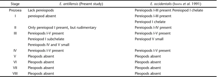
Morphological results of each zoeal stage of E. antillensis are shown in table I. A comparison of the morphological char-acteristics of E. antillensis described at the present study with previous descriptions of Enoplometopidae larvae was limited to the study of IWATAet al. (1991) for E. occidentalis. The most no-table distinction between these two species by that E. antillensis
antillensis they are segmented. In this stage, the arrangement of setation of these species is rather different in the append-ages mainly those of endopod of maxillule, scaphognathite of maxilla and telson (Tab. III).
These facts suggest that a noticeable distinction may oc-cur in the larval development of Enoplometopidae species.
Fur-Table I. Main morphological characteristics of zoeal stages of Enoplometopus antillensis.
Stages Mean carapace length
(mm)
Maxilla scaphognathite
Antenna (Exopod)
Number of natatory setae
Maxilliped Pereiopod
1st 2nd 3rd 1st 2nd 3rd 4th 5th
I 1.89 4+1 (8-9)+1 4 4 4 - - - -
-II 2.24 4+1 (8-9)+1 4 4 5 3-4 - - -
-III 1.96 18+1 22-23 6 6 8 8 8 7 2
-IV 2.23 18-21 22-23 4+2 6 8 10 8 6 4
-V 2.88 23-24+2 23-26 4-6 8 10-12 12 12 12 8 4-5
VI 3.14 38-39+5 30+1 6 8 10-12 12 16 16 8 4-5
VII 3.42 47-48 32-33 7+6 10 14 18 14-16 14-16 10-12 10-12
VIII 4.54 60+13 43 13+3 16 16 20-22 20-22 20 14-16 14-16
Table II. A comparison between the main characteristics of each zoeal stage of E. antillensis (present study) and E. occidentalis described
by IWATAet al. (1991).
Stage E. antillensis (Present study) E. occidentalis (IWATAet al. 1991)
Prezoea Lack pereiopods Pereiopods I-III present Pereiopod I chelate
I pereiopod absent Pereiopods I-III present
Pereiopod I chelate
II Only pereiopod I present, but rudimentary Pereiopods I-IV present
III Pereiopods I-V present Pereiopods I-V present
Pereiopod I subchelate Pereiopod V small
Pereiopods IV and V small
IV Pereiopods I-V present Pereiopods I-V present
V Pleopods absent Pleopods absent
VI Pleopods absent Pleopods absent
VII Pleopods absent Pleopods absent
Maxillule Endopod 3-segmented with (2+2)+2+2 setae Unsegmented, 5 setae
Maxilla Endopod 4-segmented Unsegmented, 4 setae
Scaphognathite 4+1 setae 13 setae
Maxilliped I Endopod 4-segmented Unsegmented
Exopod 4 setae Rudimentary with 2 setae
Maxilliped II Endopod 4-segmented 3-segmented
Exopod 4 setae Rudimentary
Maxilliped III Endopod 4-segmented 3-segmented
Exopod 5 setae 7 setae
Pereiopod Endopod 1-5 (1st undeveloped) 1~5 (1st chelate and well developed
Abdomen Lateral spine + (somite 6) + (somite 5)
Telson Processes 7+7 8+8
Endopod absent Absent
Exopod absent Absent
ther studies of phylogeny and/or molecular biology studies will be needed to elucidate the correct taxonomic position of the Enoplometopidae species. AHYONG & O’MEALLY (2004) reported a comprehensive study on the phylogeny of decapods using both molecular biology and morphological characters in which the genusEnoplometopus is included. However, only the species E. occidentalis was used in DNA mitochondrial analysis. Scientific data of phylogenetic study is lacking for other species.
ACKNOWLEDGMENT
The authors wish to thank Dr. Gil Penha Lopes of the Laboratório Maritimo da Guia, FCUL & IMAR for sending his oral presentation in the Marine Ornamental’ 04.
REFERENCES
AHYONG, S.T. & D. O’MEALLY. 2004. Phylogeny of the Decapoda Reptantia: Resolution using three molecular loci and mor-phology. Raffles Bulletin of Zoology 52 (2): 673-693. IWATA, Y; H. SUGITA; Y. DEGUCHI & F.I. KAMEMOTO. 1991. The early
larval development of the tropical reef lobster Enoplometopus occidentalis (Decapoda, Axiidae) reared in the laboratory.
Researches on Crustacea 20: 1-15.
KITTAKA, J. 1994. Culture of phyllosomas of spiny lobster and its application to studies of larval recruitment and aquaculture.
Crustaceana 66 (3): 257–270.
MARTIN, J.W. & G.E. DAVIS. 2001. An updated classification of the recent Crustacea. Natural History Museum of Los Angeles County, Science Series 39: 1-124.
MERINO, S.E. & J.A. LINDLEY. 2003. First record of Enoplometopus callistus (Crustacea: Decapoda: Nephropidae) in the Cape Verde Islands. Journal of the Marine Biological Association
of the United Kingdom 83: 1233-1234.
PENHA-LOPES, G.; A.L. RHYNE; J. FIGUEREDO; J. LIN & L. NARCISO. 2006. Can larvae produced from stored sperm in the ornamental crabMithraculus forceps (A. Milne Edwards, 1875) (Decapoda: Brachyura: Majidadae) be used in aquaculture? Aquaculture 257 (1-4): 282-286.
POUPIN, J. 2003. Reef lobster Enoplometopus A. Milne Edwards, 1862 from French Polynesia, with a brief revision of the genus (Crusta-cea, Decapoda, Enoplometopidae). Zoosystema 25 (4): 643-664. RHYNE, A.L.; G. PENHA-LOPES & J. LIN. 2005. Growth, development, and survival of larval Mithraculus sculpus (Lamarck) and Mithraculus forceps (A. Milne Edwards) (Decapoda: Brachyura: Majidae): economically important marine ornamental crabs.
Aquaculture 245: 183-191.