1
UNIVERSIDADE FEDERAL DE SÃO CARLOS-UFSCAr
PROGRAMA DE PÓS-GRADUAÇÃO EM FISIOTERAPIA
DEPARTAMENTO DE FISIOTERAPIA
José Carlos Tatmatsu Rocha
Efeitos da Fototerapia em Marcadores Inflamatórios, na Função
Mitocondrial e no Estresse Oxidativo em Roedores com Diabetes
Induzida
2
UNIVERSIDADE FEDERAL DE SÃO CARLOS- UFSCAr
PROGRAMA DE PÓS-GRADUAÇÃO EM FISIOTERAPIA
DEPARTAMENTO DE FISIOTERAPIA
José Carlos Tatmatsu Rocha
Efeitos da Fototerapia em Marcadores Inflamatórios, na Função
Mitocondrial e no Estresse Oxidativo em Roedores com Diabetes
Induzida
Tese apresentada ao Programa de Pós-Graduação em Fisioterapia da Universidade Federal de São Carlos, para obtenção do Título de Doutor em Fisioterapia. Área de Concentração: Processos de avaliação e intervenção em Fisioterapia do sistema músculo-esquelético.
Orientador: Prof. Dr. Nivaldo Antonio Parizotto.
Co-orientadora: Profa. Dra. Patrícia Driusso
Ficha catalográfica elaborada pelo DePT da Biblioteca Comunitária UFSCar Processamento Técnico
com os dados fornecidos pelo(a) autor(a)
R672e
Rocha, José Carlos Tatmatsu
Efeitos da fototerapia em marcadores
inflamatórios, na função mitocondrial e no estresse oxidativo em roedores com diabetes induzida / José Carlos Tatmatsu Rocha. -- São Carlos : UFSCar, 2016. 115 p.
Tese (Doutorado) -- Universidade Federal de São Carlos, 2016.
4 Apoio Financeiro
5 A Deus, pela oportunidade de viver e de compreender nossas falhas e limitações, buscando compreender também de modo fraterno as limitações do meu próximo.
À minha família, em especial minha esposa, agradeço por conseguir me suportar ao longo de 15 anos de casamento, e a meus filhos, por terem pais tão próximos mas às vezes tão longe ... Aos amigos que conseguem nos compreender e dar o alento necessário em tempo oportuno.
6
AGRADECIMENTOS
A realização desta Tese só foi possível graças ao apoio e colaboração direta ou indireta de muitas pessoas, em especial:
Ao Professor Nivaldo Parizotto, por acreditar em meu potencial e pelo seu trabalho que tornou a área de Fototerapia no Brasil respeitável mundialmente. Meus agradecimentos e afeto.
Às Professoras Patrícia Druso e Lucimar Avo e ao professor Rubens Bernardes Filho, pela paciência e pela disponibilidade em nos receber sempre que precisamos.
Aos membros da Banca por estarem dispostos a contribuir com suas considerações e sugestões que são de grande relevância para o enriquecimento deste trabalho.
Aos colegas e amigos do laboratório de Recursos Fisioterapêuticos, pelos momentos bons e ruins que passamos juntos e pela grande troca de experiências que me proporcionaram ao longo do doutorado, em especial a Carla Roberta Tim, Hueliton Kido, Patrícia Brassolatti, Lia Mara Neves, Cintia Martignago, Ana Laura Martins e Cynthia Castro.
Aos meu amigos e colegas da Harvard Medical School/Massachussetts General Hospital Cleber Ferraresi e Fernanda Freire pela troca de experiências enriquecedoras e pelos momentos de apoio mútuo, meu muito obrigado!
7 A família Fernandes, aos amigos do grupo de voluntários do Grupo Posto de Rua de São Carlos, Cauê e Taty Ribeiro, Anália Rosa Gussen, Solange e Luiz Hortenci, cuja amizade, carinho e atenção nos fizeram sentir parte de suas famílias.
A Iolanda da Silva Vilela, funcionária do Departamento de Fisioterapia, cujo trabalho dedicado faz do Departamento de Fisioterapia da UFSCAR um ambiente mais humanizado.
Aos meus pais que sempre me apoiaram nos estudos, a Fernando Brito meu cunhado que deu o suporte familiar necessário no período de ausência da família
À minha esposa Daniely Tatmatsu, por sempre estar ao meu lado me incentivando e apoiando nos momentos mais difíceis, para mim um exemplo de profissional, mãe e mulher. Muito obrigado por me escolher como seu marido e pai dos nossos amados filhos.
Aos meus filhos, Nathália Brito Tatmatsu e Benjamin Brito Tatmatsu, minhas paixões e orgulho. Obrigado por serem filhos maravilhosos que muito me orgulham e pela paciência (embora nem sempre consciente) que tiveram comigo ao longo desta jornada.
Á Fundação Cearense de Apoio ao Desenvolvimento Científico e Tecnológico – FUNCAP e à Universidade Federal do Ceará-UFC que apoiaram e financiaram minha permanência na cidade de São Carlos, o que foi essencial e decisivo para a conclusão desta pesquisa. Espero que os resultados possam contribuir para o crescimento científico e social de nosso País.
8 Ninguém cruza nosso caminho por acaso e nós não entramos na vida de alguém sem nenhuma razão.
9
ABREVIATURAS E SÍMBOLOS
ADP: Adenosine diphosphate AMP: Adenosine monophosphate ANOVA: Analysis of variance AsGa- Arsenite Galium
ATP: Adenosine triphosphate Ca 2+: Calcium ion
CCO- citochrome C oxidase COX2 – cyclooxygenase-2 DLASER - diabetic Laser treated DLASER- diabetic Laser treated
DLED-diabetic Light-emitting diode treated DMT1: DM mellitus Type 1
DMT2: DM mellitus Type 1 DNA: Deoxyribonucleic Acid DRP1- Dynamin-1-like protein ECM: extracellular matrix
FIS1 - Mitochondrial fission 1 protein GLP-1 : Glucagon-like peptide- 1 GLUT: glucose transporter GLUT4:glucose transporter 4 GTT: Glucose tolerance test
10 IL: Interleukin
iNOS- nitric oxide isozyme ITT: Insulin tolerance test Kg: kilogram
KITT: glucose decrease constant L: liter
LED – Light-emitting diode LLLT - Low level laser therapy MFN1- Mitofusin 1
MFN2- Mitofusin 2 mg: milligram mL: milliliter
MMPs: Matrix metalloproteinases
NAD- Nicotinamide adenine dinucleotide NFkappa: factor nuclear kappa B
NID – no irradiated diabetic nm: nanometer
NO – nitric oxide
NOS - nitric oxide species
11 TBARS - thiobarbituric acid
TNF- α: Tumor necrosis factor
VEGF- Vascular endothelial growth factor WHO– world health organization
12
RESUMO
O reparo tecidual em pacientes diabéticos é prejudicado por vários fatores, dentre eles a deficiência da secreção de insulina que conduz à hiperglicemia e consequente produção de radicais livres. A disfunção mitocondrial tem sido apontada como um dos prováveis fatores etiopatogênicos do DM mellitus. Sabe-se que a Fototerapia tem efeitos proliferativos em tecidos com algumas patologias. O presente estudo teve como objetivo avaliar os efeitos da Fototerapia utilizando-se Laser e LED de baixa Potência sobre a regeneração tecidual, estresse oxidativo e marcadores mitocondriais em animais diabéticos. Metodologia: 20 camundongos Swiss, pesando entre 30g , 6 semanas, para o estudo relacionado ao estresse oxidativo e 20 ratos Wistar pesando 250 g, 8 semanas, foram submetidos a indução do DM (injeção de streptozotocina 70mg/kg) e após 120 dias de seguimento, sofreram lesão cutânea e foram tratados com Terapia Laser de Baixa Intensidade e LED durante 5 dias consecutivos e no 6º
dia os animais foram mortos e retirada a pele. Um outro grupo de animais (20 ratos Wistar pesando 250 g, 8 semanas) foi submetido à irradiação Laser e LED na região do pâncreas durante 5 dias consecutivos e no 6º dia os animais foram mortos e retirado o pâncreas,
fígado e músculos gastrocnêmio. Foram divididos em quatro grupos: Sham (controle
saudável), NID (diabético não irradiado), DLED(diabético irradiado pelo LED) e DLASER
(diabético irradiado pelo LED). Mensurou-se níveis de catalase, àcido tiobarbitúrico
(TBARS), nitrito, conteúdo de colágeno, fator de crescimento endotelial vascular (VEGF),
Ciclo-oxigenase-2 (COX2) Mitofusin 2 (MFN2), Mitochondrial fission 1 protein (FIS1),
glicogênio hepático e muscular bem como testes funcionais de glicose e insulina.
13
904nm bem como níveis de TBARS e nitrito menores nos diabéticos irradiados. Além
disso, a expressão da COX-2 foi maior entre NID quando comparados aos grupos SHAM e
DLED. DLED apresentou os maiores scores em relação a MFN2 e a expressão da FIS1 foi
maior no grupo DLASER. As Terapias Laser e LED alteraram a expressão de VEGF e
COX2 e estimularam a proliferação das fibras colágenas maduras em feridas de animais
diabéticos. Os resultados relacionados à irradiação sobre o pâncreas demonstraram que no
grupo NID, o conteúdo de glicogênio hepático e muscular foram estatisticamente menores
que os grupos DLASER e DLED. Somando-se a esses dados, a densidade dos ductos pancreáticos e ilhotas pancreáticos diferiram significativamente quando comparados ao grupo diabético controle.Constatamos com esses resultados que tanto o Laser de Baixa Intensidade quanto o LED obtiveram potenciais efeitos terapêuticos sobre a pele e o pâncreas sob condições hiperglicêmicas e que ambas as terapias atuaram modulando parâmetros histológicos, angiogênicos e mitocondriais no pâncreas e pele dos animais diabéticos.
14
ABSTRACT
15 MFN2 and FIS1 expression was higher in DLASER group. Laser and LED Therapy altered the expression of VEGF and COX2 and stimulated the proliferation of mature collagen fibers in diabetic animal wounds. About irradiation over pancreas results showed that the NID group, hepatic and muscle glycogen content were statistically lower than DLASER and DLED groups. Adding to these data, the density of the pancreatic duct and pancreatic islets were significantly different when compared to NID. These results suggest that both the low-level laser LED obtained as potential therapeutic effects on the skin and pancreas under conditions hyperglycemic and that both therapies acted modulating histological, angiogenic and mitochondrial parameters on pancreas and skin of diabetic animals.
16
LISTA DE TABELAS
ESTUDO I:
Table 1.Irradiation and Treatment Parameters (AsGa laser, 904nm; model Laserpulse, handheld probe), used in contact to the mice skin ... 43 Table 2.Statistical analysis using morphometric computational systems to determine amount collagen. The significance level was set at p < 0.05. ... 48 Table 3.TBARS concentration levels in the skin of different groups: non-irradiated control (NIC), non-irradiated diabetic (NID), irradiated control (IC) and irradiated diabetic (ID).* p < 0.001 compared to each respective control group; # relative to the group ... 48
ESTUDO II:
Table. 1 Phototherapy treatment ... 65 Table. 2Statistical analysis using morphometric computational systems to determine amount collagen analyzed on 5th day by analysis of variance (ANOVA). Statistical analysis using morphometric computational systems to determine amount collagen analyzed on 5th day by analysis of variance (ANOVA) The 3th day period not showed because no significant differences were observed. Groups: non-diabetic control (Sham), diabetic non treated (NID), diabetic treated with LLLT (DLASER) and diabetic LEDT treatment (DLED). a versus Sham group; b versus NID group; c versus DLASER group. Date are showed mean + SEM. The significance level was set at p < 0.05. ... 68
ESTUDO III:
Table . 1. Device Information, Irradiation and Treatment Parameters ... 87 Table . 2.Table 2: Duct density and islet Langerhans density analysis of LLLT in
streptozotocin (STZ)-induced diabetes ... 93 Table . 3. Glycogen hepatic and muscle (μml/g) in diabetic animals treated with
17
LISTA DE FIGURAS
ESTUDO I:
Figure 1.Wound creation: the skin was cut with a carbon steel surgical blade until it reached the hypodermis with 2mm width and 2 cm long. ... 42 Figure 2.Schematic drawing showing procedure for determination of the antioxidant activity. The skin was dissected and homogenized in 1,15% KCL (1g/10nml solution). The mixture was centrifuged and the supernatant used for biochemical determinations 45 Figure 3.I. Photomicrograph showing granulation tissue (a); newly formed blood vessels (blue arrows) and fibroblasts (f) (HE 10X). Figure 3.II. Photomicrograph Masson staining showing organization and collagen deposition (c). (20X) ... 47 Figure 4.Catalase concentration levels in the skin of different groups: non-irradiated control (NIC), non-irradiated diabetic (NID), irradiated control (IC) and irradiated diabetic (ID). *p ≤ 0.001Figure 4. Catalase concentration levels in the skin of different groups: non-irradiated control (NIC), non-irradiated diabetic (NID), irradiated control (IC) and irradiated diabetic (ID). *p ≤ 0.001). ... 49 Figure 5..Nitrite concentration levels in the skin of different groups: non-irradiated control (NIC), non-irradiated diabetic (NID), irradiated control (IC) and irradiated diabetic (ID) . *p ≤ 0.001.Figure 5. Nitrite concentration levels in the skin of different groups: non-irradiated control (NIC), non-irradiated diabetic (NID), irradiated control (IC) and irradiated diabetic (ID) . *p ≤ 0.001. ... 50
ESTUDO II:
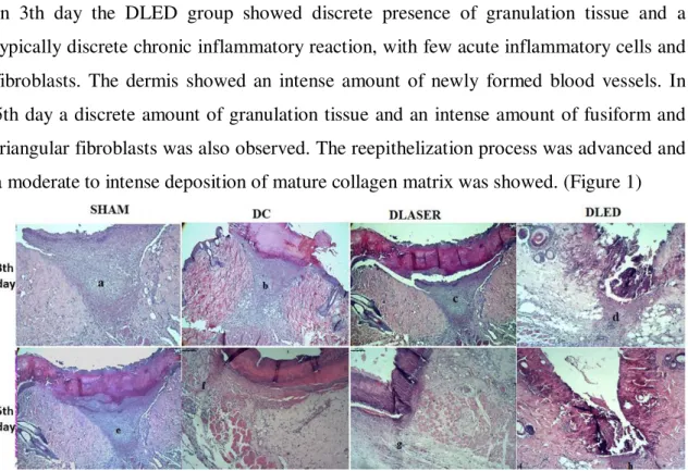
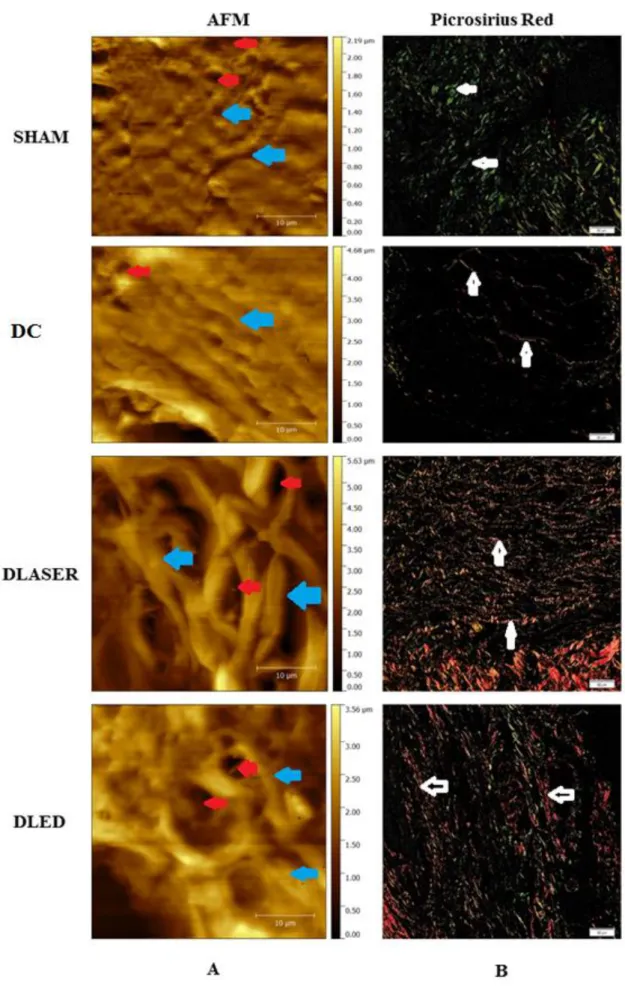
18 without any treatment, DLASER=diabetic laser group, incision irradiated group, DLED=diabetic LED group, incision LED irradiated group. ... 68 Figure . 3Histologic slides of skin diabetic mice and non-diabetics on the 5th day of post-surgical SHAM groups, NID. DLASER and DLED. A) Blades analyzed by atomic force microscopy (blue arrows indicate fibers that make up the ECM). Red arrows in show the formation of new blood vessels. B) Staining with picrosirius red (white arrows links point to collagen fibers). SHAM=incision without any treatment, diabetic group, NID=incision without any treatment, DLASER=diabetic laser group, incision irradiated group, DLED=diabetic LED group, incision LED irradiated group. ... 70 Figure . 4. Mean an SD scores for immunostaining of VEGFFigure 4. Mean an SD scores for immunostaining of VEGF. Significant differences of p = 0,00 are represented with a single asterisk; two asterisks p=0,002. SHAM=incision without any treatment; diabetic group, NID=incision without any treatment; DLASER=diabetic laser group, incision irradiated group; DLED=diabetic LED group, incision LED irradiated group. 71 Figure . 5.Mean an SD scores for immunostaining of COX-2.Figure 5. Mean an SD scores for immunostaining of COX-2. Significant differences of p = 0,001 are represented with a single asterisk; two asterisks p=0,008; three asterisks p= 0,005. SHAM=incision without any treatment, diabetic group; NID=incision without any treatment; DLASER=diabetic laser group; incision irradiated group; DLED=diabetic LED group; incision LED irradiated group. ... 72 Figure . 6Mean an SD scores for immunostaining of MFN2Figure 6. Mean an SD scores for immunostaining of MFN2. Significant differences of p = 0,00 are represented with a single asterisk and p=0,04 by two asterisks. SHAM=incision without any treatment, diabetic group; NID=incision without any treatment; DLASER=diabetic laser group; incision irradiated group; DLED=diabetic LED group; incision LED irradiated group. 73 Figure . 7.Mean an SD scores for immunostaining of FIS1Figure 7. Mean an SD scores for immunostaining of FIS1. Significant differences of p = 0,04 are represented with a single asterisk. SHAM=incision without any treatment, diabetic group; NID=incision without any treatment; DLASER=diabetic laser group; incision irradiated group; DLED=diabetic LED group; incision LED irradiated group. ... 74
ESTUDO III
20
Sumário
ABREVIATURAS E SÍMBOLOS ... 9
RESUMO ... 12
ABSTRACT ... 14
LISTA DE TABELAS ... 16
LISTA DE FIGURAS ... 17
INTRODUÇÃO... 21
CONTEXTUALIZAÇÃO ... 23
1.1 Epidemiologia do Diabetes Mellitus (DM) ... 23
1.2 Fisiopatologia do DM - Disfunção mitocondrial e Estresse oxidativo ... 23
1.3 DM, Cicatrização Tecidual e estresse oxidativo ... 27
1.3 Fototerapia, Cicatrização Tecidual e estresse oxidativo... 30
OBJETIVO ... 35
HISTÓRICO DA CONSTRUÇÃO DA TESE ... 36
ESTUDO I ... 38
ESTUDO II ... 60
ESTUDO III ... 83
CONCLUSÕES GERAIS ... 102
DIFICULDADES E PERSPECTIVAS FUTURAS ... 104
REFERÊNCIAS BIBLIOGRÁFICAS ... 106
21
INTRODUÇÃO
O Diabetes Mellitus gera altos custos financeiros ao país. O Brasil aparece como o 8.º país no mundo com maior prevalência da doença(1). Devido ao comprometimento neurovascular, os diabéticos frequentemente sofrem uma perda sensorial e pequenas feridas podem se desenvolver em áreas corporais com proeminências ósseas (calcanhar, no caso do pé diabético, por exemplo) que evoluem para úlceras mais profundas de difícil manejo (2).
Sabe-se que uma das principais patologias que afetam os diabéticos é a cicatrização tardia(3). Deficiências na angiogênese, em particular, podem conduzir ao atraso no reparo tecidual (4) e um grande esforço tem sido dedicado para produção de novas drogas e outros agentes capazes de promover uma revascularização segura e eficaz(5). Em adição à vascularização deficitária encontra-se o estresse oxidativo, que pode ocasionar falhas na geração de ATP induzida pela glicose, a diminuição da secreção de insulina e um aumento da produção de Espécies Reativas de Oxigênio (do inglês:ROS) nas mitocôndrias (6) .
22 função, a fissão é coordenada com a replicação do DNA, através das proteínas FIS1 e Dynamin-1-like protein (DRP1) .(8)
Sabe-se que a Fototerapia atua sobre a função mitocondrial, estimulando a atividade respiratória e aumentando a síntese de ATP(9). Karu sugere que os fótons no espectro da luz vermelha e infravermelha são absorvidos pelos cromóforos da cadeia respiratória mitocondrial e aumentam o potencial de membrana mitocondrial , o transporte de elétrons e consumo de oxigênio (10).
No entanto, poucos trabalhos investigaram a relação entre a Fototerapia e suas conseqüências sobre estresse oxidativo e nitrosativo em pele de ratos diabéticos.
O estresse oxidativo induzido pela hiperglicemia do DM pode causar danos nos tecidos e induzir uma aceleração da produção de radicais hidroxilo relacionados com o nível de ácido tiobarbitúrico (TBARS), que é marcador de peroxidação lipídica (18, 19).
23
CONTEXTUALIZAÇÃO
1.1 Epidemiologia do Diabetes Mellitus (DM)
A Organização Mundial da Saúde (do inglês : WHO) estima que cerca de 347 milhões de pessoas no mundo são portadores de DM e a previsão é que em 2030 a DM se tornará a 7ª principal causa de morte no mundo (11, 12). Nos Estados Unidos da América, o número de diabéticos é de 29,1 milhões de pessoas (13). No Brasil, segundo a Sociedade Brasileira de Endocrinologia 12 milhões de brasileiros já foram diagnosticados com a doença(14). Os dados são preocupantes quando a atenção volta-se para os jovens, pois de acordo com a Federação Internacional de DM (IDF) o número de crianças com DM tipo 1 em 2015 chegou a 542.000 e cerca de 86.000 novos casos irã ocorrer a cada ano (1).
Prevê-se que a prevalência de Diabetes Melito Tipo 2 (DM2) em adultos irá aumentar nas próximas duas décadas e grande parte desse aumento ocorrerá nos países em desenvolvimento sendo que a maioria dos pacientes na faixa etária entre 45 e 64 anos (15).
Esses números alarmantes relacionados a prevalência bem como as comorbidades associadas ao DM demonstram que pesquisas abordando novas estratégias terapêuticas para o tratamento do DM devem ser encorajados, bem como uma melhor compreensão acerca dos mecanismos envolvidos na fisiopatologia do DM.
1.2 Fisiopatologia do DM - Disfunção mitocondrial e Estresse oxidativo
24 resistência periférica à insulina (16). Dentre os sintomas característicos do DM estão: fome constante, poliúria (excreção excessiva de urina), alterações na visão, polidipsia (sede excessiva), e fadiga (14). As principais complicações associadas ao DM são retinopatia diabética(14), hipertensão arterial(17) amputações de membros inferiores(18, 19) glaucoma(20), e vasculopatias periféricas(21).
No DM ocorre diminuição da sensibilidade das células à insulina causada pela queda na produção de insulina, resistência insulínica, e/ou eventual incompetência das células beta do pâncreas em produzir insulina (22) alterando o transporte de glucose para músculos, fígado e adipócitos. A insulina reduz a glicemia ao promover o transporte de glicose nas células, sendo essencial para o metabolismo dos carboidratos, sintese proteica e armazenamento lipidico. Níveis inadequados de insulina e aumento da resistência à insulina produzem altos índices glicêmicos, sinal patognômico do DM (23).
Recentemente, a disfunção das células-beta tem sido apontada como uma das causas da fisiopatologia da DM2 (24). Idade e genes (25) são variáveis que influenciam diretamente sobre a homeostase das células beta. Tem sido estudado mais recentemente o polimorfismo rs7903146 no gene TCF7L2, que codifica um fator de transcrição importante no desenvolvimento das ilhotas pancreáticos e a adipogénese e que hipoteticamente poderia estar relacionado à etiologia do DM (26).Outros fatores, como a hipersecreção de polipeptideo amilóide das ilhotas, que é co-segregado com a insulina pode levar a progressiva falha dessas células(27).
25 pelas Mitofusinas 1 e 2 (MFN1 e MFN2) na membrana externa da mitocôndria e OPA1 na membrana interna(29). A fissão mitocondrial é regulada pela proteína citoplasmática e pela proteína Mitochondrial fission 1 protein (FIS1), que funciona presumivelmente como o adaptador de DRP1 na membrana mitocondrial externa(30). Avanços acerca da função mitocondrial e possíveis mecanismos de controle sobre a homeostasia mitocondrial das células beta (28, 31-33) apresentam dados que nos induzem a crer numa forte relação etiológica entre a disfunção mitocondrial e o DM (32). Um estudo utilizando modelo de disfunção mitocondrial experimental resultou na promoção de resistência insulínica em tecidos periféricos, como músculos, fígado e adipócitos (34). Em outra vertente, um estudo com modelo de resistência insulinica experimental estimulou a disfunção mitocondrial (35), o que era esperado, uma vez que a sinalização de insulina é fundamental para a biogênese e metabolismo mitocondrial (34).
26 Além da disfunção das células beta, outros fatores são fundamentais no desenvolvimento do DM, como perturbações no metabolismo lipídico e da glicose no fígado, contribuindo para a resistência insulinica e dislipidemia / hiperlipidemia, levando ao DM e hepatopatias e caracterizando a sindrome metabólica (39). O papel do fígado na resistência insulínica e a superprodução de lipoproteína foram demonstrados em modelos animais diabéticos geneticamente modificados (40) em que a produção de lípidos e de glicose pelo fígado foi elevada e a depuração prejudicada (41). Portanto, não só o fígado desempenha um papel importante nas dislipidemias, como também atua no desenvolvimento de resistência à insulina (42) através de desequilíbrios no estado de energia. Fatores como resistência à insulina, aumento da sensibilidade hepática ao glucagon, hiperglucagonemia, glicotoxicidade e lipotoxicidade aumentam a gluconeogênese hepática. Entretanto, algumas moléculas atuam sobre este defeito metabólico, como a metformina, que atua na redução da neoglicogênese, diminuição da absorção de glicose no trato gastrointestinal e aumento da sensibilidade à insulina (43), e o GLP-1 (Glucagon-like peptide- 1 ), incretina derivada do produto da transcrição do gene pró-glucagon, que dentre outras ações aumenta a secreção de insulina do pâncreas dependente de glicose (44).
27 unidades motoras responsáveis pelas contrações musculares, que podem ser relacionadas com a desnervação das fibras musculares e / ou aumento da gordura intramuscular (46). A indústria farmacêutica tem utilizado esse conhecimento acerca do papel do tecido muscular sobre o DM e dentre os medicamentos para DM, surgiram as glitazonas, que aumentam a sensibilidade à insulina, interferindo na liberação de sinais que agem no tecido muscular e no fígado, como a adiponectina e o fator de necrose tumoral alfa (TNFa)(47).
A partir dos dados supracitados acerca da fisiopatologia do DM, compreender como o DM prejudica o reparo tecidual e a interferência do estresse oxidativo sobre esse processo pode favorecer a assimilação entre os diversos estudos até então realizados.
1.3 DM, Cicatrização Tecidual e estresse oxidativo
O processo de cicatrização tecidual requer uma integração de eventos biológicos e moleculares complexos que envolve migração celular, proliferação celular, e deposição da matriz extracelular (ECM) (48). O reparo tissular perpassa por 3 fases a saber: inflamação, proliferação e remodelação (49).
Na fase inflamatória, ocorre uma modulação celular com aumento da população de células inflamatórias e liberação de fatores de crescimento (50). Na fase proliferativa, ocorre aumento da proliferação de fibroblastos, angiogênese (51) e deposição de colágeno (52) contribuindo para formação de tecido de granulação. Na última fase, a remodelação do tecido ocorre com intensa neovascularização e deposição de colágeno com orientação elevada e fibras maduras (53).
28 níveis plasmáticos de óxido nítrico (NO). Todavia, a ação do óxido nítrico sobre o reparo tem um papel dúbio, às vezes benéfico, às vezes prejudicial, pois ao mesmo tempo que está envolvido no relaxamento vascular e protege os vasos sanguíneos, o excesso dessa molécula leva à lesão endotelial(55).
Os radicais livres podem combinar-se rapidamente com o NO, formando peroxinitrito, podendo reduzir a quantidade de NO disponível (56). Além disso, o estresse oxidativo inibe a cadeia respiratória, permitindo a transferência de elétrons para o oxigénio molecular, formando superóxido e bloqueando as enzimas envolvidas no metabolismo de glicose (gliceraldeído-3-fosfato desidrogenase a partir da glicólise), o que conduz a uma redução do ATP / ADP e liberação insulínica deficitária (57).
Adicionalmente, a hiperglicemia desencadeia o estresse oxidativo por meio do aumento da produção de ânion superóxido mitocondrial e do aumento da glicosilação não-enzimática de proteínas, assim como por meio da ativação de vários fatores de transcrição celular (58). Apesar da enorme quantidade de pesquisas sobre cicatrização de feridas no DM, ainda não há resposta clara acerca da sua patogênese, muito embora existam fortes evidências relacionando distúrbios da microvasculatura nos diabéticos com angiogênese inadequada(59).
29 Além disso, o peroxinitrito tem sido implicado na patogênese de muitas doenças crônicas, incluindo o DM. Cuzzocrea et al. (61) ao analisar a catálise da decomposição do peroxinitrito através de um catalisador de decomposição específico, observou a redução da incidência e da gravidade do DM mellitus em ratos submetidos a múltiplas doses baixas de estreptozotocina, fortalecendo as evidências de que o excesso das ROS provocaria alterações fisiológicas importantes no processo de cicatrização tecidual (61).
O mecanismo pelo qual níveis elevados de glicose causam lesões vasculares e resultam em alterações estruturais e funcionais em vários tecidos pode ser multifatorial, e dentre os mais importantes estão o estresse oxidativo, o aumento da síntese/acúmulo de diacilglicerol, a ativação da proteína quinase C, o aumento da ativação da via do sorbitol do metabolismo glicídico, a glicosilação não-enzimática das proteínas e as alterações relativas ou absolutas na produção de substâncias vasoativas, tais como endotelina, prostaglandinas e subprodutos de óxido nítrico (62). Ademais, a susceptibilidade da catalase pelos radicais livres gerados pelo DM (63) contribui para a progressão da doença.
30
1.3 Fototerapia, Cicatrização Tecidual e estresse oxidativo
LLLT é a aplicação de um laser ou diodo emissor de luz no intervalo de 1 mW - 500 mW a determinada condição patológica para promover a regeneração dos tecidos, reduzir a inflamação e aliviar a dor (67).
A LLLT estimula a atividade eletroquímica mitocondrial e aumenta a síntese de ATP (9). Esse tipo de irradiação exerce efeito em cascata sobre a sinalização celular, promovendo uma proliferação celular e citoproteção (65). Chung et al. sugeriram que na Fototerapia vias de sinalização são ativadas, conduzindo a uma cascata de eventos que promovem a sobrevivência celular, proliferação celular, citoproteção, migração celular e a reparação de tecidos (68).
Apesar dos efeitos benéficos já observados pela LLLT, estes dispositivos requerem alta energia e os custos para sua produção ainda são relativamente altos. Atualmente, o uso de Diodos Emissores de Luz (LEDs) ou Light-Emitting Diode Therapy (LEDT) tem sido apontados como uma alternativa nova e barata de Fototerapia (69, 70). LEDs emitem uma luz não-coerente e tem sido apresentados como alternativa terapêutica aos Lasers, já que é o comprimento de onda da luz o mais importante na fototerapia e não a coerência ou a falta da mesma. LEDs são pequenos e robustos dispositivos que emitem radiação eletromagnética que varia em comprimento de onda ultravioleta ao infravermelho, normalmente gerando luz de baixa intensidade na faixa miliwatt (71). Os primeiros estudos com LEDs demonstraram promover o alívio da dor(72), melhorar o desempenho muscular (73) minimizar a fadiga muscular (74) e estimular a cicatrização de feridas (71).
31 como com outras formas de medicamentos, a LLLT tem os seus princípios ativos (parâmetros de irradiação ou “remédio”) e uma "dose" (o tempo de irradiação) (67). A Tabela 1 lista as principais variáveis que definem os parâmetros de irradiação:
Tabela 1. Variáveis envolvidas na determinação dos parâmetros da LLLT
(Adaptado de Huang & Hamblin,2009)
Irradiação Unidade de medida Comentário
Comprimento
de onda
nm Luz é uma energia eletromagnética
transportada em “pacotes” que também tem
propriedade semelhante a onda e é visível entre 400-700nm
Irradiância W/cm2 Muitas vezes chamado de intensidade ou densidade de potência e é calculado: Irradiância = Potência (W) / Área (cm2)
Pulso Pico de energia(W) Frequencia de pulso (Hz) Largura do pulso(s) ciclo de trabalho (%)
Se o feixe é pulsado, a energia deve ser a média.
Potência média (W) = pico de potência (W) × largura de pulso (s) x Frequencia pulso(Hz)
Coerência Comprimento da banda de coerência depende da largura de banda espectral.
Luz coerente produz salpicos do laser, que tem sido postulados desempenhando um papel na interação com fotobiomodulação das células e organelas
Polarização Polarização linear ou circular
32
Tempo segundos o tempo de irradiação poderia ser definido como "dose"
Densidade de
Energia
J/cm2 Possui uma relação de reciprocidade entre irradiância e tempo.
Intervalo de
Tratamento
Horas, dias ou semanas Os efeitos de diferentes intervalos de tratamento são pouco explorados
embora haja evidências suficientes para sugerir que este é um parâmetro importante.
No que se refere ao mecanismo de ação da luz sobre os tecidos biológicos, algumas pesquisas sugerem que fótons de baixa energia são absorvidos nos cromóforos da cadeia respiratória da mitocôndria. Esses estudos (75-77) sustentam a hipótese de que a absorção destes fótons aparentemente incrementa o potencial de membrana, podendo estar associada com um aumento da produção de ATP na célula. Esse fato pode representar um mecanismo fundamental a respeito dos efeitos fotomoduladores da LLLT (75). Entretanto, os mecanismos envolvidos nas respostas teciduais à estimulação LLLT ainda permanecem obscuros, principalmente no que se refere aos efeitos na cadeia respiratória mitocondrial e sobre os marcadores de estresse oxidativo (78).
33 cadeia respiratória e inibem a síntese mitocondrial de ATP (23). Em adição a isto, o NO-CCO pode ser quebrado por energia luminosa visível e infravermelho (24) para restaurar a função mitocondrial para a síntese de ATP (24, 25).
Alguns trabalhos comprovam essa ação da Fototerapia sobre marcadores de estresse oxidativo , como Rubio et al.(79) , que observou diminuições significativas nos níveis plasmáticos de fibrinogênio, NO, L-citrolina e nitrotirosina em animais irradiados pela LLLT (He-Ne 640nm, 8 J/cm2, 3x/dia), demonstrando uma modulação na resposta oxidativa pela LLLT.
Outro possível mecanismo de ação do LLLT sobre o estresse oxidativo é através do aumento dos radicais superóxido (O2-) e peróxido de hidrogênio (H2O2), que conduzem a regulação do metabolismo celular, em equilíbrio com algumas enzimas antioxidantes (9). Karu demonstrou efeito biomodulador da LLLT sobre o metabolismo oxidativo ao verificar que a produção do radical superóxido e da atividade da enzima catalase foram aumentadas, levando ao incremento da síntese protéica em cultura de células de levedura (9).
34 As mitocôndrias não são estáticas e numerosos eventos de fusão e / ou de fissão ocorrem periodicamente. A fissão e a fusão são acompanhadas por variações no tamanho, número e a massa mitocondrial, acionados por uma variedade de estímulos fisiológicos. Karu (10) afirma que o NO• via NO•- sistema citocromo c oxidase é um importante mensageiro-chave para ativar o programa de biogênese mitocondrial em vários tipos de células. Experiências realizadas com células de levedura (84, 85) concluíram que irradiação com LLLT He-Ne (632,8 nm) influenciou de um modo dose- dependente a ultraestrutura mitocondrial ao lado de mudanças na taxa de proliferação celular por sete passagens sucessivas. Além disso, mitocôndrias bipartidas e hipertrofiadas foram observadas após a irradiação de fígado de rato com LLLT He-Ne (86).
35
OBJETIVO
Diante do exposto, o presente estudo teve como objetivo avaliar o efeito da Fototerapia Laser e LED sobre a regeneração tecidual e do estresse oxidativo em animais diabéticos e não diabéticos.
Objetivos Especificos
Investigar se a Fototerapia foi capaz de melhorar os parâmetros de estresse oxidativo e nitrosativo durante o processo de cicatrização de feridas em ratos diabéticos mensurando-se a quantidade de colágeno, concentração de catalase, nitrito e TBARS.
Analisar os efeitos da Fototerapia (Laser e LED) sobre a cicatrização da pele em roedores diabéticos e possíveis mecanismos envolvidos. Para tanto, parâmetros histológicos, estudo comparativo do colágeno pela coloração picrosirius e microscopia de força atômica, imunoexpressão de VEGF, COX2, FIS1 e MFN2 foram observados.
36
HISTÓRICO DA CONSTRUÇÃO DA TESE
A presente Tese de Doutorado é composta por três artigos originais. Além disso, um quarto artigo será escrito com resultados colhidos a partir da experiência do doutorando no estágio sandwich realizado no Massachussetts General Hospital,
vinculado a Harvard Medical School na cidade de Boston-EUA. Esses dados não puderam compor o escopo desta Tese devido ao protocolo de sigilo dos dados assinados pelo discente.
38
ESTUDO I
Low-Level Laser Therapy (904nm) Can Increase Collagen and Reduce Oxidative and Nitrosative Stress in Diabetic Wounded Mouse Skin
José Carlos Tatmatsu Rocha*, MsC1,2,5, Cleber Ferraresi, PhD 1,5,6, Michael R. Hamblin, PhD 5, Flávio Damasceno Maia, PhD 3, Nilberto Robson Falcão do Nascimento, PhD 4, Patricia Driusso, PhD 1, Nivaldo Antonio Parizotto, PhD.1
1 – Physical Therapy Department, Federal University of Sao Carlos, Sao Carlos, SP, Brazil.
2- Physical Therapy Department, Federal University of Ceara, Fortaleza, Ce, Brazil. 3- Pharmacology Department, Federal University of Ceara, Fortaleza, CE, Brazil.
4-Ceara State University, Superior Institute of Biomedicine, Laboratory of Renal and Cardiovascular Pharmacology, Fortaleza, Ceará, Brazil.
5-Department of Dermatology, Harvard Medical School, Boston, MA, USA. Wellman Center for Photomedicine, Massachusetts General Hospital, Boston, MA, USA.
* Corresponding Author
39
ABSTRACT
Background and Objective: Over the last decade we have seen an increased interest in the use of Low-Level Laser Therapy (LLLT) in diseases that involve increased oxidative stress. It is well established that hyperglycemia in diabetes elicits a rise in reactive oxygen species (ROS) production but the effect of LLLT remains unclear. This study aimed to investigate whether LLLT was able to improve oxidative/nitrosative stress parameters in the wound healing process in diabetic mice. Study Design/Materials and Methods: Twenty Swiss male mice, 30 weight, 6 months, were divided into four groups: irradiated control (NIC), irradiated control (IC), non-irradiated and diabetic (NID), non-irradiated and diabetic (ID). Diabetes was induced by administration of streptozotocin. Wounds were created 120 days after the induction of diabetes in groups IC and ID and these groups were irradiated daily for 5 days (superpulsed 904 nm laser, average power 40 mW, 60 sec). All animals were sacrificed 1 day after the last irradiation and histology, collagen amount, catalase activity, nitrite and TBARS were measured. Results: Histology showed collagen fibers were more organized in IC and ID when compared to NID group and significant differences in collagen content were found in group ID versus NID. Catalase activity was higher in IC group compared to other groups (p < 0.001). TBARS levels were higher in IC vs NIC, but were lower in ID vs NID (p<0.001). Nitrite was lower in both irradiated groups vs the respective non-irradiated groups (p < 0.001). Conclusions: Increased production of collagen and photobiomodulation over oxidative and nitrosative stress suggests that LLLT may be a viable therapeutic alternative in diabetic wound healing.
40
1.0INTRODUCTION
Diabetes mellitus (DM) is characterized by elevated blood glucose levels, an impaired blood supply, and increased production of reactive oxygen species (ROS) (1). Chronic ROS generation is implicated in the pathogenesis of many illnesses, including atherosclerosis and inflammation (2, 3). The hyperglycemia caused by diabetes elicits an increase in ROS production, due to overproduction of superoxide by the mitochondrial electron-transport chain(4). In addition to this oxidative stress, the inflammatory response induced by epidermal injury in wounds provokes the migration and accumulation of neutrophils and macrophages, which also produce ROS via their respiratory burst. When there is increased production of ROS coupled with decreased antioxidant defenses, oxidative stress occurs, and the ROS interacts with cellular molecules and enhances the process of lipid peroxidation (LPO), causing DNA damage and/or inducing protein and nucleic acid turnover (5).
41 using photobiomodulation. These parameters include wavelength, power density, energy, time, and frequency of application. Based on these facts, our hypothesis was to test if near-infrared laser (904 nm) could reduce nitrosative/oxidative stress parameters in diabetic mice and thus accelerate the tissue repair process.
2.0 MATERIALS AND METHODS
2.1 Animals
CEPA/UFC (IACUC) approved the study under protocol number 01/2013. All experiments were performed in accordance with the National Institutes of Health Guide for Care and Use of Laboratory Animals. The animals used were bred in the Department of Pathology, Faculty of Medicine, Federal University of Ceara and kept in an environment with a constant temperature of 24oC and light/dark cycle of 12 hours. We used 20 male Swiss mice, weighing 25-30 g at baseline. The animals were randomly allocated into four groups: irradiated control (NIC), irradiated control (IC), non-irradiated and diabetic (NID) and non-irradiated diabetic (ID). Ten animals were induced into a diabetic state by administration of streptozotocin and 10 animals remained euglycemic (control).
2.2 Diabetes induction
For induction of diabetes we used a previously described methodology (11). The mice were subjected to fasting for 6 hours and then anesthetized by thiopental (40mg/kg) and kept in a prone position for an intraperitoneal injection of streptozotocin (solution 70mg/kg - STZ) dissolved in citrate buffer solution (pH 4.5). Six hours after administration, the animals had their water supply replaced by an aqueous solution of glucose (10%) for 24 hours. Glucose levels were determined in blood samples taken from the tail vein and measured on a glucose meter (AccuChek, Roche Diagnostics,
Indianapolis, IN).
2.3 Wound creation
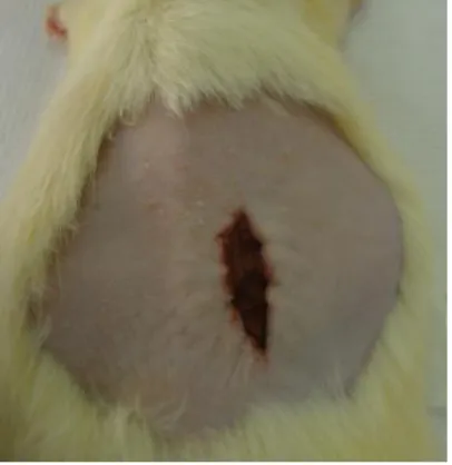
42 until the depth reached the hypodermis. In order to standardize the lesions, we used a surgical field with 2mm width and 2 cm long, with reference to the animal's posterior iliac crest, which was labeled with an alcohol soluble marker (see figure 1).
Figure 1.Wound creation: the skin was cut with a carbon steel surgical blade until it reached the hypodermis with 2mm width and 2 cm long.
2.4 Low-level laser therapy (LLLT)
43
Table 1: Laser Parameters
Parameter [unit] value
Center wavelength [nm] 904
Operating mode pulsed
Frequency [Hz] 9500
Pulse duration [ns] 60
Duty cycle [%] 20
Energy per pulse [J] 42 nJ
Peak power [W] 70
Average power [mW] 39.9
Polarization No
Spot size [cm2] 0.1309
Beam shape elliptical
Beam profile Gaussian
Irradiance at target [mW/cm2] 304.8
Exposure duration [sec] 60
Radiant exposure [J/cm2] 18.288
Total Radiant energy [J] 2.394
Number of points irradiated 1
Area irradiated [cm2] 0.1309
Application technique Contact
Number and frequency of treatment sessions 1 x day / 5 days
44 2.5 Histopathological analysis
The skin samples were fixed in 10 % buffered formalin (Merck, Darmstadt, Germany) for 24 h. Afterwards, they were dehydrated and embedded in paraffin blocks. Three sections (5μm) from each specimen were sectioned (Microtome Leica Microsystems SP 1600, Nussloch, Germany) and stained with hematoxylin and eosin (H.E. stain, Merck, Darmstadt, Germany). The morphological description of the healing characteristics was performed with optical microscopy (Olympus Optical Co., Tokyo, Japan) and histological analysis was made by a pathologist in a double-blind manner. Five slides from each specimen were analyzed and the following criteria were scored as absent, discrete, moderate or intense: granulation tissue, inflammatory process, area of fibrosis and fibroblast number.
2.6 Collagen Analysis by Masson Staining/
Sections stained with Masson were examined microscopically and the images used for analysis were captured by microscopy using a 20X objective (Carl Zeiss, Germany) and a capture system consisting of a camera (Olympus Optical Co., Tokyo, Japan) equipped with AxioVision 4.7.2.0 software. The processing and image analysis were performed with the public domain software ImageJ 1.36 version (National Institutes of Health, Bethesda, USA; http://rsbweb.nih.gov/ij/) using plugin color deconvolution and
percentage of area occupied in relation to the total area was measured as described in previous studies (12-14).
2.7 Procedure for determination of the antioxidant activity.
45 Figure 2.Schematic drawing showing procedure for determination of the antioxidant activity. The skin was dissected and homogenized in 1,15% KCL (1g/10nml solution). The mixture was centrifuged and the supernatant used for biochemical determinations
2.7.1 Concentration of thiobarbituric acid reactive substances (TBARS)
Lipid peroxidation was assessed by measuring the concentration of thiobarbituric acid reactive substances (TBARS). In this process, two molecules of thiobarbituric acid react with a molecule of malondialdehyde to form a pink pigment with maximum absorbance in acid solution at 532-535 nm. TBARS was determined in the skin homogenates that had been diluted to 10% (w/v). Then, 250 µL of homogenate were incubated in a water bath at 37°C for 1 hour, followed by addition of 400 µL of 35% perchloric acid to precipitate the protein. The mixture was centrifuged at 14,000 rpm at 4 ° C for 10 minutes and then the supernatant was added to 200 µL of 1.2% thiobarbituric acid. Next, the mixture was again incubated in a water bath at 100°C for 30 minutes. After cooling on ice, samples were read in a spectrophotometer at 535 nm.(15)
2.7.2 Determination of catalase activity.
The skin of mice was homogenized in a 0.1 M sodium phosphate buffer solution, pH 7.0, at an equivalent volume of 200 times its weight. Then, the homogenate was centrifuged at 5,800 rpm, 10 min at 4°C, the upper layer was discarded and the bottom layer was used for spectrophotometric measurements of H2O2 at 230 nm (28). In the
cuvette, were added 980 μL of the reaction medium (15% H2O2, in 1 M Tris-HCL
buffer), containing 5 mM EDTA, pH 8.0, and 20 μL of the sample diluted in the Tris-HCl buffer. Initial and final absorbance was recorded at 230 nm, after 1 and 6 min, respectively. A standard curve was established using purified catalase (Sigma, MO, USA). Results were expressed in mmol/min/mg protein. Protein was determined by the Lowry method.(16)
2.7.3 Nitrite concentration
46 (1% sulfanilamide, N-(1-naphthyl)-ethylenediamine 0.1%, 1.0% phosphoric acid and distilled water in the ratio of 1:1:1:1 ) were added to 250 µL of homogenate (supernatant) and the mixture was maintained at room temperature for 10 minutes. Thereafter, the absorbance of the samples was determined by spectrophotometry at 560 nm and the blank prepared by adding 250 µL of the Griess reagent to 250 µL of 50 mM phosphate buffer, pH 7.4. The absorbance values were interpolated on a calibration curve containing NaNO2 concentrations ranging from 0.75 to 100 µM.(17)
2.8 Statistical Analysis
Data were expressed as mean and standard deviation and statistically analyzed by analysis of variance (ANOVA) one-way with Bonferroni post hoc test. The significance level was set at p< 0.05. We used the statistical software package SPSS, version 18.0.
3.0 RESULTS
3.1 Histopathological analysis
47 Figure 3.I. Photomicrograph showing granulation tissue (a); newly formed blood vessels (blue arrows) and
fibroblasts (f) (HE 10X). Figure 3.II. Photomicrograph Masson staining showing organization and collagen deposition (c). (20X)
3.2 Collagen amount
48
GROUPS % Collagen
(Mean+SE)
Angiogenesis
(Mean+SE)
NIC (a) 3.82 + 0.18 (b,d) * 10.18 + 1.09
IC (b) 4.99 + 0.01 7.52 + 1.32
NID (c) 3.90 + 0.01(b,d)* 7.38 + 0.99(d)* ID (d) 4.09 + 0.01(a,c)* 15.34 + 1.14 (a,b,c)*
Table 2.Statistical analysis using morphometric computational systems to determine amount collagen. The significance level was set at p < 0.05.
3.3 Concentration of TBARS
As seen in Table 3, the average concentration of TBARS in the four groups was significantly different (F = 12.888, P ≤ 0.001). The Bonferroni test showed significant differences between the groups: ID had a decreased concentration of TBARS compared to NID (p< 0.05) and IC had an increased concentration of TBARS compared to NIC (p< 0.05). In addition, group ID showed significantly lower levels of TBARS than group IC (p< 0.05).
TBARS
Mean + DP ANOVA
T
B
AR
S
NIC
2.38 ± 0.29
≤ 0,001 IC
5.05 ± 1.52* NID
3.57 ± 1.37 ID
1.65 ± 1.57*#
49
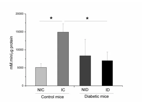
3.4 Catalase activity
As seen in Figure 4, the levels of catalase in the four groups were significantly different (p ≤ 0.001). An increase in catalase activity was observed for IC mice, when their results were compared with those obtained from NIC and ID mice. The Bonferroni test (post hoc) showed that group IC had higher catalase activity compared to NIC group (p ≤ 0.05). Other comparisons between groups were not statistically different (p> 0.05).
Figure 4.Catalase concentration levels in the skin of different groups:non-irradiated control (NIC), non-irradiated
diabetic (NID), irradiated control (IC) and irradiated diabetic (ID). *p ≤ 0.001Figure 4. Catalase concentration levels in the skin of different groups: non-irradiated control (NIC), non-irradiated diabetic (NID), irradiated control (IC) and
irradiated diabetic (ID). *p ≤ 0.001).
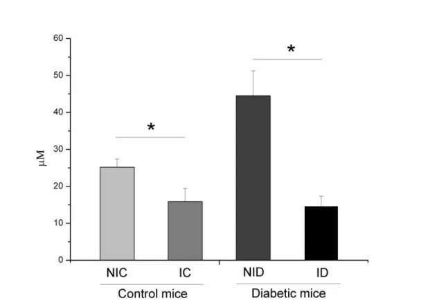
3.5 Nitrite concentration
50 Figure 5..Nitrite concentration levels in the skin of different groups:non-irradiated control (NIC), non-irradiated diabetic (NID), irradiated control (IC) and irradiated diabetic (ID) . *p ≤ 0.001.Figure 5. Nitrite concentration levels in the skin of different groups: non-irradiated control (NIC), non-irradiated diabetic (NID), irradiated control (IC) and irradiated diabetic (ID) . *p ≤ 0.001.
4.0 DISCUSSION
51 normal part of aerobic metabolism and ROS are produced naturally at a certain level, but at greatly elevated levels as a result of some pathological disease states. Antioxidants are substances that fight ROS and free radicals, and can act enzymatically, such as glutathione peroxidase, catalase and superoxide dismutase, or can act non-enzymatically for example: ascorbate, vitamin E, histidine peptides, and some iron-sequestering proteins (ferritin and transferrin). Inflammatory processes can generate ROS and reactive nitrogen species (RNS). The RNS include nitric oxide (NO•) in large amounts, and peroxynitrite (ONOO-). The presence of nitric oxide in biological systems promotes RNS production such as peroxynitrite (formed by spontaneous reaction between NO and superoxide) that reacts with tyrosine residues in proteins to form nitrotyrosine(27). Besides, nitrites and nitrates that circulate in the blood are produced from NO oxidation.
Photobiomodulation can produce NO through photodissociation of NO that is bound to cytochrome c oxidase (CCO). CCO is a photoacceptor located in the inner mitochondrial membrane whose function is to catalyze the oxidation of cytochrome c and the reduction O2 to water, resulting in the pumping of protons out of the mitochondrial matrix. CCO has two heme centers (aa and a3) and two copper centers (CuA and CuB), of which the heme iron of cytochrome a3 and CuB together forms the O2 binding site (28). Thus, NO may compete with oxygen to bind to the iron-sulfur complex and to the iron and copper centers in the respiratory chain and inhibit the mitochondrial ATP synthesis (29). It is proposed that the NO-CCO bond can be broken by visible and NIR photon absorption (30) to restore mitochondrial function and increase ATP synthesis (30, 31). Our results showed a significant reduction of nitrite levels by LLLT in the skin homogenate of wounded mice (Figure 5). This could be explained due to a possible protective effect that LLLT could have on the microvasculature because some studies (32) have suggested that hyperglycemia is a factor which naturally leads to injury of blood vessels and causes long-term microvascular and macrovascular complications. The excessive production of NO may also lead to endothelial injury. Volpe et al demonstrated that the hyperglycemia typical of diabetes exacerbated in vitro inflammatory responses (33). Their group observed that
52 NF-kB activity, and increased levels ofcytokines, chemokines, and circulating adhesion molecules (34-36). We believe that one important factor to explain our results may be mediated by NFkB, because a previous report (10) suggested that LLLT not only enhanced mitochondrial respiration, but also activated the redox-sensitive NF-kB transcription factor via brief generation of ROS. NF-kB regulates immune and inflammatory responses in endothelial cells, vascular smooth muscle cells, and macrophages (37) as well as production of inflammatory cytokines, TNF-alpha and NO (through iNOS expression) (38). Our findings agree with Kandolf-Sekulovic et al (24)
who used LLLT (904 nm, irradiance 60mW/cm2, fluence 3.6 J/ cm2) in a model of contact hypersensitivity (CHS in albino Oxford rats) and observed a reduced release of nitric oxide by the inflammatory cells. These authors suggested that lower levels of NO were caused by fewer inflammatory cells in the dermis, and a possible lower capacity for NO production by skin resident cells (keratinocytes and Langerhans cells). These cells have been suggested to be the NO-producing cells in the CHS skin reaction (39). Eduardo et al (40) analyzed peroxynitrite formation in zymosan-induced arthritis in rats, and found inhibition of joint hyperalgesia that correlated with decreased nitric oxide levels and nitrotyrosine levels in the joint exudates, when compared to control rats,. Taken together, these results point towards a homeostatic role of low levels of NO derived from the constitutive NOS enzymes. Moreover other studies utilizing different experimental models have suggested that LLLT is able to induce SOD expression, decreasing the available concentration of superoxide anion and, as a result, reduce peroxynitrite production (24).
53 effect of LLLT could be on pyruvate levels associated with a metabolic signaling pathway depending on the oxidizing profile of the target cell. Pyruvate is associated with protection of different cells against oxidative damage through non-enzymatic scavenging of ROS including H2O2 (48). Therefore, pyruvate could selectively cause reduction of H2O2 to prevent the generation of the hydroxyl radical (OH•). On the other hand, Karu(49) reported that laser photobiomodulation decreased production of superoxide anion and also increased catalase activity (antioxidant), leading to an increased protein synthesis in a culture of yeast cells.
An important factor to consider is that during oxidative stress, membrane lipids are continuously subjected to lipid peroxidation, shown by an increase in TBARS. In diabetes, lipid peroxidation is a possible factor that influences insulin resistance. Previous studies (41) found high TBARS levels in the blood and in the lung tissue in diabetes mellitus, suggesting that lipid peroxidation occurred in the first 60 days after onset of the disease. In this context, Silveira et al.(31) demonstrated a significant reduction of lipid peroxidation in rats treated with 2 J/cm2 and 4 J/cm2, suggesting that LLLT stimulated antioxidant mechanisms that protected against oxidative damage in lipid membranes. In our study, LLLT significantly reduced the amount of MDA generated in the skin of the mice (Table. 2). Many studies in experimental models have shown that LLLT can modulate ROS/RNS by lowering lipid peroxidation (TBARS and MDA) (50, 51) and reducing RNS by inhibiting synthesis of iNOS(51), and increasing the activity of respiratory (41) chain for increased ATP synthesis. However, it is important to emphasize that the effects of LLLT could depend on the total energy, power density, timing and frequency of irradiation, and the wavelength and other characteristics of the laser or light source used (7).
5.0 CONCLUSIONS
In this work our findings in the wounds of diabetic animals indicated a possible protective effect that 904 nm laser could have on the microvasculature, with lowered levels of nitrite, and increased protection against oxidative damage in lipid membranes. Besides, the better-organized and increased amount of collagen fibers demonstrated that LLLT could be effective in clinical practice with poorly healing diabetic wounds.
55
7.0REFERENCES
1.Santos N, dos Santos J, dos Reis J, Oliveira P, de Sousa A, Carvalho C, et al. Influence of the use of laser phototherapy (λ660 or 790 nm) on the survival of cutaneous flaps on diabetic rats. Photomedicine and laser surgery [Internet]. 2010; 28(4):[483-8. pp.].
2.Ames BN, Shigenaga MK, Hagen TM. Oxidants, Antioxidants, And the Degenerative Diseases of Aging. Proceedings of the National Academy of Sciences of the United States of America. 1993;90(17):7915-22.
3.Wiseman H, Halliwell B. Damage to DNA by reactive oxygen and nitrogen species: Role in inflammatory disease and progression to cancer. Biochemical Journal. 1996;313:17-29.
4.Brownlee M. Biochemistry and molecular cell biology of diabetic complications. Nature. 2001;414(6865):813-20.
5.Dean RT, Fu SL, Stocker R, Davies MJ. Biochemistry and pathology of radical-mediated protein oxidation. Biochemical Journal. 1997;324:1-18.
6.Stocker R, Keaney JF, Jr. Role of oxidative modifications in atherosclerosis. Physiol Rev. 2004;84(4):1381-478.
7.Gupta A, Avci P, Sadasivam M, Chandran R, Parizotto N, Vecchio D, et al. Shining light on nanotechnology to help repair and regeneration. Biotechnology advances. 2013;31(5):607-631.
8.Karu T. Primary and secondary mechanisms of action of visible to near-IR radiation on cells. J Photochem Photobiol B. 1999;49(1):1-17.
9.Chen AC-H, Huang Y-Y, Arany PR, Hamblin MR. Role of reactive oxygen species in low level light therapy. Proc SPIE. 2009;7165:doi: 10.1117/12.814890.
10.Chen AC, Arany PR, Huang YY, Tomkinson EM, Sharma SK, Kharkwal GB, et al. Low-Level Laser Therapy Activates NF-kB via Generation of Reactive Oxygen Species in Mouse Embryonic Fibroblasts. PLoS ONE. 2011;6(7):e22453.
11.Farias VX, Macedo FH, Oquendo MB, Tome AR, Bao SN, Cintra DO, et al. Chronic treatment with D-chiro-inositol prevents autonomic and somatic neuropathy in STZ-induced diabetic mice. Diabetes Obes Metab. 2011;13(3):243-50.
12.Collins TJ. ImageJ for microscopy. Biotechniques. 2007;43(1):25-40.
56 14.Miot HA, Brianezi G. Morphometric analysis of dermal collagen by color clusters segmentation. Anais Brasileiros De Dermatologia. 2010;85(3):361-4.
15.Nobre-Junior HV, Oliveira RA, Maia FD, Nogueira MAS, de Moraes MO, Bandeira MAM, et al. Neuroprotective Effects of Chalcones from Myracrodruon urundeuva on 6-Hydroxydopamine-Induced Cytotoxicity in Rat Mesencephalic Cells. Neurochemical Research. 2009;34(6):1066-75.
16.Michiels C, Raes M, Toussaint O, Remacle J. Importance of SE-Glutathione peroxidase,catalase and Cu/Zn-SOD for cell-survival against oxidative stress. Free Radical Biology and Medicine. 1994;17(3):235-48.
17.Green LC, Tannenbaum SR, Goldman P. Nitrate synthesis in the germ-free and conventional rat. Science. 1981;212(4490):56-8.
18.Brownlee M. Biochemistry and molecular cell biology of diabetic complications. Nature. 2001;414(6865):813-20.
19.Francis-Goforth KN, Harken AH, Saba JD. Normalization of diabetic wound healing. Surgery. 2010;147(3):446-9.
20.Adams SB, Jr., Sabesan VJ, Easley ME. Wound healing agents. Foot and ankle clinics. 2006;11(4):745-51.
21.Demidova-Rice TN, Hamblin MR, Herman IM. Acute and Impaired Wound Healing: Pathophysiology and Current Methods for Drug Delivery, Part 2: Role of Growth Factors in Normal and Pathological Wound Healing: Therapeutic Potential and Methods of Delivery. Adv Skin Wound Care. 2012;25(8):349-70.
22.Demidova-Rice TN, Hamblin MR, Herman IM. Acute and Impaired Wound Healing: Pathophysiology and Current Methods for Drug Delivery, Part 1: Normal and Chronic Wounds: Biology, Causes, and Approaches to Care. Adv Skin Wound Care. 2012;25(7):304-14.
23.Tatmatsu Rocha JC, Jansem de Almeida Catanho MT, da Mota DL. Application of the Laser Radiation in Patients of Pressure Ulcers: Clinical and Histomorphometric Analysis of the Derm. Brazilian Archives of Biology and Technology. 2008;51:231-4. 24.Kandolf-Sekulovic L, Kataranovski M, Pavlovic MD. Immunomodulatory effects of low-intensity near-infrared laser irradiation on contact hypersensitivity reaction. Photodermatology Photoimmunology & Photomedicine. 2003;19(4):203-12.
57 26.Winiarska K, Drozak J, Wegrzynowicz M, Fraczyk T, Bryla J. Diabetes-induced changes in glucose synthesis, intracellular glutathione status and hydroxyl free radical generation in rabbit kidney-cortex tubules. Molecular and Cellular Biochemistry. 2004;261(1-2):91-8.
27.Pacher P, Liaudet L, Soriano FG, Mabley JG, Szabo E, Szabo C. The role of poly(ADP-ribose) polymerase activation in the development of myocardial and endothelial dysfunction in diabetes. Diabetes. 2002;51(2):514-21.
28. Ferraresi, Cleber; Pires de Sousa, Marcelo Victor; Huang, Ying-Ying; et al. Time response of increases in ATP and muscle resistance to fatigue after low-level laser (light) therapy (LLLT) in mice. Lasers in Medical Science.2015;30(4): 1259-1267. 29.Huang Y-Y, Sharma SK, Carroll J, Hamblin MR. Biphasic dose response in low level light therapy- an update. Dose-Response. 2011;9(4):602-18.
30.Vladimirov YA, Osipov AN, Klebanov GI. Photobiological principles of therapeutic applications of laser radiation. Biochemistry-Moscow. 2004;69(1):81-90.
31.Silveira PC, Silva LA, Fraga DB, Freitas TP, Streck EL, Pinho R. Evaluation of mitochondrial respiratory chain activity in muscle healing by low-level laser therapy. J Photochem Photobiol B. 2009;95(2):89-92.
32.Bagasra O, Michaels FH, Zheng YM, Bobroski LE, Spitsin SV, Fu ZF, et al. Activation of the inducible form of nitric oxide synthase in the brains of patients with multiple sclerosis. Proceedings of the National Academy of Sciences of the United States of America. 1995;92(26):12041-5.
33.Oliveira Volpe CM, Machado Abreu LF, Gomes PS, Gonzaga RM, Veloso CA, Nogueira-Machado JA. The Production of Nitric Oxide, IL-6, and TNF-Alpha in Palmitate-Stimulated PBMNCs Is Enhanced through Hyperglycemia in Diabetes. Oxidative Medicine and Cellular Longevity. 2014.2014:12-24.
34. Kolb H, Mandrup-Poulsen T. An immune origin of type 2 diabetes? (vol 48, pg 1038, 2005). Diabetologia. 2005;48(8):1677-1688.
35.Tripathy D, Mohanty P, Dhindsa S, Syed T, Ghanim H, Aljada A, et al. Elevation of free fatty acids induces inflammation and impairs vascular reactivity in healthy subjects. Diabetes. 2003;52(12):2882-7.
58 37.Sena CM, Pereira AM, Fernandes R, Santos-Silva D, Faustino A, Ceica R. Novel therapeutic approach to target endothelial dysfunction in type 2 diabetes. Cardiovascular Research. 2014;103: S102–S141.
38.Suganami T, Tanimoto-Koyama K, Nishida J, Itoh M, Yuan X, Mizuarai S, et al. Role of the Toll-like receptor 4/NF-kappa B pathway in saturated fatty acid-induced inflammatory changes in the interaction between adipocytes and macrophages. Arteriosclerosis Thrombosis and Vascular Biology. 2007;27(1):84-91.
39.Ross R, Gillitzer C, Kleinz R, Schwing J, Kleinert H, Forstermann U, et al. Involvement of NO in contact hypersensitivity. International Immunology. 1998;10(1):61-9.
40.Eduardo FP, Mehnert DU, Monezi TA, Zezell DM, Schubert MM, Eduardo CP, et al. Cultured epithelial cells response to phototherapy with low intensity laser. Lasers Surg Med. 2007;39(4):365-72.
41.Dhaunsi GS, Bitar MS. Antioxidants attenuate diabetes-induced activation of peroxisomal functions in the rat kidney. J Biomed Sci. 2004;11(5):566-70.
42.Urbich C, Dernbach E, Aicher A, Zeiher AM, Dimmeler S. CD40 ligand inhibits endothelial cell migration by increasing production of endothelial reactive oxygen species. Circulation. 2002;106(8):981-6.
43.Mowat AG, Baum J. Chemotaxis of Polymorphonuclear Leukocytes from Patients with Diabetes Mellitus. New England Journal of Medicine. 1971;284(12):621-9.
44.Collison KS, Parhar RS, Saleh SS, Meyer BF, Kwaasi AA, Hammami MM, et al. RAGE-mediated neutrophil dysfunction is evoked by advanced glycation end products (AGEs). Journal of Leukocyte Biology. 2002;71(3):433-44.
45.Chanchamroen S, Kewcharoenwong C, Susaengrat W, Ato M, Lertmemongkolchai G. Human Polymorphonuclear Neutrophil Responses to Burkholderia pseudomallei in Healthy and Diabetic Subjects. Infection and Immunity. 2009;77(1):456-63.
46.Lin J-C, Siu LK, Fung C-P, Tsou H-H, Wang J-J, Chen C-T, et al. Impaired phagocytosis of capsular serotypes K1 or K2 Klebsiella pneumoniae in type 2 diabetes mellitus patients with poor glycemic control. Journal of Clinical Endocrinology & Metabolism. 2006;91(8):3084-7.
59 48.Moriguchi N, Hinoi E, Tsuchihashi Y, Fujimori S, Iemata M, Takarada T, et al. Cytoprotection by pyruvate through an anti-oxidative mechanism in cultured rat calvarial osteoblasts. Histology and Histopathology. 2006;21(9):969-77.
49.Karu T, Pyatibrat L, Kolyakov S, Afanasyeva N. Absorption Measurements of Cell Monolayers Relevant to Mechanisms of Laser Phototherapy: Reduction or Oxidation of Cytochrome c Oxidase Under Laser Radiation at 632.8 nm. Photomedicine and Laser Surgery. 2008;26(6):593-9.
50.Luo L, Sun Z, Zhang L, Li X, Dong Y, Liu TC-Y. Effects of low-level laser therapy on ROS homeostasis and expression of IGF-1 and TGF-beta 1 in skeletal muscle during the repair process. Lasers in Medical Science. 2013;28(3):725-34.
60
ESTUDO II
Phototherapy Laser and LED acts over Mitochondrial Function and
estimulates collagen organization in diabetic animals
José Carlos Tatmatsu Rocha1,2*, Carla Roberta Tim3, Rubens Bernardes Filho4, Lucimar Avo5, Patrícia Brassolatti6, Nivaldo Antonio Parizotto1
1 – Physical Therapy Department, Federal University of Sao Carlos, Sao Carlos, SP, Brazil 2- Physical Therapy Department, Federal University of Ceara, Fortaleza, Ce, Brazil 3- Department of Bioscience, Federal University of São Paulo, Santos, SP, Brazil 4- Brazilian Enterprise of Agriculture Research, EMBRAPA, São Carlos, SP, Brazil. 5- Medicine Department, Federal University of Sao Carlos, Sao Carlos, SP, Brazil
6- Department of Physiotherapy, Post-Graduate Program of Biotechnology, Federal University of São Carlos (UFSCar), São Carlos, SP, Brazil.)
* Corresponding Author