The Neotropical genus Proceratophrys Miranda-Ribeiro, 1920 currently consist of 36 species distributed in Brazil, Argen-tina, and Paraguay (FROST 2014). The species of Proceratophrys
have been clustered in species groups or complexes based on overall morphological similarity (LYNCH 1971, IZECKSOHNet al. 1998, GIARETTAet al. 2000, KWET & FAIVOVICH 2001, PRADO & POMBAL 2008). Species without palpebral appendages are assigned to the Proceratophrys bigibbosa and P. cristiceps species groups. Species of the Proceratophrys cristiceps group occur in open and dry environments of Cerrado and Caatinga. They are character-ized by the absence of post-ocular swellings (GIARETTAet al. 2000, CRUZet al. 2012, GODINHOet al. 2013). The thirteen species
in-cluded in the group are: P. aridus Cruz, Nunes & Juncá, 2012; P. bagnoi, Brandão, Caramaschi, Vaz-Lima & Campos, 2013; P. branti, Brandão, Caramaschi, Vaz-Lima & Campos, 2013; P. caramaschii Cruz, Nunes & Juncá, 2012; P. carranca Godinho, Moura, Lacerda & Feio, 2013; P. concavitympanum Giareta, Bernarde & Kokubum, 2000; P. cristiceps (Müller, 1883); P. cururu Eterovick & Sazima, 1998; P. dibernardoi, Brandão, Caramaschi, Vaz-Lima & Campos, 2013; P. goyana (Miranda-Ribeiro, 1937); P. huntingtoni Ávila, Pansonato & Strüssmann, 2012; P. moratoi (Jim & Caramaschi, 1980); P. rotundipalpebra, Martins & Giaretta, 2013; P. strussmannae Ávila, Kawashita-Ribeiro & Morais, 2011; and P. vielliardi Martins & Giaretta, 2011.
Proceratophrys bigibbosa group is found in southern and southeastern Brazil, Argentina, and Paraguay. The species in this group are characterized by a blunt and short snout, by the
presence of a post-ocular swelling, and a large marginal row of tubercles on the eyelid (KWET & FAIVOVICH 2001). Currently four
species are assigned to the group: P. avelinoi Mercadal de Barrio & Barrio, 1993; P. bigibbosa (Peters, 1872); P. brauni Kwet & Faivovich, 2001; and P. palustris Giaretta & Sazima, 1993.
Species with a long and single palpebral appendage are placed in the Proceratophrys appendiculata and P. boiei groups (IZECKSOHNet al. 1998, PRADO & POMBAL 2008, CRUZ & NAPOLI 2010,
DIASet al. 2013a). The species of the P. boiei group occur primarily
in the Atlantic Forest, from Paraíba to Santa Catarina states (PRADO
& POMBAL 2008): P. boiei (Wied-Neuwied, 1824); P. paviotii Cruz, Prado & Izecksohn, 2005; and P. renalis (Miranda-Ribeiro, 1920). Proceratophrys appendiculata species group is found only in Atlantic Forest, from Bahia to Santa Catarina states (IZECKSOHN et al. 1998, CRUZ & NAPOLI 2010, DIASet al. 2013a). The ten in-cluded species, listed below, are characterized by the presence of a triangular rostral appendage: P. appendiculata (Günther, 1873); P. belzebul Dias, Amaro, Carvalho-e-Silva & Rodrigues, 2013; P. izecksohni Dias, Amaro, Carvalho-e-Silva & Rodrigues, 2013; P. laticeps Izecksohn & Peixoto, 1981; P. melanopogon (Miranda-Ribeiro, 1926); P. moehringi Weygoldt & Peixoto, 1985; P. phyllostomus Izecksohn, Cruz & Peixoto, 1998; P. sanctaritae Cruz & Napoli, 2010; P. subguttata Izecksohn, Cruz & Peixoto, 1998; and, P. tupinamba Prado & Pombal, 2008.
In addition, Proceratophrys schirchi (Miranda-Ribeiro, 1937), P. rondonae Prado & Pombal, 2008 (which have a short single multi-cuspidate palpebral appendage) (PRADO & POMBAL
The tadpole of
Proceratophrys izecksohni
(Amphibia: Anura: Odontophrynidae)
Pedro H. dos Santos Dias
1,2, Ana M.P.T. de Carvalho-e-Silva
2& Sergio P. de Carvalho-e-Silva
31 Departamento de Zoologia, Instituto de Biociências, Universidade de São Paulo. Rua do Matão 101, 05508-090 São Paulo,
SP, Brazil. Corresponding author. E-mail: [email protected]
2 Laboratório de Biossistemática de Anfíbios, Departamento de Zoologia, Universidade Federal do Estado do Rio de Janeiro.
Urca, 22290-240 Rio de Janeiro, RJ, Brazil.
3 Instituto de Biologia, Universidade Federal do Rio de Janeiro. Avenida Brigadeiro Trompowsky, Bloco A, Ilha do Fundão,
21941-590 Rio de Janeiro, RJ, Brazil.
ABSTRACT. We describe the external morphology of the tadpole of Proceratophrys izecksohni Dias, Amaro, Carvalho-e-Silva & Rodrigues, 2013, its internal oral features, and chondrocranial anatomy, based on specimens collected at the type locality. The tadpole has short and oval body, spiracle with inner wall fused to the body, and oral formula 2/3(1). The oral cavity of P. izecksohni is typical of stream-dwelling tadpoles, with several papillae and pustulations. The chon-drocranium is longer than wide and the suprarostral corpora are free ventromedially. The palatoquadrate has a well developed processus pseudopterygoideus. We also compare the tadpole of P. izecksohni with tadpoles those of other species of the genus, emphasizing the usage of larval morphology to assist in the systematic of the genus.
182 P.H. dos S. Dias et al.
ZOOLOGIA 31 (2): 181–194, April, 2014
2008, NAPOLIet al. 2011) and P. minuta Napoli, Cruz, Abreu &
Del-Grande, 2011 and P. redacta Teixeira, Amaro, Recoder, Dal Vechio & Rodrigues, 2012 (which has a series of small append-ages on the eyelid) are not associated with any species group. The classification of Proceratophrys into species groups has been used due the morphological similarity among adults. In stipe of the practical utility of these groups, recent molecu-lar analysis has not recovered them as monophyletic units (AMAROet al. 2009, TEIXEIRAet al. 2012, DIASet al. 2013a).
Larval morphology and bioacustic data have not been used in assessing evolutionary relationships within the genus (DIAS et al. 2013b,c). Larval morphology, however, has been successfully used in taxonomic and phylogenetic studies (LARSON
& DE SÁ 1998, MAGLIAet al. 2001, HASS 2003, PÚGENERet al. 2003, GRANTet al. 2006, CANDIOTI 2008) and seems to be a very useful
tool in the classification of Odontophrynidae frogs (DIASet al.
2013b, NASCIMENTO et al. 2013). Nevertheless, the absence of
data on larval morphology, especially on internal morphology, limits the use of such characters in broader analyses.
Within Proceratophrys, only 15 species have their tadpoles described (FATORELLIet al. 2010, NASCIMENTOet al. 2010, NAPOLIet al. 2011, PROVETEet al. 2013). The internal oral morphology is known for eight species (WASSERSUG & HEYER 1988, DE SÁ & LANGONE 2002, VIEIRAet al. 2007, NASCIMENTOet al. 2010, PROVETE et al. 2013), and chondrocranial data for five species (DIASet al. 2013b). Herein we provide a description of the tadpole of Proceratophrys izecksohni including the internal oral features and chondrocranial morphology.
Proceratophrys izecksohni is a small to medium sized spe-cies (SVL 32.1-54.1 mm in males) occurring in the southeast-ern portion of the sate of Rio de Janeiro (DIASet al. 2013a). It
can be found ca 200 m above sea level, at least in its type local-ity – Reserva Rio das Pedras, Mangaratiba municipallocal-ity (CARVALHO-E-SILVAet al. 2008).
MATERIAL AND METHODS
Tadpoles were collected at the type locality Reserva Rio das Pedras, municipality of Mangaratiba, Rio de Janeiro state Brazil (22°59’29"S, 44°06’01"W, ca. 200 m above sea level). Voucher specimens are deposited in the Amphibians Collec-tion of the Laboratório de Biossistemática de Anfíbios of Universidade Federal do Estado do Rio de Janeiro (UNIRIO). The tadpole illustration provided herein is based on a speci-men on GOSNER (1960) stage 36, deposited with the catalog number UNIRIO 4010.
The following dimensions of ten tadpoles (stages 35-36) were measured according to ALTIG & MCDIAMIRD (1999) and ALTIG
(2007): total length (TL), body length (BL), tail length (TAL), body width (BW), body height (BH), tail height (TH), nostril to snout distance (NSD), eye to snout distance (ESD), interor-bital distance (IOD), eye to nostril distance (END), internarial distance (IND), oral disc width (ODW) and eye diameter (ED).
Colors patterns are standardized according with SMITHE’s (1975)
catalog. Developmental stages were determined following GOSNER’s (1960) staging table. Some individuals were raised through metamorphosis to confirm species identifications (UNIRIO 4218). Proceratophrys izecksohni is not sympatric with any other Proceratophrys species in Mangaratiba municipality. For analysis of the internal oral morphology, six tadpoles (Gosner 34-35) were dissected according with WASSERSUG (1976) and the oral features were stained with methylene blue solu-tion. Terminology follows WASSERSUG (1976, 1980) and
WASSERSUG & HEYER (1988). We also analyzed the internal oral
features of eight tadpoles (Gosner stages 35-36) of a close re-lated species, P. appendiculata, collected in its type locality, Serra dos Órgãos (DIASet al. 2013a). The description of the chondro-cranium was based on 19 tadpoles between Gosner stages 30-39 cleared and double stained following DIASet al. (2013b);
illustration is based on a specimen on Gosner stage 33 (UNIRIO 4010-B). Chondrocranial terminology follows LARSON & DE SÁ
(1998) and HAAS (2003).
Material examined (given in lots). Proceratophrys izecksohni. BRAZIL, Rio de Janeiro: Mangaratiba (Reserva Rio das Pedras),
UNIRIO 368, 4010, 4203, 4216. Proceratophrys appendiculata. BRA
-ZIL, Rio de Janeiro: Teresópolis (Parque Nacional da Serra dos Órgãos), UNIRIO 2676, 3592, 3852, 4027, 4030, 4036.
RESULTS
Description of the tadpole (stage 35-36 n = 10)
(Figs 1-9, Table I)
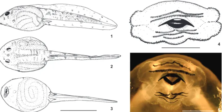
Tadpole with an oval body and a rounded snout (Figs 1-4); height of body representing 14.4% of the total length. The eyes are in a latero-dorsal position, separated by a distance approximately twice the eye’s diameter. The nostrils are reni-form and positioned on the dorsal side, separated by approxi-mately 1.5 times the diameter of an eye; eye diameter representing 15% of the body width and 7.2% of body length. Internarial distance about 74.2% of interorbital distance; eye to nostril distance corresponding to 33% of eye to snout dis-tance; eye to snout distance corresponding to 20% of body length and 7.4% of total length. The lateral line system is not distinguished nor in living or fixed tadpoles.
The mouth is ventral and has a dermal fringe on its con-tour; presence of a single row of sub-marginal papillae on the lower labium, bearing two folds; keratizned denticles in two upper series and three lower series, with the first lower series being interrupted [2/3(1)]; A-1 = A-2, P-1 = P-2>P-3 (Fig. 5); upper jaw large than lower jaw; jaws serrated; upper jaw arch shaped and lower jaw “V” shaped. Oral disc width correspond-ing to 27.2% of body width.
Tail muscles distinct ending in a rounded tip; the tail length represents 63.2% of the total length, with its height slightly greater than the height of the body (TH/BH 98.2%). Dorsal fin originates on the posterior third of the body. Maxi-mum tail height (at its medial portion) representing 15.2% of total length; dorsal and ventral fins about the same height; they slightly decrease in height, ending in rounded tip.
Color in life. Dorsum of a light brown color (fawn color), covered with small darker brown spots, giving a granite-like appearance. A small brown spot is found between the eyes (am-ber color). The overall coloration of the lateral portion of the body is similar to that described for the dorsum. Ventral skin is translucent, with a brownish tone owing to the gut’s con-tent. The iris has a golden color (clay color). At the end of the body there are two very clear small round spots (cream color). Two other small spots of the same color can be seen, one on the dorsum, parallel to the spots at the end of the body and another near the top of the tail fin (Figs 6-9). Light brown spots are also found. There is an overall silver coloration on the lateral side of the fins (pearl gray). The tail is pale with a pinkish color in muscles due to the circulatory system; it has four small dark brown stripes (hair brown).
Color of the tadpole in preservative. Translucent skin, central and tail areas of a pale yellow coloration (cream color), with small spaced spots of a dark brown tone (tawny).
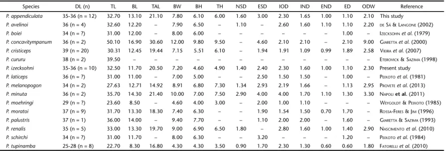
Internal oral morphology (Fig. 10-11)
Buccal roof (BF). The buccal roof is triangular in shape. It is longer than wider; its width corresponding to 45% of the buccal roof length. The prenarial arena is trapezoidal in shape, possessing six to twelve pustulations; it also bears a reduced transverse triangular ridge. The internal nares are elliptical and placed transversally to the main axis, forming a 60° angle. There are four to five postnarial papillae on each side of the postnarial arena, oriented parallel to the nares; the narial velve is well Figures 1-5. Tadpole of Proceratophrys izecksohni, UNIRIO 4010, stage 36: (1) dorsal; (2) lateral; (3) ventral views; (4) oral disc morpho-logy; (5) detail of the oral disc – oral formula 2/3(1). Scale bars: 1-3 = 10.0 mm, 4-5 = 1.0 mm.
Table I. Measurements of Proceratophrysizecksohni larvae. Measurements for the illustrated tadpole (UNIRIO 4010, stage 35) with mean ± standard deviation (SD) are provided below as well as the range among the tadpoles used in the description (n = 10, stages 35-36).
Drawn tadpole Mean ± SD Range
Total length 33.0 32.5 ± 1.3 (29.9-33.4)
Body length 13.0 11.7 ± 1.0 (10.4-13.5)
Tail length 20.2 20.5 ± 0.3 (20.2-21.0)
Body width 9.4 7.2 ± 1.5 (5.8-9.8)
Body height 6.5 4.6 ± 1.0 (3.4-6.6)
Tail height 5.3 4.9 ± 0.7 (41-6.1)
Nostril to snout distance 1.8 1.4 ± 0.2 (1.2-1.8)
Eye to snout distance 2.6 2.4 ± 0.3 (2.1-2.9)
Interorbital distance 2.5 2.3 ± 0.1 (2.2-2.6)
Eye to nostril distance 1.1 1.0 ± 0.08 (0.8-1.1)
Internarial distance 1.4 1.6 ± 0.1 (1.4-1.8)
Oral disc width 2.1 2.3 ± 0.1 (1.9-2.8)
Eye diameter 1.0 1.09 ± 0.1 (0.9-1.2)
1
2
3
4
184 P.H. dos S. Dias et al.
ZOOLOGIA 31 (2): 181–194, April, 2014
developed. The postnarial papillae are conical in shape and orientated toward the postnarial arena; the surface of each papillae is covered by pustulations. The postnarial arena is tri-angular in shape and has a thick lateral ridge papilla, which is divided into five conical projections covered by pustulations; the two most anterior projections are longer than the others. The median ridge is low and triangular, with six to nine short papillae around it. The buccal roof arena is an inverted “U”. It is laterally delimitated by 14 to 18 approximately uniform coni-cal papillae on each side, which face the center of the bucconi-cal roof arena. The buccal roof arena is densely covered with pus-tulations (over 300). Also, about 10-12 papillae are located on the posterior third of the postulation area of the buccal roof
arena interspersed with the pustulations. Posteromedially, the buccal roof arena is delimitate by 10 to 13 conical papillae with varied sizes near the vellum, which is smooth.
Buccal floor (BF). The buccal floor is ovoid in shape. It has four infralabial papillae, a medial pair and two lateral. The medial ones are hand-shaped, covered with pustulations, un-equal in size. The lateral papillae are short and multi-branched, covered with pustulations. The lingual bud is elliptic and pos-ses two pairs of lingual papillae. The lingual papillae are paral-lel to infralabial papillae; they are hand-shaped, bifurcated, with pustulations on their surface. The buccal floor arena (BFA) is triangular, delimitated by approximately 75 conical papil-lae; apices of papillae point to the center of buccal floor arena. Figures 6-9. Living specimens of Proceratophrys izecksohni: (6) UNIRIO 4216 stage 25; (7) specimen in its natural habitat; (8) specimen raised in laboratory stage (45); (9) froglet UNIRIO 4218.
6
7
The buccal floor arena is also covered with pustulations (over 350). The buccal pockets are shallow. The ventral vellum pre-sents irregular edge.
Chondrocranial morphology (stage 33, UNIRIO
4010-B) (Figs 12-16)
Until stage 33 the chondrocranium is entirely cartilagi-nous; by stage 34 the parasphenoid begins to ossify and at stage 35 the frontoparietals and the exoccipitals are visible. The chon-drocranium is longer than wide; its greatest width (at the level of the palatoquadrate) is approximately 80% and its greatest height (at the level of the processus muscularis) is about 25% of its length.
Neurocranium
Ethmoidal region. The upper jaw sheets are supported by the paired suprarostral cartilages. The suprarostral (Fig. 15) con-sists of a central corpus and lateral alae. These elements are dor-sally fused. The corpora are free on its medial region; they are thin, sub rectangular shaped; in frontal view it shows a “V”. The lateral alae are wide, flattened, and rectangular in shape, with rounded ventral surface. Each ala curves posteriorly from its point of fusion with the corpora. The posterodorsal margin of each ala possesses a well-developed processus posterior
dor-salis. The cornua trabeculae articulate with the suprarostral at the point of junction of the corpora and the alae.
The cornua trabeculae originate from the planum trabe-culare anticum and represent 40% o the chondrocranium length. They diverge forming a “V” and are curved ventrally. They are uniform along their extension. The planum trabeculare anticum is continuous with the planum ethmoidale that forms the ante-rior wall of the braincase. At the point of confluence of the cor-nua trabeculae, the septum nasi is visible as a small strip of cartilage.
Orbitotemporal region. The fenestra basicranialis is closed by a thin sheet of cartilage, the planum intertrabeculare, which also closes the central area of the cranial floor. The chondro-cranial floor is pierced by two pairs of foramina. The anterior, smaller pair is the foramina craniopalatina, and the posterior, larger pair is the foramina carotica primaria.
The lateral walls of the braincase are formed by the or-bital cartilages, which are posteriorly connected to the otic capsule. This connection forms the dorsal margin of the fora-men prooticum, a large forafora-men located between the anterior edge of the otic capsule and the posterior margin of the orbital cartilage. Other two foramina are visible in the orbital carti-lage: the foramen opticum and the foramen oculomotorium.
10 11
PNA IP
TR
N
PONP
PONA PONAP MR BRA
DV LP
LB
BFA
BFAP
BP
VV BRAP
186 P.H. dos S. Dias et al.
ZOOLOGIA 31 (2): 181–194, April, 2014
12
14
15
13 16
The frontoparietal fontanelle is large and ovoid in shape; its width represents 50% of its length. This fontanelle repre-sents 50% of the chondrocranium length. Laterally, it is bor-dered by the taenia tecti marginales, anteriorly by the planum ethmoidale, and posteriorly by the tectum synoticum.
Otoocipital region. The otic capsules are rhomboid in shape and represents about 25% of the chondrocranium length. In dorsal view, it bears a distinct anterolateral triangular pro-jection, the larval crista parotica. The tectum synoticum bridges the two otic capsules and forms the dorsal roof of the foramen magnum. Each arcus occipitalis is continuous with the tectum synoticum dorsally and with the planum basale ventrally, form-ing the foramen magnum and the foramen jugulare. The fora-men perilymphaticum is found laterally to the forafora-men jugulare.
Visceral components
Palatoquadrate. In dorsal view the palatoquadrate is C shaped, wider in its medial region. Posteriorly it connects to the braincase through the processus ascendens, a thin rod-like car-tilage. The processus ascendens attaches just posterior of the oculomotorium foramen (intermediate condition sensu SOKOL
1981). The processus ascendens is almost perpendicular to the main axis of the chondrocranium, forming an angle of 90°.
Anteriorly the palatoquadrate connects to the braincase through the commissura quadratocranialis anterior. The com-missura quadratocranialis anterior extends between the palatoquadrate, at the level just posterior to the pars articularis quadrati, and the floor of the neurocranium. The anterior margin of the commissura bears a triangular process, the pro-cessus quadratoethmoidalis. A triangular-shaped propro-cessus pseudopterygoideus is present on the posterior margin of the commissura quadratocranialis.
Each palatoquadrate bears two distinct processes: the processus muscularis quadrati and the pars articularis quad-rati. The broad and flat processus muscularis quadrati extend dorsally from the lateral edges from the palatoquadrate at the level posterior to the pars articularis quadrati. The edges of the processus muscularis quadrati curves to attach the processus antorbitalis forming the commissura quadratoorbitalis.
Below the processus muscularis and ventrolaterally on the palatoquadrate there is a process, the facies articularis hyalis that serves as the point of articulation of the ceratohyal to the palatoquadrate. This feature is partially formed by the hyoquadrate process, a ventral enlargement of the palatoqua-drate. The pars articularis quadrati, the anterior tip of the palatoquadrate, articulates with the posterior margins of the Meckel’s cartilage.
Meckel’s cartilage and cartilage labialis inferior. Together with the infrarostral cartilages, the Meckel’s cartilage forms the mandible. Meckel’s cartilage is sigmoid shaped and represents 20% of the chondrocranial length. It is wider on its posterior edge, narrowing towards the rostral portion. Meckel’s cartilage is oriented almost perpendicular to the main axis of the
chon-drocranium, positioned ventrally to the cornua trabeculae. Rostrally, it articulates to the cartilage labialis inferior and pos-teriorly with the pars articularis quadrati trough the processus retroarticularis.
The paired infrarostral cartilages provide support for the lower horny beak. They are almost rectangular. They are bounded by connective tissue, and slightly curved towards main chondro-cranial axis, forming a “V” shape structure in ventral view.
Hyobranchial apparatus.The ceratohyalia are wide, flat, and subtriangular shaped cartilages; they are dorsally twisted to articulate with the facies articularis of the palatoquadrate. Each ceratohyal bears two distinct processes: the processus anterior hyalis and the processus anterolateralis hyalis. Both processes are almost triangular in shape. Additionally, they possess a well-developed triangular processus posterior hyalis. The ceratohyalia are bounded together by the pars reuniens, which bears a developed processus urobranchialis. The plana hypobranchiales are wide sheets of cartilage that serve as the point of attachment of the ceratobranchials.
The branchial basket has four ceratobranchials that are distally continuous via the commissurae terminales. Proximally, ceratobranchial I attaches to the planum hypobranchiale by a wide strip of cartilage. Ceratobranchials I, II and III bear a dor-sally projecting spiculum near their point of attachment to the planum hypobranchiale.
Natural History. Adults and tadpoles were found at least 200 m above sea level in a dense, undisturbed, forest. Tadpoles occur in moderately deep rocky stream with sandy bottom. This is also true of a closely related species P. appendiculata (PEIXOTO & CRUZ 1980, DIAS & CARVALHO-E-SILVA 2012). The
tad-poles of P. izecksohni are benthonic and cryptic with the sand (Fig. 7); at the first sign of disturbance of the water, the tad-poles swim down the rocks or bury in the debris. They were found together with tadpoles of Hylodes asper (Müller, 1924), Hylodes phyllodes (Heyer & Cocroft, 1986), and Phasmahyla cruzi Carvalho-e-Silva, Siva & Carvalho-e-Silva, 2009.
Comparisons with other tadpoles
Data for species in comparison are given in parenthesis; major morphological variation is summarized in the Tables II-IV. External morphology. The tadpole of P. izecksohni can be promptly differentiated from tadpoles of the following groups of by having the second row of denticles without interruption (second row of denticles interrupted in the species enumerated below): P. bigibbosa [P. avelinoi (DE SÁ & LANGONE 2002), P. palustris
(GIARETA & SAZIMA 1993)], P. cristiceps groups [P. concavitympanum
(GIARETTAet al. 2000), P. cristiceps (VIEIRAet al. 2007), P. cururu (ETEROVICK & SAZIMA 1998), P. moratoi (ROSSA-FERES & JIM 1996)]
and P. boiei [P. boiei (IZECKSOHNet al. 1979), P. renalis (NASCIMENTO et al. 2010)], as well of P. laticeps (PEIXOTO et al. 1981), P. melanopogon (PROVETEet al. 2013), P. moehringi (WEYGOLDT & PEIXOTO
188
P.H. dos S. Dias
et al.
ZOOLOGIA 31 (2): 181–194, April, 2014
Table II. Variation of the described tadpoles of Proceratophrys (measurements in millimeters). (DL) Developmental stage (GOSNER 1960), (n) number of examined specimens, (TL) Total length, (BL) body length, (TAL) tail length, (BW) body width, (BH) body height, (TH) tail height, (NSD) nostril to snout distance, (ESD) eye to snout distance, (IOD) interorbital distance, (END) eye to nostril distance, (IND) internarial distance, (ODW) oral disc width, (ED) eye diameter.
Species DL (n) TL BL TAL BW BH TH NSD ESD IOD IND END ED ODW Reference
P. appendiculata 35-36 (n = 12) 32.70 13.10 21.10 7.80 6.10 6.00 1.60 3.00 2.30 1.65 1.00 1.10 2.10 This study
P. avelinoi 36 (n = 4) 32.60 12.20 – 7.90 6.50 – 1.10 – 2.60 1.60 1.10 1.10 2.20 DE SÁ &LANGONE (2002)
P. boiei 34 (n = ?) 31.00 12.00 – 8.00 6.00 – – – – – – 1.00 – IZECKSOHNet al. (1979)
P. concavitympanum 36 (n = 2) 50.10 16.90 30.60 12.00 9.80 9.50 – 4.60 2.10 2.10 – 2.10 9.00 GIARETTAet al. (2000)
P. cristiceps 39 (n = 20) 30.31 12.45 19.44 7.15 5.51 6.10 – 1.94 1.91 1.09 0.99 1.89 2.58 VIEIRAet al. (2007)
P. cururu 38 (n = 2) 39.50 – – – – – – – – – – – – ETEROVICK &SAZIMA (1998)
P. izecksohni 35-36 (n = 10) 32.50 11.70 20.50 7.20 4.60 4.90 1.40 2.40 2.30 1.60 1.00 1.10 2.30 Present study
P. laticeps 36 (n = ?) 31.00 11.00 – 7.00 5.00 – – 2.50 1.50 1.50 – 1.00 – PEIXOTOet al. (1981)
P. melanopogon 34 (n = 2) 27.63 12.71 14.92 8.91 6.80 7.30 1.34 2.93 2.19 1.66 – 1.13 2.95 PROVETEet al. (2013)
P. minuta 36 (n = 2) 35.70 14.30 21.40 10.00 7.00 7.50 2.90 4.00 4.00 1.70 1.10 1.30 3.30 NAPOLI et al. (2011)
P. moehringi 29 (n = ?) 23.60 8.50 – 4.60 4.00 3.00 – 2.00 1.00 1.10 – – – WEYGOLDT &PEIXOTO (1985)
P. moratoi 37 (n = 9) 31.70 13.30 18.30 7.40 6.30 – – 1.90 1.54 1.50 0.70 1.70 – ROSSA-FERES &JIM (1996)
P. palustris 37 (n = 1) 36.00 14.00 – 9.40 7.70 – – 1.10 2.00 2.00 – 1.60 – GIARETTA &SAZIMA (1993)
P. renalis 35 (n = 5) 33.00 13.30 19.70 9.00 6.90 6.50 1.80 – 2.80 1.60 1.00 1.40 2.90 NASCIMENTOet al. (2010)
P. schirchi 34 (n = ?) 31.00 11.70 – 8.00 6.30 – – 3.20 – – – 1.20 – PEIXOTOet al. (1984)
P. tupinamba 25-28 (n = 8) 22.70 8.30 16.80 4.30 4.30 3.50 0.90 1.70 2.30 1.30 0.60 0.60 1.80 FATORELLIet al. (2010)
Table III. Morphological variation of Proceratophrys species. Characteristics followed by an * were inferred from the illustrations, which can be found in correspondent reference. The # refers to observations of NASCIMENTOet al. (2010) for P. boiei from Camanducaia, Minas Gerais state, Brazil.
Sepcies/Structure Nostril shape Spiracle Origin of dorsal fin Tip of the tail Labium emargination Oral formula Reference
P. appendiculata reniform fused to the body posterior third of the body rounded two folds 2/3(1) Present study
P. avelinoi rounded – junction body/tail rounded two folds 2(2)/3(1) DE SÁ &LANGONE (2002)
P. boiei reniform free# posterior third of the body rounded none 2(2)/3(1) IZECKSOHNet al. (1979)
P. convavitympanum rounded free* posterior third of the body acute none 2(2)/3(1) GIARETTAet al. (2000)
P. cristiceps rounded fused to the body posterior third of the body rounded none 2(2)/3(1) VIEIRAet al. (2007)
P. cururu – – posterior third of the body acute* none 2(2)/3(1) ETEROVICK &SAZIMA (1998)
P. izecksohni reniform fused to the body posterior third of the body rounded two folds 2/3(1) Present study
P. laticeps reniform – posterior third of the body acute two folds 2(2)/3(1) PEIXOTOet al. (1981)
P. melanopogon oval free posterior third of the body rounded none 2(2)/3(1) PROVETEet al. (2013)
P. minuta reniform free junction body/tail rounded none 2(2)/3(1) NAPOLIet al. (2011)
P. moehringi rounded – – rounded two folds 2(2)/3(1) WEYGOLDT &PEIXOTO (1985)
P. moratoi rounded fused to the body junction body/tail rounded none 2(2)/3(1) ROSSA-FERES &JIM (1996)
P. palustris – fused to the body junction body/tail acute two folds 2(2)/3(1) GIARETTA &SAZIMA (1993)
P. renalis reniform free posterior third of the body acute two folds 2(2)/3(1) NASCIMENTOet al. (2010)
P. schirchi reniform fused to the body* posterior third of the body acute two folds 2(2)/3(1) PEIXOTOet al. (1984)
melanopogon, P. minuta, and P. moratoi by presenting two folds in the mar-gin of the lower labium (no folds). Additionally, P. izecksohni tadpoles can be distinguished from the tadpoles of P. concavitympanum, P. cururu, P. laticeps, P. palustris, P. renalis, P. schirchi, and P. tupinamba (FATORELLIet al. 2010) by a
rounded tail tip (acute in those species). The posterodorsal orientation of the spiracle distinguishes the tadpole of P. izecksohni from those of P. cristiceps and P. cururu (spiracle oriented dorsally). The reniform-shaped nostrils also set the tadpole of P. izecksohni apart from the tadpoles of P. avelinoi, P. concavitympanum, P. cristiceps, P. moehringi, P. moratoi, P. palustris P. tupinamba (rounded) and P. melanopogon (oval). Proceratophrys izecksohni tadpoles dif-fer from the tadpoles of P. concavitympanum, P. melanopogon, P. minuta and P. renalis by presenting the inner wall of the spiracle fused to the body wall (free); from the tadpoles of P. avelinoi, P. minuta, P. moratoi, P. palustris and P. tupinamba by having the tail fins originating on the posterior third of the body (tail fins originated at the tail/body junction).
Proceratophrys izecksohni tadpoles can be distinguished from the tad-poles of Proceratophrys tupinamba by presenting a pair of cream dots in the lateral portion of the body (absent) and several silvered areas along the body (uniform brown coloration of the body) and by having submar-ginal papillae in the oral disc (absent).
From the tadpole of P. appendiculata, P. izecksohni tadpoles can be distinguished by presenting a lower body height in relation to total length (BH/TL 14% in P. izecksohni and 19% in P. appendiculata), by the lower tail height in relation to total length (TH/TL 15% in P. izecksohni and 19% in P. appendiculata), by the larger internarial distance in relation to interorbital distance (IND/IOD 74% in P. izecksohni and 72% in P. appendiculata), by the smallest eye to nostril distance in relation to body length (END/BL 7.5% in P. izecksohni and 9% in P. appendiculata), by the smallest body width in relation to total length (BW/TL 24% in P. izecksohni and 22% in P. appendiculata) and by the overall coloration; tail with silver dots (silver dots absent) and a single cream dot on the dorsum (absent).
Internal oral morphology. The internal oral features of P. izecksohni tadpoles differ from those of of P. boiei (WASSERSUG & HEYER 1988) by the larger number of papillae in the buccal floor arena (about 75 papillae in P.
Ta b le I V . M a jo r v ariation on in te rn al or al f ea tur es o f Pr o ce ra top h ry s lar va e . Bucca l fl oo r ( B F) ; b u ccal fl oo r ar en a ( B FA ), b u ccal r oof ( B R) , b u ccal r oof ar en a ( B RA) , infralab ial p a p illa e (I L) , li n g ua l p apill a e ( LP) , p o st n aria l a rena ( P ONA) . C h ar ac te ri st ics followed b y a n * we re inf e rr ed from the i llustra tions, which ca n b e found in co rresp o n d e nt re fe re nce . Se p ci e s B R s h ap e PON A pa pill ae BRA s h ap e BRA pa pi lla e BR A pu st u la ti o n s BF s h ap e IL pa pi lla e LP pa pil la e BF A sh ap e BF A pa pilla e BF A pu st u la tio n s Re fe re n ce P. ap pe nd ic ul at a tr ia n g ul ar 5 t o 10 in ve rt e d U 30-36 2 3 0 o vo id 4 4 tr ia n g ul ar 30 -5 0 270 -32 0 Pr es e n t st u d y P. a vel in oi se mi-cir cu la r 5 to 6 ro u nde d 3 0 – tr iangu lar 4 4 U -s hape d 2 0 -2 5 – DE S Á & L ANGO NE (20 02) P. b o ie i ov oi d * – U -s h ap e d 3 0 – tri an g ul ar* 2 4 o va l 5 0 – W AS S ER S UG & H EY ER ( 198 8) P. cr is ti cep s tr iangu lar 4 se m i-ci rc u lar 1 8 -2 0 – cir cu lar 4 3 tr iangu lar 2 8 -3 4 – V IE IRA et a l. ( 200 7) P. c u ru ru he xag o nal 1 2 e lip tical 2 8 8 0 -9 0 e lip soid 2 4 h e x ag onal 5 0 4 0 P RO V ET E et a l. (2 013) P. izecks oh n i triang ular 8 to 1 0 inv e rte d U 1 4 to 1 8 ab o u t 3 6 0 o vo id 4 4 triang ular 7 5 ab out 3 7 0 Pr es e n t s tu d y P. m el a n o p o g o n tr ap e zoi d 1 0 t o 1 2 q u ad ra te 14 to 3 0 10 0-200 tr ia n g ul ar 4 5 ro un d ed 5 0 1 80 P RO V ET E et a l. (2 013) P. m o ra to i re ct an g u la r 6 ro un d ed 1 5 40-50 tr ia n g ul ar 2 4 d ia m o n d 40-50 50 P RO V ET E et a l. (2 013) P. r en a lis cir cu lar 5 cir cu lar 2 4 – tr iangu lar 2 6 U -shape d 4 2 -4 6 – N AS CIME NT O et a l. ( 201 0)
Figure17. Hypothesis of phylogenetic relationships within Proceratophrys (DIAS et al. 2013a). The evolutionary transition of the oral formula 2(2)/3(1) to 2/ 3(1) is a putative synapomorphy for this clade. The phylogenetic position of
190 P.H. dos S. Dias et al.
ZOOLOGIA 31 (2): 181–194, April, 2014
izecksohni and 50 papillae in P. boiei), the presence of 4-5 papil-lae in the prenarial arena (a single papilla) and by the inverted U buccal roof arena (U-shaped).
Proceratophrys izecksohni tadpoles differs from those of P. cristiceps (VIEIRAet al. 2007) by the ovoid buccal floor (circular),
by having four lingual papillae (three), by the larger number o papillae in the buccal floor arena (about 75 papillae in P. izecksohni and 28-34 papillae in P. cristiceps), by the presence of papillae only on the posterior part of postulation area of the buccal floor arena (30-40 papillae homogeneously spread in the buccal floor arena), by the larger number of papillae in the median ridge (6-9 in P. izecksohni and 4-5 in P. cristiceps), and by presenting about 10-12 papillae located on the posterior part of the postulation area of the buccal roof arena (20-30 papillae well distributed on the postulation area of buccal roof in P. cristiceps).
Proceratophrys izecksohni larvae differ from those of P. renalis (NASCIMENTOet al. 2010) by two pairs of infralabial
papil-lae (single pair), two pairs of lingual papilpapil-lae (three pairs), an overall triangular buccal roof (circular), by the presence of pustulations on the postnarial arena (pustulations absent), “U” shaped buccal roof arena (circular), triangular buccal floor arena (“U” shaped), larger number of papillae surrounding the buc-cal floor arena, i.e., approximately 75 (42-46), posterior part of buccal floor arena only with papillae (papillae spread within postulation buccal floor).
Proceratophrys izecksohni tadpoles differ from those of P. avelinoi (DE SÁ & LANGONE 2002) by triangular buccal roof (semi-circular), triangular buccal floor arena (“U” shaped), and by the 8-10 postnarial papillae (5-6).
Larvae of Proceratophrys izecksohni differ from those of P. cururu (PROVETEet al. 2013)by the triangular buccal roof
(hex-agonal), by the angle of the nares (60° in P. izecksohni and 45° in P. cururu), by the U-shaped buccal roof (elliptical), by the smaller number of buccal roof arena papillae (14-18 in P izecksohni and 28 in P. cururu), by a greater number of pustula-tions in the buccal roof arena (360 in P. izecksohni and 80-90 in P. cururu), by the ovoid buccal floor shape (ellipsoid), by hav-ing four infralabial papillae (two), by the triangular shaped buccal floor arena (hexagonal), by the absence of papillae in the center of the buccal floor arena (10 present), by the greater number of buccal floor arena papillae (75 in P. izecksohni and 50 in P. cururu) and by the greater number of pustulations in the buccal floor arena (370 in P. izecksohni and 40 in P. cururu). It differs from the tadpoles of P. moratoi (PROVETEet al. 2013) by the triangular buccal roof (rectangular), by the larger number of pustulations in the prenarial arena (6-12 in P. izecksohni and 3 in P. moratoi), by the angle of the nares (60° in P. izecksohni and 30° in P. moratoi), by the larger number of postnarial papillae (8-10 in P. izecksohni and 6 in P. moratoi), by the smaller number of lateral ridge (a single papilla in P. izecksohni and two rows of papillae in P. moratoi), by the in-verted U buccal roof arena (rounded), by the larger number of pustulations in buccal roof (360 in P. izecksohni and 40-50 in P.
moratoi), by the ovoid buccal floor (triangular), by the triangu-lar buccal floor arena (diamond shaped), by the triangu-larger number of buccal floor papillae (75 in P. izecksohni and 40-50 in P. moratoi), and by the larger number of pustulations in the buc-cal floor arena (about 370 in P. izecksohni and 50 in P. moratoi). The tadpoles of Proceratophrys izecksohni can be distin-guished from those of P. melanopogon (PROVETEet al. 2013)by the shape of the buccal roof (triangular in P. izecksohni and trapezoid in P. melanopogon), by the larger number of pustula-tions in the prenarial arena (6-12 in P. izecksohni and two in P. melanopogon), by the inverted U shaped buccal roof arena (quad-rate), by the larger number of pustulations in the buccal roof arena (about 360 in P. izecksohni and 100-200 in P. melanopogon), by the ovoid buccal floor (triangular), by having four lingual papillae (five), by triangular buccal floor arena (rounded), by the larger number of papillae in the buccal floor arena (75 in P. izecksohni and 50 in P. melanopogon), and by the larger number of pustulations in the buccal floor arena (about 370 in P. izecksohni and 180 in P. melanopogon).
The internal oral morphology of the tadpoles of P. izecksohni seems very similar to that of P. appendiculta. We found as the major distinction between them the larger number of papillae delimitating the buccal floor arena (75 in P. izecksohni and 30-50 in P. appendiculata), the smaller number of papillae in the buccal roof arena (14-18 in P. izecksohni and 30-36 in P. appendiculata), the larger number of pustulations in the buccal roof arena (360 in P. izecksohni and 230 in P. appendiculata) and in the buccal floor arena (370 in P. izecksohni and 270-320 in P. appendiculata). Our observations for P. appendiculata differ in some points from that made by WASSERSUG & HEYER (1988); this
is probably due to the sample size – WASSERSUG & HEYER (1988)
observed only one specimen also collected in Teresópolis mu-nicipality, Rio de Janeiro state, while in this work nine speci-mens were analyzed.
Chondrocranium morphology. The tadpoles of P. izecksohni differs from those of P. appendiculata, P. cristiceps, P. laticeps, P. tupinamba and P. boiei from Rio de Janeiro state (DIAS et al. 2013b) by the morphology of the suprarostral cartilage. The central elements – the corpora – are not fused ventromedi-ally (fused in those species). The tadpole of P. izecksohni also differs from those of P. boiei from Minas Gerais state by not pre-senting either the taenia tecti transversals or mediales (present), and by for not ventrally free suprarostral corpus from the alae joined ventrally (joined); from the tadpoles of P. boiei from Paraná state by the morphology of the ventral surface of the hypobran-chial plate (smooth in P. izecksohni and acute in P. boiei).
DISCUSSION
Proceratophrys tadpoles – taxonomy, phylogeny,
and perspectives
Previous studies have suggested that the tadpoles of Proceratophrys possess a highly conserved morphology. For ins-tance, de SÁ & LANGONE (2002) enumerated the following set of
external characteristics as representative for the genus: 1) oval body; 2) low caudal fins; 3) ventral mouth; 4) sinistral spiracle with midlateral opening; 5) dextral vent tube’s opening; 6) la-bial tooth row formula 2(2)/3(1) or 2/3(1); 7) oral disk emarginated and with two folds of the posterior labium; 8) single row of marginal papillae with a large rostral gap; and 9) serrated, keratinized jaw sheaths.
The internal oral morphology also seems to be conserved, with several papillae and pustulations in the buccal roof and floor arenas, hand-like lateral ridge papillae. WASSERSUG & HEYER
(1988) – classified Proceratophrys within other stream-dwelling species – classification corroborated by other studies (DE SÁ & LANGONE 2002, VIEIRAet al. 2007, NASCIMENTOet al. 2010, PROVETEet al. 2013, present study). The morphology of the chondrocra-nium also proved to be very conservative, at least within spe-cies that possess a long and unique palpebral appendage (DIASet al. 2013b).
Several other morphological traits (e.g., tail tips, nostril shape, folds on the lower labium) vary considerably among the known tadpoles (PROVETE et al. 2013). The absence of data for
more than 50% of the species limits our understanding of the variation in this genus. Moreover, several characteristics reported as variable have been constantly inferred from rudimentary de-scriptions (IZECKSOHNet al. 1979, PEIXOTOet al. 1984, WEYGOLDT &
PEIXOTO 1985) or from illustrations (some illustrations are
par-ticularly questionable). SANCHEZ (2013), analyzing tadpoles from
Dendrobatoidea Cope, pointed out that coding data from illus-trations can be problematic, once the interpretations of the il-lustrator might not be congruent with the actual morphology of the animal, as well as the interpretation of the reader.
Another problem is that the distribution of some mor-phological traits among tadpoles are not consistent with avail-able phylogenies (AMAROet al. 2009, PYRON & WIENS 2011, TEIXEIRA et al. 2012, DIASet al. 2013a), but rather with phenetic groups
(DIASet al. 2013b). A solution to this problem is the
incorpora-tion of multiple sources of evidence (i.e., internal and external morphological data, from adults and tadpoles, DNA sequences, behavioral and bioacoustics) in a broad and extensive phylo-genetic reconstruction.
A first step to recover such phylogenies, leastwise in rela-tion to larval morphology, would be a broad sampling of speci-mens, especially from unknown and from rare tadpoles – most tadpoles have been described based on one or few specimens (e.g., PEIXOTOet al. 1981, 1984, WEYGOLDT & PEIXOTO 1985, GIARETTA &
SAZIMA 1993, ETEROVICK & SAZIMA 1998, GIARETTAet al. 2000, DE SÁ &
LANGONE 2002). New samples are rare in herpetological collections.
The second step is the standardization of the terminol-ogy from the observed morphological features and a survey of internal morphological data. Including the results of the present study, the internal oral morphology is known from only nine species (Table IV), chondrocranium for only six spe-cies (DIASet al. 2013b, present study), and the myology is
com-pletely unknown – in other words, our knowledge of the internal aspects of these animals is restricted to less than 30% of the species of the genus.
Proceratophrys appendiculata complex vs
P. appendiculata clade
IZECKSOHNet al. (1998) defined a species complex within Proceratophrys in which all members presented a single, long, unicuspidate palpebral appendage, and a rostral fleshy appenda-ge. They included the following species in this complex: Procera-tophrys appendiculata, P. laticeps, P. melanopogon, P. moehringi, P. phyllostomus, and P. subguttata. Subsequently, P. tupinamba, P. sanctaritae, P. belzebul and P. izecksohni were added to it.
Although supported by the overall adult morphology, molecular data indicates that the P. appendiculata complex is not monophyletic. In the phylogenies referred to below, P. laticeps is frequently recovered as the sister species of the clade P. cururu – P. renalis (AMAROet al. 2009, TEIXEIRAet al. 2012); or all species
are inserted in a broad clade composed of P. cururu, P. renalis, P. moratoi, P. concavitympanum, P. boiei and P. goyana (DIASet al. 2013a). PYRON & WIENS (2011), however, argued that P. laticeps is sister to the clade P. renalis – P. boiei. The other species (from which tissue sample is available) form a well supported clade composed of P. izecksohni, P. tupinamba, P. appendiculata and P. belzebul; the sister group of this clade is composed by several evolutionary lineages associated with P. melanopogon (DIASet al. 2013a). The phylogenetic position of P. moehringi, P. phyllostumus, P. subguttata and P. sanctaritae was never accessed.
The larval morphology provides support for a P. appendi-culata clade, composed of P. izecksohni, P. tupinamba, P. appendiculata and P. belzebul. The oral formula 2/3(1) is known from all those species (with exception of P. belzebul, whose tad-poles is unknown). It might represent a putative synapomorphy for this clade (Fig. 17). The internal oral morphology and the morphology of the chondrocranium of P. appendiculata and P. izecksohni (DIASet al. 2013b, present study) reinforce the close relationships between them.
The chondrocranium morphology of P. laticeps resembles that of P. appendiculata and P. izecksohni (DIASet al. 2013b; this
study), but the external morphological features of the tadpoles, such as tail tips and the oral formula do not fit with that clade. This is expected, once the adult of P. laticeps differs from P. izecksohni, P. tupinamba, P. appendiculata and P. belzebul by the absence of a preocular cutaneous crest (IZECKSOHNet al. 1998,
192 P.H. dos S. Dias et al.
ZOOLOGIA 31 (2): 181–194, April, 2014
Adults of P. moehringi are extremely similar to those of P. izecksohni, P. tupinamba, P. appendiculata, and P. belzebul (IZECKSOHNet al. 1998, PRADO & POMBAL 2008, DIASet al. 2013a). Moreover, the advertisement call of P. moehringi is more simi-lar to that of P. appendiculata – even more than that of P. melanopogon – and both species share several behavioral traits, such as calling from under large rocks and during all day (WEYGOLDT & PEIXOTO 1985, DIASet al. 2013c). However, the oral formula of of P. moehringi tadpole is 2(2)/3(1) and specimens present a rounded nostril.
The internal morphology of the tadpole of P. moehringi is unknown. Furthermore, this species is sympatric with sev-eral other Proceratophrys species, such as P. boiei, P. laticeps, P. paviotii, and P. schirchi (PRADO & POMBAL 2008), some of which
are morphologically similar to P. moehringi. The tadpole de-scribed by WEYGOLDT & PEIXOTO (1985) might not correspond to
this species.
The corroboration of the existence of a monophyletic P. appendiculata clade might be attested with the survey of new data, specifically with the descriptions of the tadpoles of P. phyllostomus, P. subguttata, and P. sanctaritae and tissues from DNA analysis. New samples of tadpoles of P. moehringi are necessary.
ACKNOWLEDGMENTS
We are very thankful to Daniel B. de Góes and other col-leagues for their field assistance. We also thank the Coordenação de Aperfeiçoamento de Pessoal de Nível Superior and Fundação de Amparo à Pesquisa do Estado de São Paulo for P.H.S. Dias fellowship and the Conselho Nacional de Desenvolvimento Científico e Tecnológico for S.P. Carvalho-e-Silva fellowship. We thank ICMBio for the collecting permit number 33288-2. We also are very thankful to Gustavo S. de Miranda for critically reading the manuscript, although all errors are our responsibil-ity. We are very grateful to Rafael O. de Sá for reviewing the manuscript; his suggestions greatly increased the quality of the text. We are also thankful to Ana L. Prudente and an anony-mous reviewer for their valuable comments on the manuscript.
LITERATURE CITED
ALTIG, R. 2007. A primer for the morphology of anuran tadpole.
Herpetological Conservation Biology2: 71-74.
ALTIG, R. & R.W. MCDIARMID. 1999. Body plan. Development
and Morphology, p. 24-51. In: W.W. MCDIARMID R. & ALTIG
(Eds). Tadpoles, the biology of anuran larvae. Chicago, The University of Chicago Press.
AMARO, R.C.; D. PAVAN & M.T. RODRIGUES. 2009. On the generic
identity of Odontophrynus moratoi Jim and Caramaschi, 1980 (Anura; Cycloramphidae). Zootaxa 2071: 61-68.
CANDIOTI, M.F.V. 2008. Larval anatomy of Andean tadpoles of
Telmatobius (Anura: Ceratophryidae) from Northwestern
Argentina. Zootaxa 1938: 40-60.
CARVALHO-E-SILVA, A.M.P.T.; G.R. SILVA & S.P. CARVALHO-E-SILVA. 2008.
Anuros da Reserva Rio das Pedras, Mangaratiba, RJ. Biota Neotropica 8: 198-209.
CRUZ, C.A.G. & M.F. NAPOLI. 2010. A new species of smooth
horned frog, genus Proceratophr ys Miranda-Ribeiro (Amphibia: Anura: Cycloramphidae), from the Atlantic Rain Forest eastern Bahia, Brazil. Zootaxa 1660: 57-67. CRUZ, C.A.G.; I. NUNES & F.A. JUNCÁ. 2012. Redescription of
Proceratophrys cristiceps (Müller, 1883) (Amphibia, Anura, Cycloramphidae), with description of two new species without eyelid appendages from northeastern Brazil. South America Journal of Herpetology 7: 110-122. doi.org/ 10.2994/057.007.0201
DE SÁ, R.O. & J.A. LANGONE. 2002. The tadpole of Proceratophrys avelinoi (Anura: Leptodactylidae). Journal of Herpetology 36: 490-494.
DIAS, P.H.S. & A.M.P.T. CARVALHO-E-SILVA. 2012. Records of
abnormalities in Proceratophrys appendiculata (Günther, 1873) (Anura; Cycloramphidae; Alsodinae). Herpetology Notes 5: 197-199.
DIAS, P.H.S.; R.C. AMARO; A.M.P.T. CARVALHO-E-SILVA & M.T.
RODRIGUES. 2013a. Two new species of Proceratophrys Miranda-Ribeiro, 1920 (Anura; Odontophrynidae) from the Atlantic Forest, with taxonomic remarks on the genus. Zootaxa 3682: 277-304. doi: 10.11646/zootaxa.3682.2.5
DIAS, P.H.S.; A.M.P.T. CARVALHO-E-SILVA & S.P. CARVALHO-E-SILVA.
2013b. Larval chondrocranium morphology of Five species
of Proceratophrys Miranda-Ribeiro (Amphibia; Anura;
Odontophrynidae). Zootaxa 3683: 427-438. doi: 10.11646/ zootaxa.3683.4.6
DIAS, P.H.S.; F.S.F. HEPP; A.M.P.T. CARVALHO-E-SILVA & S.P. CARVA -LHO-E-SILVA. 2013c. Breeding biology and advertisement call
of the horned leaf-frog Proceratophrys appendiculata (Amphibia: Anura: Odontophrynidae). Zoologia 30: 388-396. doi: 10.1590/S1984-46702013000400004
ETEROVICK, P. & I. SAZIMA. 1998. New species of Proceratophrys
(Anura: Leptodactylidae) from southeastern Brazil. Copeia 1998: 159-164.
FATORELLI, P.; P.N. COSTA; R.C. LAIA; M. ALMEIDA-SANTOS; M. VAN
SLUYS & C.F.D. ROCHA. 2010. Description, microhabitat and
temporal distribution of the tadpole of Proceratophrys
tupinamba Prado and Pombal, 2008. Zootaxa 2684: 57-62.
FROST, D.R. 2014. Amphibia Species of the World: an online reference. New York, American Museum of Natural History, version 6.0. Avalaible online at: http://research.amnh.org/ herpetology/amphibia/index.html [Accessed: February 18, 2013].
GIARETTA, A.A. & I. SAZIMA. 1993. Nova espécie de Proceratophrys Mir. Rib. do Sul de Minas Gerais, Brasil (Amphibia, Anura, Leptodactylidae). Revista Brasileira de Biologia 53: 13-19. GIARETTA, A.A.; P.S. BERNARDE & M.N.C. KOKUBUM. 2000. A new
GOSNER, K.L. 1960. A simplified table for staging anurans embryos
and larvae with notes on identifications. Herpetologica 16: 183-190.
GODINHO, L.B.; M.R. MOURA; J.V.A. LACERDA & R.N. FEIO. 2013. A
new species of Proceratophrys (Anura: Odontophrynidae) from the middle São Francisco River, southeastern Brazil.
Salamandra 49: 63-73.
GRANT, T.; D.R. FROST; J.P. CALDWELL; R. GAGLIARDO; C.F.B. HADDAD; P.J.R. KOK; D.B. MEANS; B.P. NOONAN; W.E. SCHARGEL & W.C. WHEELER.
2006. Phylogenetic systematics of dart-poison frogs and their relatives (Amphibia: Athesphatanura: Dendrobatidae). Bulletin of the American Museum of Natural History 299: 1-252. HAAS, A. 2003. Phylogeny of frogs as inferred from primarily
larval characters (Amphibia: Anura). Cladistics 19: 23-89. doi: 10.1016/S0748-3007(03)00006-9
IZECKSOHN, E.; C.A.G. CRUZ & O.L. PEIXOTO. 1979. Notas sobre o
girino de Proceratophrys boiei (Wied) (Amphibia, Anura, Lepto-dactylidae). Revista Brasileira de Biologia 39: 233-236. IZECKSOHN, E.; C.A.G. CRUZ & O.L. PEIXOTO. 1998. Sobre
Procera-tophrys appendiculata e algumas espécies afins (Amphibia: Anura: Leptodactylidae). Revista da Universidade Rural, Série Ciências da Vida,20: 37-54.
IZECKSOHN, E.; S.P. CARVALHO-E-SILVA & I. DEISS. 2005. O osteocrânio de Proceratophrys boiei (Wied-Neuwied), P. appendiculata (Günther), P. melanopogon (Miranda-Ribeiro) e P. laticeps Izecksohn & Peixoto (Anura, Leptodactylidae). Revista Bra-sileira de Zoologia 22 (1): 225-229. doi: 10.1590/S0101-81752005000100028.
KWET, A. & J. FAIVOVICH. 2001. Proceratophrys bigibbosa species group (Anura,Leptodactylidae), with Description of a New Species. Copeia 2001: 203-215.
LARSON, P.M. & R.O. DE SÁ. 1998. Chondrocranium morphology
of Leptodactylus larvae (Leptodactylidae; Leptodactylinae): its utility in phylogenetic reconstruction. Journal of Morphology 238: 287-305.
LYNCH, J.D. 1971. Evolutionary relationships, osteology, and
zoogeography of leptodactyloid frogs. Miscellaneous Publications of the University of Kansas Museum of Na-tural History 53: 1-238.
MAGLIA, A.M.; L.A. PÚGENER & L. TRUEB. 2001. Comparative
development of anurans: using phylogeny to understand ontogeny. American Zoologist 41: 538-551.
NAPOLI, M.F.; C.A.G. CRUZ; R.O. ABREU & M.L. DEL-GRANDE. 2011. A new species of Proceratophrys Miranda-Ribeiro (Amphibia: Anura: Cycloramphidae) from the Chapada Diamantina, State of Bahia, northeastern Brazil. Zootaxa 3133: 37-49. NASCIMENTO, F.A.; B.S. LISBOA; G.O. SKUK & R.O. DE SÁ. 2010.
Description of the tadpole of Proceratophrys renalis (Miranda-Ribeiro, 1920) (Anura: Cycloramphidae). South American Journal of Herpetology 5 (3): 241-248. doi: 10.2994/ 057.005.0309
NASCIMENTO, F.A.C.; T. MOTT; J.A. LANGONE; C.A. DAVIS & R.O. DE
SÁ. 2013. The genus Odontophrynus (Anura:
Odontophry-nidae): a larval perspective. Zootaxa 3700: 140-158. doi: 10.11646/zootaxa.3700.1.5
PEIXOTO, O.L. & C.A.G. CRUZ. 1980. Observações sobre a larva de Proceratophrys appendiculata (Günther, 1873) (Amphibia, Anura, Leptodactylidae). Revista Brasileira de Biologia 41: 83-86.
PEIXOTO, O.L.; E. IZECKSOHN & C.A.G. CRUZ. 1981. Notas sobre o girino de Proceratophrys laticeps Izecksohn and Peixoto (Amphibia, Anura, Leptodactylidae). Revista Brasileira de Biologia 41: 553-555.
PEIXOTO, O.L.; C.A.G. CRUZ; E. IZECKSOHN & S.P. CARVALHO-E-SILVA.
1984. Notas sobre o girino de Proceratophrys precrenulata (Amphibia, Anura, Leptodactylidae). Arquivos da Univer-sidade Federal Rural do Rio de Janeiro 7: 83-86. PRADO, G.M & J.P. POMBAL. 2008. Espécies de Proceratophrys
Miranda-Ribeiro, 1920 com apêndices palpebrais (Anura; Cycloramphidae). Arquivos de Zoologia 39: 1-85. PROVETE, D.B.; L.S.O. MELO; M.V. GAREY; F.B.R. GOMES; I.A. MARTINS
& D.C. ROSSA-FERES. 2013. Larvae of Proceratophrys melanopogon
(Amphibia: Anura), with emphasys on internal oral morpho-logy and comparisons with P. cururu and P. moratoi.
Herpetologica 69: 163-174.
PÚGENER, L.A.; A.M. MAGLIA & L. TRUEB. 2003. Revisiting the contribution of lar val characters to an analysis of phylogenetic relationships of basal anurans. Zoological Journal of the Linnean Society 139: 129-155. doi: 10.1046/ j.1096-3642.2003.00075.x.
PYRON, R.A. & J.J. WIENS. 2011. A large scale phylogeny of Amphibia including over 2800 species, and a revised classifi-cation of extant frogs, salamanders, and caecilians. Molecu-lar Phylogenetics and Evolution 61: 543-583. doi: 10.1016/ j.ympev.2011.06.012.
ROSSA-FERES, D.C. & J. JIM. 1996. Tadpole of Odontophrynus moratoi (Anura, Leptodactylidae). Journal of Herpetology 30: 536-539.
SANCHEZ, D.A. 2013. Larval morphology of dart-poison frogs
(Anu-ra: Dendrobatoidea: Aromatidae and Dendrobatidae).
Zootaxa 3637: 569-591. doi: org/10.11646/zootaxa.3637.5.5. SMITHE, F.B. 1975. Naturalist’s Color Guide. New York, American
Museum of Natural History.
SOKOL, O.M. 1981. The larval chondrocranium of Peleodytes puncatuts, with a review of tadpole chondrocranial. Journal of Morphology 169: 161-183.
TEIXEIRA, M.; R.C. AMARO; R.S. RECODER; F. DAL VECHIO & M.T.
RODRIGUES. 2012. A new dwarf species of Proceratophrys
Miranda-Ribeiro, 1920 (Anura, Cycloramphidae) from highlands of Chapada Diamantina, Bahia, Brazil. Zootaxa 3551: 25-42.
VIEIRA, W.L.S.; K.S. VIEIRA & G. GOMES-SANTANA. 2007. The tadpole
of Proceratophrys cristiceps (Anura: Cycloramphidae,
Odontophrynini). Zootaxa 1397: 17-24.
194 P.H. dos S. Dias et al.
ZOOLOGIA 31 (2): 181–194, April, 2014
of the Museum of Natural History of University of Kansas 48: 1-23.
WASSERSUG, R.J. 1980. Internal oral features of larvae from eight anuran families: functional, systematic, evolutionary and ecological considerations. Miscellaneous Publications of the University of Kansas Museum of Natural History 68: 1-146.
Submitted: 21.IX.2013; Accepted: 25.II.2013. Editorial responsibility: Ana Lúcia Prudente
WASSERSUG, R.J. & W.R. HEYER. 1988. A survey of internal oral
features of leptodactyloid larvae (Amphibia: Anura).
Smithsonian Contributions to Zoology 457: 1-99. WEYGOLDT, P. & O.L. PEIXOTO. 1985. A new species of horned