D e p a rtamento de Neurologia da Faculdade de Medicina da Universidade de São Paulo, São Paulo, Brasil (FMUSP): 1P ro f e s s o r
Associado; 2Doutor; 3Acadêmica da FMUSP; 4Professor Titular. This work was supported by FAPESP (1998/16599-7) and CNPQ.
Received 6 January 2005, received in final form 8 April 2005. Accepted 27 May 2005.
Dra. Um b ert in a Co nt i Reed - Neu ro lo g ia HCFM USP - Av. En éas d e Carvalh o A g u iar 255/5oan d ar sala 5131 - 05430-900 São Pau lo SP - Brasil. E-m ail: u co n t ireed @h cn et .u sp .b r
DYSTROPHIN-GLYCOPROTEINS ASSOCIATED IN
CONGENITAL MUSCULAR DYSTROPHY
Immunohistochemical analysis of 59 Brazilian cases
Lu cio Go b b o Fer reira
2, Su ely Kazu e M arie
1, En n a Crist in a Liu
3,
M aria Bern ad et e Du t ra Resen d e
2, M ary So u za Car valh o
2,
M ilb ert o Scaf f
4, Um b ert in a Co n t i Reed
1ABSTRACT - The congenital muscular dystrophies (CMD) are heterogeneous muscular diseases with early and dystrophic pattern on muscle biopsy. Many diff e rent subtypes have been genetically identified and most phenotypes not yet identified belong to the merosin-positive (MP) CMD subgro u p . Object ive: To analyze the immunohistochemical expression of the main proteins of the dystro p h i n - g l y c o p roteins associa-ted complex in muscle biopsy of patients with diff e rent CMD phenotypes, for investigating a possible corre-lation with clinical and histopathological data. M et hod: Fifty-nine patients with CMD had clinical, histo-pathological and immunohistochemical data evaluated: 32 had MP-CMD, 23 CMD with merosin deficien-cy (MD-CMD), one Ullrich phenotype and three Wa l k e r- Wa r b u rg disease. Results: D y s t rophin and dysfer-lin were normal in all; among the patients with MD-CMD, merosin deficiency was partial in nine who sho-wed the same clinical severity as those with total deficiency; the reduced expression of α- s a rcoglycan (SG) and α- d y s t roglycan (DG) showed statistically significant correlation with severe MD-CMD phenotype. C o n -c l u s i o n :T h e re is a greater relationship between merosin and the former proteins; among MP-CMD patients, no remarkable immunohistochemical/phenotypical correlations were found, although the reduced expre s-sion of β-DG had showed statistically significant correlation with severe phenotype and marked fibro s i s on muscular biopsy.
KEY WORDS: congenital muscular dystro p h y, merosin, dystro p h i n - g l y c o p roteins associated complex, sarc o g l y-can complex, dystroglyy-can complex.
Complexo distro f i n a - g l i c o p roteínas associadas na distrofia muscular congênita: análise imuno-histoquímica em 59 casos
RESUMO - A distrofia muscular congênita (DMC) é doença muscular heterogênea, de início precoce e padrão histopatológico de distrofia. Diversos subtipos foram geneticamente identificados e os fenótipos ainda não identificados pertencem em geral ao subgrupo de DMC merosina-positiva (MP). Objet ivo: Analisar a e x p ressão imuno-histoquímica das principais proteínas do complexo distro f i n a - g l i c o p roteínas associadas na biópsia muscular de pacientes com diferentes fenótipos de DMC, a fim de investigar uma eventual cor-relação com o quadro clínico e histopatológico. M ét odo: Cinqüenta e nove pacientes com DMC foram ava-liados clinicamente e sua biópsia muscular, histopatologica e imuno-histoquimicamente: 32 eram MP, 23 m e rosina-deficiente (MD), um mostrava fenótipo Ullrich e três síndrome de Wa l k e r- Wa r b u rg . R e s u l t a d o s : D i s t rofina e disferlina foram normais em todos; nove pacientes MD apresentavam déficit parcial de mero s i-na, porém com a mesma gravidade clínica daqueles com deficiência total. Conclusão: A hipoexpressão de α -s a rcoglicana (SG) and α- d i s t roglycan (DG) se correlacionou estatisticamente com o grave fenótipo MD, as-sim indicando maior correlação entre a merosina e as referidas proteínas; entre os pacientes MP, apesar da hipoexpressão de β-DG ter se correlacionado significativamente com fenótipo e histopatologia mais g r a-ve, não houve correlação clínica/imuno-histoquímica valorizável.
Congenital muscular dystrophy (CMD) re p res e n t s a heterogeneous group of diseases characterized b y early onset of hypotonia and weakness (neonatal or during the first year of life), and non specific m u s-cular dystrophic pattern1 , 2. The disorder can be
lim-ited to the muscles or associated with the central n e rvous system (CNS) and/or eye abnorm a l i t i e s . D i ff e rent specific phenotypes have been described, many of them defined on a molecular basis3. The
most common form, CMD1A, accounting for about 40% of the cases, is due to mutations in LAMA2 gene (6 q2), which codes for α2-laminin2 ( m e ro s i n )1 -4. Other defined although rarer forms are: Ullrich
CMD (COL6A1, COL6A2, COL6A3 genes, 21q22, 2q 37) with α-1, α-2 or α-3 collagen VI deficiency4 - 5;
CMD with rigid spine (SPT1 gene, 1p35) with sele-n o p roteisele-n N 1 deficiesele-ncy6; CMD1C (FKRP gene, 1 9 q 1 )
with fukutin related protein deficiency7; CMD1D
(LARGE gene, 22q12) with acetylglucosaminyltrans-ferase-like protein deficiency8; Fukuyama CMD
(FCMD gene, 9q31) with fukutin deficiency9;
mus-cle-eye-brain (MEB) disease (POMGnT1 gene, 1p33) with O-mannose β- 1 , 2 - N - a c e t y l g l u c o s a m i n y l t r a n s f e-rase deficiency1 0and Walker Wa r b u rg (WW) CMD
(POMT1 gene, 9q34) with deficiency of O-manno-s y l t r a n O-manno-s f e r a O-manno-s e1 1. Cases with no identified genetic
defects belong to the merosin-positive (MP) CMD s u b g roup. In 1998, an early onset muscular dystro-phy with diaphragmatic involvement, early re s p i r a-t o ry failure, calf or generalized muscle hypera-t rop h y and secondary alpha2 laminin deficiency was as-signed to 1q42 and named CMD 1B1 2but a
specif-ic gene or protein has not been defined yet. The above classification of CMD forms was in p a rt facilitated by the recent knowledge about d e-fined or putative glycosiltransferases that inter-f e re with the glycosilation ointer-f dystroglycan (DG) inter-f ro m the muscular membrane. The muscle-eye-brain forms, i.e. Fukuyama CMD, MEB disease and WW s y n d rome, and other forms of CMD with norm a l m e rosin or partial deficiency (CMD1B, 1C and 1D) have been associated with glycosilation defects of a - d y s t ro g l i c a n3 , 1 3 , 1 4which currently re p resent a
b road field for re s e a rchers. In spite of the continu-ous advances, within the MP-CMD subgroup there a re some apparently specific clinical phenotypes t h a t could re p resent new genetic and biochemical sub-types not yet identified.
Our objective was to analyse the expression of the components of the dystro p h i n - g l y c o p roteins a s-sociated complex (DGA) in muscle biopsy of pat i e n t s with diff e rent forms of CMD, in an attempt of cor-relating such findings with clinical and histopatho-logic data.
METHOD
F rom January 1990 to June 2002 we have followed 5 9 patients aged 0 to 15 years with a diagnosis of CMD ba-sed on clinical, i.e.early onset of muscle weakness and h y-potonia, as well as a histopathological criteria that de-fine dystrophic pattern on muscle biopsy, i.e. fiber size v a-r i a b i l i t y, endo/pea-rimysial fiba-rosis and fatty infilta-ration ( Ta-bles 1 and 2). All patients have been examined periodi-cally by one of us.
Muscle samples were obtained from the biceps bra-chial, rapidly frozen in liquid nitrogen and processed by routine histological techniques. The intensity or amount of the above histopathological changes were graded as follows: - absent; + mild; ++ moderate; +++ marked; ++-++ severe and widespread.
The immunohistochemical study was perf o rmed on muscle sections by means of immunofluorescency15 or i m m u n o p e roxidase methods, using the following prima-ry antibodies to: merosin, 80 kDa, (monoclonal, Life Te-chnologies), diluted 1/1000; laminin α2 chain (mero s i n ) , 300 kDa (Mer 3/22B2, Novocastra), diluted 1/800; dystro-phin, carboxy terminus (Novocastra, Dy8/6C5), diluted 1/1000; α- s a rcoglycan (Novocastra, Ad1/20A6), diluted 1/100-200; β- s a rcoglycan (Novocastra, bSarc/5B1), dilut-ed 1/100-200; γ- s a rcoglycan (Novocastra, 35DAG/21B5), diluted 1/100-200; δ- s a rcoglycan (Novocastra, d S a rc 3 / 1 2 C I ) , diluted 1/100-200; β-dystroglycan (Novocastra, g43DA-G1/8D5), diluted 1/100-200; dysferlin (Novocastra), dilut-ed 1/20; collagen type VI (Development studies Hybrido-ma Bank), diluted 1/100 e α- d y s t roglycan (kindly given b y D r. Stephan Kroger), diluted 1/2000. FITCconjugated a n t i -mouse was used as a secondary antibody. The immuno-reactivity evaluation, perf o rmed by two of the authors, followed Hayashi methodology1 6: negative (-); minimal
(±), positive or normal (P) and decreased or irre g u l a r (weak).
The following clinical features were evaluated: age at onset, maximal motor ability, serum creatine kinase (CK) level, mental status and brain neuroimaging chan-ges (Tables 1 and 2).
A statistical analysis was perf o rmed utilizing the Pearson chi-squared test17 for testing the possible asso-ciation or independence between each component of the clinical, histopathological and immunohistochemic a l variables categories. All results were considered as mean ± standard deviation and expressed as a level of signifi-cance of 0.05 (α= 5%).
RESULTS
After clinical evaluation, immunohistochemi-cal test for merosin and brain neuroimaging pro c e-d u res, 32 patients were classifiee-d as MP, 23 as MD, one as Ullrich CMD and three as WW syndro m e (Tables 1 and 2).
nev-er acquired independent walking (Table 1). One child presented marked cervical weakness. Thre e had minor mental re t a rdation (MR), one modera t e MR and two, among 22 who were submitted to magnetic resonance imaging (MRI), had focal and nonspecific white matter abnormalities. Other a b n o rmal findings on MRI were brain cortical atro-phy in two patients and cerebellar atroatro-phy in one. CK was normal in 9 patients, 2 to 5 times up fro m
the normal in 13, up to 5 times the normal in 7 a n d not available in three patients. Two children had cataracts and one had Type 1 diabetes mellitus.
All the 23 MD patients (11 male, 12 female) ma-nifested symptoms at birth and only three acquire d independent walking (that was after lost in two). One of them re c o v e red independent walking for a period while receiving deflazacort. All patients had n o rmal intelligence and presented white matter
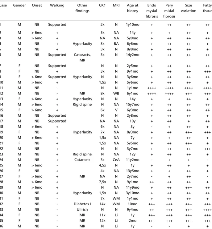
Table 1. Clin ical an d h ist o p at h o lo g ic asp ect s in 32 p at ien t s w it h M P-CM D, o n e w it h Ullrich CM D an d t hree w it h Wa l k e r- Wa r b u rg syn d ro m e.
Case Gender Onset Walking Other CK↑ MRI Age at Endo Pery Size Fatty findings biopsy mysial misial variation tissue
fibrosis fibrosis
1 M NB Supported 2x N 1y10mo + ++ ++ ++
2 M > 6mo + 5x NA 14y + + ++ +
3 M > 6mo + NA NA 5y9mo + ++ ++ ++
4 M NB + Hyperlaxity 3x BA 6y6mo + ++ ++ +
5 M NB + 3x N 8y8mo + ++ ++ +
6 M NB Supported Cataracts, 3x N 14y2mo + ++ ++ ++
MR
7 F NB Supported N N 2y5mo + ++ ++ ++
8 F NB + 3x N 9y1mo + ++ ++ +++
9 F > 6mo Supported Hyperlaxity N N 3y6mo + ++ ++ ++
10 M > 6mo + 1,5x N 5y6mo + ++ ++ +
11 M NB - N N 1y1mo ++++ ++++ ++++ ++++
12 M NB - MR 8x WB 6y1mo ++++ ++++ +++ +++
13 F > 6mo + Hyperlaxity N N 14y + + ++ +
14 M > 6mo + Rigid spine N NA 15y7mo + ++ ++ ++
15 F > 6mo + 6x V 6y3mo + ++ ++ ++
16 M NB Supported N N 2y8mo + ++ ++ +
17 M NB Supported NA NA 10y + ++ + ++
18 M > 6mo + 6x NA 3y - + ++ +
19 F NB + Hyperlaxity 7x NA 8y3mo + ++ +++ +++
20 M > 6mo + 1,5x NA 7y + + ++ +
21 F NB + 1,5x NA 5y5mo + ++ +++ +
22 M NB + N N 3y7mo + ++ ++ +++
23 M NB + Rigid spine N NA 12y + ++ ++ +
24 M NB + Cataracts 3x CeA 11y2mo - + +
-25 M > 6mo - 4,5x N 1y + ++ + +
26 F NB + 4x NA 13y5mo + + ++ +
27 F > 6mo + MR NA N 2y7mo - + ++
-28 M > 6mo + 7,5x N 9y1mo ++ ++ ++ +
29 M > 6mo + N NA 11y9mo + ++ +++ ++
30 M NB + Hyperlaxity 1,5x N 3y10mo + ++ ++ ++
31 F NB - 7x WM 1y1mo + ++ ++ +
32 F NB - Diabetes I 14x WM 10mo +++ +++ +++ +++
33 M NB - Ullrich N N 9y4mo ++ +++ +++ ++++
34 F NB - MR 11x Li 1y +++ +++ +++ +++
35 F NB - MR 12x Li 2mo +++ +++ +++ +++
36 M NB - MR N Li 1y - - + +
a b n o rmalities on brain MRI. CK was normal in one patient, increased until 5 times in 10, above 5 times in 11 patients, and not available in one (Table 2).
The only patient with Ullrich phenotype, confir-med as UCMD by molecular analysis1 8had
symp-toms at birth and never acquired walking. He had normal intelligence and normal CK level.
All the 3 children with WW syndrome (1 male, 2 female) manifested symptoms at birth, were neve r able to walk, presented severe MR and neuronal m i-gration defects (lissencephaly) on brain MRI. The C K level was normal in one patient and increased abo-ve 10 times in the remaining two.
Eight patients (Cases 12, 32, 34, 35, 36, 39, 40 a n d 55) died due to re s p i r a t o ry interc u rrences. Three o f them (Cases 34, 35, and 36) were diagnosed as WW s y n d rome, three (Cases 39, 40, and 55) had MD-CMD and among the two MP-MD-CMD patients who died, one had a severe MR (Case 12) and the other had Type 1 diabetes mellitus (Case 32).
Im m u n o h ist o ch em ical d at a (Tab le 3)
D y s t rophin and dysferlin expression were nor-mal in all patients. Among the 23 MD patients, the
deficiency was partial in 9 (detected with the anti-body 80 Kda in two, 300 Kda in 5, and both in two).
C o n c e rning α- s a rcoglycan (SG) and γ-SG expre s-sion, each one was reduced in 5 (15.6%) of the 32 MP patients, as well as in 18 (78.2%) and 8 (34.7%) of the 23 MD patients, respectively. The δ-SG and
β-SG expression was evaluated in 31 MP and in 23 MD patients and resulted normal in all, except in one MP patient, who presented a reduced expre s-sion of β-SG.
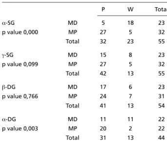
The β-DG reaction resulted weak in 7 patients (22.5%) among 31 with MP and in 6 (26%) among 23 with MD. The a-DG reaction, evaluated in 22 M D and in 22 MP patients, resulted weak in 11 (50%) and in two (10%) patients, re s p e c t i v e l y. Add i t i o n-a l l y, in two pn-atients with WW n-a wen-ak strn-ain wn-as o b-served in each one of β-SG and γ-SG, respectively. The reduction of the expression of a-SG and α -DG showed statistically significant corre l a t i o n ( Table 4) with the diagnosis of MD-CMD (p = 0,00 e p = 0,003, respectively)
Among MP-CMD patients, the reduced expre s-sion of the β-DG had statistically significant corre l a-tion with both, a severe phenotype (walking abili-Tab le 2. Clin ical an d h ist o p at h o lo g ic asp ect s in 23 p at ien t s w it h M D-CM D.
Case Gender Walking CK↑ Age at Endo Pery Size Fatty biopsy mysial misial variation tissue
fibrosis fibrosis
37 F - NA 4y1mo ++++ +++ +++ ++++
38 F Supported NA 1y5mo +++ +++ +++ +++
39 F - 27x 10y7mo ++ +++ +++ ++++
40 M - 14x 9y3mo ++ ++ ++ ++
41 M - 2x 3y +++ +++ +++ +++
42 F - 1.5x 1y4mo +++ +++ +++ ++++
43 F - 6x 1y9mo +++ ++++ +++ +++
44 M - 5x 2y ++++ ++++ +++ ++
45 M - 13x 11mo +++ ++++ +++ +++
46 M - 23x 1y3mo +++ +++ +++ +++
47 F +/lost 5x 10y5mo ++ + ++ +++
48 F - 35x 1y2mo +++ +++ +++ +++
49 M - 12x 2y5mo +++ ++++ +++ ++
50 M - 3x 3y6mo ++ +++ +++ +++
51 M - 4x 1y4mo +++ +++ +++ +++
52 M - 6x 5y5mo +++ ++++ +++ ++
53 F + 9x 1y10mo ++ ++ +++ +
54 F +/lost 4x 4y +++ +++ +++ +++
55 F - 3x 3y8mo +++ ++ +++ ++
56 M - 12x 2y8mo +++ ++ +++ +
57 F - 7x 2y4mo +++ +++ +++ ++
58 M - 1,5x 4y1mo + ++ +++ ++
59 F - 1,5x 4y3mo ++ +++ +++ +++
Tab le 3. Resu lts o f im m u no h ist o chem ical analysis usin g d iff e rent an t ibo d ies o n mu scu lar samp les of 59 CM D p at ient s. Case α2-LM α2-LM Col VI DYS-C α-SG β-SG γ-SG δ-SG α-DG β-DG
80 kd 300 kd
1 P P NA P P P P P P P
2 P P NA P P P P P P P
3 P P P P P P P P NA P
4 P P P P P P P P NA P
5 P P P P P P P P P P
6 P P P P P P P P P P
7 P P NA P P P P P NA P
8 P P NA P P P P P P P
9 P P NA P P P P P P P
10 P P NA P P P P P NA P
11 P P NA P P NA P NA NA NA
12 P P P P W P W P P W
13 P P NA P P P P P P P
14 P P NA P P P W P NA P
15 P P NA P W P P P P P
16 P P NA P P P P P P P
17 P P P P P P W P P P
18 P P P P P P P P P P
19 P P P P P P P P P W
20 P P P P W W P P P W
21 P P NA P W P W P P P
22 P P P P P P P P P P
23 P P P P P P P P P W
24 P P P P P P P P NA P
25 P P NA P P P P P W P
26 P P P P P P P P NA P
27 P P P P P P P P NA P
28 P P P P P P P P NA W
29 P P P P P P P P P P
30 P P P P P P P P P P
31 P P NA P W P W P P W
32 P P NA P P P P P W W
33 P P - P P P P P P P
34 P P P P P W P P P P
35 P P P P W P P P P P
36 P P P P P P P P P P
37 -/+ -/+ P W P P P NA P P
38 - - P W P W P W W W
39 - - P P P P P W P P
40 - W P P P P P P P P
41 - W P W P P P P P P
42 - - P W P P P P P P
43 - - P W P P P P P P
44 - - P W P W P W P P
45 - - P W P W P W W W
46 -/+ - P W P P P P P P
47 - W P P P P P P P P
48 - - P W P W P W P P
49 -/+ - P W P W P P W W
50 -/+ W P W P W P W P P
51 - - P W P P P P P P
52 - W P W P W P P W W
53 - - P P P P P W P P
54 - - P W P W P P W W
55 - - P P P P P P P P
56 - - P W P P P W P P
57 - - P W P P P W P P
58 - - P W P P P W W W
59 -/+ -/+ P W P P P W P P
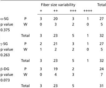
ty) (p = 0.017) and a marked fibrosis on muscle biopsy (p = 0.035) (Tables 5 and 6). The re d u c e d e x p ression of the α-SG, γ-SG and β-DG had no sta-tistically significant correlation with fat infiltra-tion (p = 0.874, p = 0.939 and p = 0.051, re s p e c-tively) and fiber size variability (p = 0.375, p = 0.263 e p = 0.073, respectively) on muscle biopsy (Ta b l e s 7 and 8).
DISCUSSION
R e g a rding the MD patients, our aim was to eva-luate the influence of the primary deficiency of me-rosin on the expression of the main proteins of the DGA complex, comparing their expression with that observed in MP patients. In the MP patients o u r intention was to select possible clinical particula-rities according to the protein expression.
Tab le 4. St at ist i cal co rrelat io n b et w een α-SG, γ-SG, β-DG, α- D G e x p r essi o n an d p r esen ce o r ab sen ce o f m e ro si n in p at i en t s’ m u scu lar b io p sy.
Crosstabulation count α-SG, γ-SG β-DG,α-DG X merosin status
P W Total
α-SG MD 5 18 23
p value 0,000 MP 27 5 32
Total 32 23 55
γ-SG MD 15 8 23
p value 0,099 MP 27 5 32
Total 42 13 55
β-DG MD 17 6 23
p value 0,766 MP 24 7 31
Total 41 13 54
α-DG MD 11 11 22
p value 0,003 MP 20 2 22
Total 31 13 44
Significance: 0.05 (α=5%), SG, sarcoglycan; DG, dystroglycan; MD, mero-sin deficient; MP; meromero-sin positive; P, positive; W, weak.
Tab le 5. St at ist ical co rrelat io n b et w een α-SG, γ-SG βDG expre s -sio n an d w alk in g h ab ilit y in M P p at ien t s.
C rosstabulation count: α-SG, γ-SG β-DG expression X walking hability
Walking with Independent No walking support walking
α-SG P 6 18 3 27
p value W 0 3 2 5
0.183
Total 6 21 5 32
γ-SG P 5 19 3 27
p value W 1 2 2 5
0.249
Total 6 21 5 32
β-DG P 6 17 1 24
p value W 0 4 3 7
0.017
Total 6 21 4 31
Significance: 0,05 (α=5%); SG, sarcoglycan; DG, dystroglycan; MP, mero s i n positive.
Table 6. St at ist ical co rrelat io n b et w een α-SG, γ-SG, βDG exp re s -sio n an d m o d erat e en d o -p erim ysial f ib ro sis in M P p at ien t s C rosstabulation count: α-SG, γ-SG, β-DG expression X moder-ate endo-perimysial fibrosis
Moderate fibrosis Total 0 .5 1.0 1.5 2.0 3.0 4.0
α-SG P 0 3 3 18 1 1 1 27
p value W 0 0 1 3 0 0 1 5
0.698
Total 0 3 4 21 1 1 2 32
γ-SG P 0 3 4 17 1 1 1 27
p value W 0 0 0 4 0 0 1 5
0.01
Total 0 3 4 21 1 1 2 32
β‚-DG P 0 3 3 18 0 0 0 24
p value W 0 0 1 3 1 1 1 7
0.035
Total 0 3 4 21 1 1 1 31
Significance: 0,05 (α=5%), SG, sarcoglycan; DG, dystroglycan; MP, mero s i n positive; P, present; W, weak
Table 7. St at ist ical co rrelat io n b et w een α-SG, γ-SG‚ βDG exp re s -sio n an d f at t y in f ilt rat io n in M P p at ien t s.
Crosstabulation count: α-SG, γ-SG, β-DG expression X fatty infiltration
Fatty infiltration Total
- + ++ +++ ++++
α-SG P 2 11 9 4 1 27
p value W 0 3 1 1 0 5
0.874
Total 2 14 10 5 1 32
γ-SG P 2 12 8 4 1 27
p value W 0 2 2 1 0 5
0.939
Total 2 14 10 5 1 32
β-DG P 2 10 10 2 24
p value W 0 4 0 3 7
0.051
Total 2 14 10 5 31
M erosin - In 9 patients among 23 merosin defi-ciency was partial. All manifested the abnorm a l white matter on brain neuroimaging and clinical s e-verity that characterize the MD patients with total absence of mero s i n1 9 - 2 1. Although patients with
CMD due to mutations in other genes, part i c u l a r-ly those related with abnormal gr-lycosilation of α -d y s t roglycan can present a secon-dary partially - defi-cient merosin3,13,14, until the moment the
associa-tion between widespread white matter changes a n d p a rtial merosin deficiency has only been described in patients with mutations in the LAMA2 gene. In DMC1C (FKRPgene)7, the partial deficiency of
me-rosin is secondary to the marked deficiency of α -d y s t roglycan an-d is not associate-d with brain white matter changes. In the muscle-eye-brain forms of CMD, like Fukuyama and MEB, a secondary defic i e n-cy of merosin may occur; however, such forms are easily distinguished from classic MD-CMD with par-tial deficiency by their characteristics ocular and brain changes1-3.
The adequate identification of the partial defi-ciency of merosin depends on the use of diff e re n t antibodies that recognize diff e rent fragments of the pro t e i n2 2 , 2 3. The most useful antibodies are
tho-se reacting to the merosin fragments of 80 and 300 k D a2 3. In two of our 9 patients with partial
deficien-c y, we defined that the defideficien-ciendeficien-cy was partial only after the utilization of the antibody against 300kDa, as with the antibody against 80 kDa, the mero s i n
seemed totally absent. This result, indicating the bet-ter sensibility of the antibody against 300 kDa, is in a g reement with other studies2 4 , 2 5. The possibility that
not yet identified subtypes of CMD may also pre s-ent secondary partial deficiency of merosin re i n-f o rces the need on-f a caren-ful investigation on-f mero s i n status in the muscular biopsy of CMD patients.
Even presenting a total deficiency of merosin, two CMD patients (Cases 53 and 54) acquired inde-pendent walking, and the youngest of them still maintains it. This finding is not re p o rted in the ca-suistics about CMD patients with total absence of m e ro s i n2 0 , 2 6. One of these three totally MD patients
(Case 54) deserves a special comment, as her abili-ty for walking independently, that had been lost a t 4 years of age, was re c o v e red and maintained for two more years under steroid therapy. In conclus i o n in our MD patients, the ability to walk independ-ently was not fully related with the partial or total m e rosin deficiency and the degree of clinical severi-ty was not related to any particular immunohisto-chemical finding.
D y s t rop h in – The expression of dystrophin in the patients’ samples was normal in all, independ-ently of the merosin status and of the degree of histopathological dystrophic changes. Therefore, the severe clinical and muscular involvement ob-s e rved in CMD patientob-s iob-s apparently not due to an abnormal interaction between laminin and dys-t rophin. However, idys-t musdys-t be emphasized dys-thadys-t we evaluated the dystrophin expression by using anti-bodies against the C-terminal domain only and n o t against the N-terminal or the central region do-mains, that could interf e re with the correct inter-p retation of the interaction between laminin and all the human dystrophin domains. In fact, Fard e a u et al.1 9, using antibodies against the central re g i o n
and N-terminal dystrophin dominium, demonstrat-ed that the primary merosin deficiency can induce a secondary dystrophin deficiency.
Sarco g lycan co m p lex – The correct association among the α, β, γ, d and εs a rcoglycans within the s a rcoglycan complex has been widely discussed and t h e re is not a perfect agreement among diff e re n t re s e a rc h e r s2 7 - 2 9. More studies about the assembly
and interactions of sarc o g l y c a n / s a rcospan complex will contribute to better clarify the muscle function. In our CMD patients, the normal βand δ- s a rc o g l y-can expression agrees with the literature3 0. A
statis-tically significant decrease of α- s a rcoglycan expre s-sion was observed in our MD patients (Table 4), pro-bably indicating a stronger interaction between t h a t Table 8. St at ist ical correlat ion b et w een α-SG, γ-SG, βDG exp re s
-sio n an d f ib er size variab ilit y in M P p at ien t s.
Crosstabulation count: α-SG, γ-SG, β-DG expression X fiber size variability
Fiber size variability Total
+ ++ +++ ++++
α-SG P 3 20 3 1 27
p value W 0 3 2 0 5
0.375
Total 3 23 5 1 32
γ-SG P 2 21 3 1 27
p value W 1 2 2 0 5
0.263
Total 3 23 5 1 32
β-DG P 3 19 2 24
p value W 0 4 3 7
0.073
Total 3 23 5 31
p rotein and merosin, through their link with β- d y s-t roglycan. In any case, s-the greas-ter dyss-trophic chan-ges seen in MD patients could also interf e re with the a-sarcoglycan expression. We also found, a de-c rease of γ- s a rcoglycan expression, mainly in the MD patients (34.7%), but without statistical signifi-cance (Table 4). This finding could also suggest a closer connection between γ- s a rcoglycan and me-rosin; however, both MD and MP patients, who pre-sented severe dystrophic pattern, also prepre-sented a g reat, but not clinically significant reduction of γ -s a rcoglycan (Table 4). There f o re, future -studie-s about the relation between sarcoglycan expre s s i o n and dystrophic pattern would be necessary.
In relation to WW, the sarcoglycans and me-rosin expression is variable. In general there is a s e c o n d a ry deficiency in the expression of mero s i n and α- s a rc o g l y c a n3 1. In three patients we observ e d
n o rmal merosin and a reduction of β-SG in one a n d of γ-SG in another patient.
D y s t roglycan co m p lex – The dystroglycan gene codes for a proteic precursor that processed by a p rotease results in αe b-DG3 2 , 3 3. The β-DG has a t r a n s
membrane domain and is directly connected to the cystein portion of the dystrophin by the C-ter-m i n a l3 2. The αand β-DG interact directly in the
ex-tracellular matrix.
Although a normal expression of β-DG in both MD and MP patients has been re p o rt e d3 4, as the
β-DG interacts with the merosin through the α- D G connection, it could be expected that in the pri-m a ry absence of the pri-merosin, the β-DG could also be reduced. In fact, we observed such reduction i n some patients, but the percent of MP and MD pa-tients presenting reduction in the β-DG staining w a s almost the same (22.5% and 26%, re s p e c t i v e l y ) . Besides, in the MP patients the β-DG reduction was statistically correlated to the intensity of the dystro-phic pattern (Table 6). This seems to suggest that the partial reduction of the β-DG in CMD patients is probably more related to the dystrophic pattern and other correlated factors than to the absence o f m e rosin. Finally, the possibility that mutations in genes not yet defined encoding other proteins c o u l d also justify this secondary β-DG reduction should be stressed.
As demonstrated in other studies, the α-DG ex-p ression can be variable in MD ex-patients3 0. In our c
a-ses, the α-DG was reduced in 50% of the MD pa-tients and in only 10% of the MP papa-tients. This dif-f e rence was statistically signidif-ficant (Table 4) and w a s
dependent on the closer relation between mero s i n and α-DG, already emphasized by others7,30,35.
A b n o rmalities in the α-DG glicosilation have been considered important in the pathogenesis of many forms of CMD: Fukuyama, MEB, WW, 1C and 1 D3,7,8,13, 30,35-37. In most of these cases a secondary d
e-ficiency in merosin has been detected. We only fo-und two MP patients with reduced expression of
α-DG and their clinical phenotype did not corre-spond to the clinical description re p o rted in the new CMD forms with α-DG glicosilation defects7 , 8.
One of these patients has a marked involvement of the cervical musculature (Case 25) and the othe r (Case 32) presented focal changes in brain white matter and Type 1 diabetes mellitus. Both had nor-mal expression of the merosin. A phenotype very s i-milar to that observed in Case 32, except by the la-ck of Type 1 diabetes mellitus, was found in Case 3 1 ; however this patient had normal α-DG expre s s i o n . It is important to emphasize that those cases with brain white matter changes, like our Cases 31 and 32, would need the study of the LAMA2 gene for ruling out any mutation that according to Tezak e t a l .3 8could lead to retention of large fragments of
m e rosin. However, in the routine attendance the homogeneously clinical severity and the wide-s p read brain white matter changewide-s typical of CMD-1A, in addition to the low availability of molecu-lar studies of the LAMA2 gene, lead to the diagno-sis of MD-CMD.
The phenotype of our WW patients was mar-kedly homogeneous and similar to that reported in the literature3 9. A neonatal severe involvement
of muscles and CNS (type II lysencephaly) leading t o death in the first two years of life was the rule. Re-cently mutations in the O-mannosyltransferase 1 have been identified4 0, suggesting that the
O-ma-nose glicosilation would be implicated in the neu-ronal migration process at least in one part of the WW patients. Both α-DG and merosin expre s s i o n , w e re found to be reduced in the CMD with glyco-silation defects7 , 3 5 , 4 1; however we did not find the
f o rmer abnormalities, perhaps because not all the WW cases are associated to O-mannosyltransferase gene (POMT1)40.
Co llag en VI – We tested the collagen VI expre s-sion in 7 MP patients with marked distal joints hy-perlaxity and variable degrees of muscular involve-ment. In two, the collagen expression was absent and one of them was later diagnosed as Bethlem myopathy and taken off from the present series1 8.
ca-suistics who was diagnosed as Ullrich CMD by mo-lecular analysis1 8. The Ullrich phenotype,
charac-terized by distal joints hyperlaxity associated with p roximal joints contractures is clinically and geneti-cally hetero g e n e o u s4 2 , 4 3and corresponds to Ullrich
CMD, i.e. associated to mutations in one of the t h re e collagen VI genes in about 40% of the patients4 2.
Clo sing remarks – Unlike MD patients who pre s-ent homogeneous clinical severity and neuro i m a g-ing changes, our MP patients had a variable motor i m p a i rment and in 6 of them we found CNS in-volvement, which we had in part already discussed in a previous work4 4. The CNS involvement was m
a-nifested by MR (Cases 6, 11, 12, 24), brain and cere-bellar atrophy (Cases 4 and 12, respectively), as w e l l as by focal and non specific changes of brain white matter (Cases 12, 31, 32). As has already been s t re s-sed, the finding of focal changes of the brain white matter in two MP patients (Cases 31, 32) not asso-ciated to other neurological abnormalities would suggest the need for molecular analysis of the LAMA2 gene for ruling out MD-CMD. However, the homogeneous and typical clinical/neuro i m a g-ing picture, as well as the use of two antibodies, against 80 and 300 kDa fragments of mero s i n , seems sufficient for defining the diagnosis of MD or MP-CMD. Two MP patients (Cases 6 and 24) had cataracts in addition to MR and this association had also been previously re p o rted by us4 5.
Concer-ning the MP patient with Type I diabetes mellitus (Case 32, already deceased) it is difficult to define whether that association was fortuitous or geneti-cally determined. There f o re, we did not find in the MP patients who manifested the above men-tioned uncommon findings of CNS and/or ocular involvement, as well as of type I diabetes mellitus, any specific change in the expression of any of the p roteins from the DGA complex. Still within the MP-CMD subgroup, two patients had a rigid spine phenotype (Cases 14 and 23), and would need ge-netic confirmation to eventually be included in the subgroup of CMD with rigid spine linked to c h romosome 1p6. Although the majority of our
MP patients present a less severe phenotype and maintain the capacity to walk, two our patients ( C a-ses 11 and 12) presented an intense motor and re s-piratory impairment, similar to those observed in the MD group. In these patients, the immunohisto-chemical analysis of the proteins from the DGA complex did not detect any abnormality.
In conclusion, although our study did not char-acterize any remarkable
clinical-immunohistochem-ical correlation, we consider that an immunohisto-chemical analysis as complete as possible, should be perf o rmed, for establishing the diff e rential d i a g-nosis with other forms of children myopathies, w h i l e we await more accessible molecular methods. In addition, the analysis of the immunohistochemi-cal expression of the proteins from muscle and ex-tracellular matrix with a number of already availa-ble antibodies, is an easy pro c e d u re, that can con-tribute for a better understanding of the pathoge-nesis of the dystrophic muscle, as well as for select-ing a particular molecular study.
Acknowledgements – We are grateful to Dr. Cars-ten G. Bonnemann for the molecular diagnosis of Case 33 to Dr. Stephan Kroger who kindly donated the anti-body to α-DG.
REFERENCES
1. Tomé F. The saga of congenital muscular dystro p h y. Neuro p e d i a t r i c s 1999;30:55-65.
2. Voit T. Congenital muscular dystrophies: 1997 update. Brain Dev 1998; 20:65-74.
3. Muntoni F, Voit T. The congenital muscular dystrophies in 2004: a centu-ry of exciting progress. Neuromuscul Disord 2004;14:635-649. 4. H e l b l i n g - L e c re rc A, Zhang X, Topaloglu H, et al. Mutations in the
lami-nin α2-chain gene (LAMA2) cause merosin-deficient congenital muscu-lar dystrophy. Nature Gen 1995;11:216-218.
5. Camacho Vanegas O, Bertini E, et al. Ullrich scleroatonic muscular dys-t rophy is caused by recessive mudys-tadys-tions in collagen dys-type VI. Proc Nadys-tl Acad Sci USA 2001;98:7516-7521.
6. Moghadaszadeh B, Petit N, Jaillard C, et al. Mutations in SEPN1 cause congenital muscular dystrophy with spinal rigidity and restrictive re s-piratory syndrome. Nat Genet 2001;29:17-18.
7. B rockington M, Yuva Y, Prandini P, et al. Mutations in the fukutin-re l a t-ed protein gene (FKRP) identify limb girdle muscular dystrophy 2I as a milder allelic variant of congenital muscular dystrophy MDC1C. Hum Mol Genet 2001;10:2851-2859.
8. Longman C, Brockington M, To relli S, et al. Mutations in the human LARGE gene cause MDC1D, a novel form of congenital muscular dys-trophy with severe mental retardation and abnormal glycosylation of alpha-DG. Hum Mol Genet 2003;12:2853-2861.
9. Toda T, Segawa M, Nomura Y, et al. Localization of a gene for Fu-kuyama type congenital muscular dystrophy to chromosome 9q31-33. Nature Genet 1993;5:283-286.
10. Yoshida A, Kobayashi K, Manya H, et al. Muscular dystrophy and neu-ronal migration disorder caused by mutations in a glycosyltransferase, POMGnT1. Dev Cell 2001;1:717-724.
11. B e l t r a n - Va l e ro De Bernabe D, Currier S, Steinbrecher A, et al. Mutations in the O-mannosyltransferase gene POMT1 give rise to the severe neuro-nal migration disorder Walker-Warburg syndrome. Am J Hum Genet 2002;71:1033-1043.
12. B rockington M, Sewry CA, Herrmann R, et al. Assignment of a form of congenital muscular dystrophy with secondary merosin deficiency to chromosome 1q42. Am J Hum Genet 2000;66:428-435.
13. Muntoni F. Journey into muscular dystrophies caused by abnormal glycosylation. Acta Myol 2004;23:79-84.
14. Muntoni F, Va l e ro de Bernabe B, Bittner R, et al. 114th ENMC Inter-national Workshop on Congenital Muscular Dystrophy (CMD). Neuro-musc Disord 2003;13:579-588.
15. Vainzof M, Zubrzycka-Gaarn EE, Rapaport MR, et al. Immuno-f l u o rescence dystrophin study in Duchenne dystrophy through the concomitant use of two antibodies directed against the carboxi-termi-nal and the amino-termicarboxi-termi-nal region of the protein. J Neurol Sci 1991; 101:141-147.
17. Agresti A. Categorial data analysis. New York: Wiley, 1990:588. 18. Pan TC, Zhang RZ, Sudano DG, Marie SK, Bonnemann CG, Chu ML.
New molecular mechanism for Ullrich congenital muscular dystro p h y : a heterozygous in-frame deletion in the COL6A1 gene causes a severe phenotype. Am J Hum Genet 2003;73:355-369.
1 9 . F a rdeau M, Tomé FMS, Helbling-Lecre rc A, et al. Dystrophie musculai-re congénitale avec déficience en mérosine: analyse clinique, histopatho-logique, immunocytochimique et génétique. Rev Neurol 1996;152:11-19. 20. Jones KJ, Morgan G, Johnston H, et al. The expanding phenotype of laminin alpha2 chain (merosin) abnormalities: case series and review. J Med Genet 2001;38:649-657.
21. Reed UC, Marie SK, Vainzof M, et al. Congenital muscular dystrophy with cerebral white matter hypodensity: correlation of clinical feature s and merosin deficiency. Brain Dev 1996;18:53-58.
22. Cohn RD, Herrmann R, Sorokin L, Wewer UM, Voit T. Laminin α2 chain-deficient congenital muscular dystrophy: variable epitope expre s-sion in severe and mild cases. Neurology 1998;5:94-101.
2 3 . Sewry CA, Uziyel Y, To relli S, et al. Diff e rential labelling of laminin al-pha 2 in muscle and neural tissue of dy/dy mice: are there isoforms of the laminin alpha 2 chain? Neuropathol Appl Neurobiol 1998;24:66-72. 24. Sewry CA, Philpot J, Mahony D, Wilson LA, Muntoni F, Dubowitz V. E x p ression of laminin subunits in congenital muscular dystro p h y. Neuromusc Disord 1995;5:307-316.
25. Morandi L, Di Blasi C, Farina L, et al. Clinical correlations in 16 patients with total or partial laminin a2 deficiency characterized using antibodi-es against 2 fragments of the protein. Arch Neurol 1999;56:209-215. 26. Allamand V, Guicheney P. Merosin-deficient congenital muscular
dys-t ro p h y, audys-tosomal recessive (MDC1A, MIM#156225, LAMA2 gene cod-ing for alpha2 chain of laminin). Eur J Hum Genet 2002;10:91-94. 27. Blake DJ, Weir A, Newey SE, Davies KE. Function and genetics of
dys-trophin and dysdys-trophin-related proteins in muscle. Physiol Rev 2002; 82:291-329.
28. Vainzof M, Passos-Bueno MR, Canovas M, et al. The sarcoglycan com-plex in the six autosomal recessive limb-girdle muscular dystrophies. Hum Mol Genet 1996;5:1963-1969.
29. Shi W, Chen Z, Schottenfeld J, Stahl RC, Kunkel LM, Chan YM. Specific assembly pathway of sarcoglycans is dependent on beta- and delta-sarcoglycan. Muscle Nerve 2004;29:409-419.
30. Muntoni F, Bertini E, Bonnemann C, et al. 98th ENMC International Workshop on Congenital Muscular Dystrophy (CMD), 7th Workshop of the International Consortium on CMD, 2nd Workshop of the MYO CLUSTER project GENRE. Neuromusc Disord 2002;12:889-896. 31. Villanova M, Sabatelli P, He Y, et al. Immunofluorescence study of a
muscle biopsy from a 1-year-old patient with Wa l k e r- Wa r b u rg syndro-me. Acta Neuropathol (Berl) 1998;96:651-654.
3 2 . I b r a g h i m o v - B e s k rovnaya O, Milatovich A, Ozcelik T, et al. Human DG: skeletal muscle cDNA, genomic stru c t u re, origin of tissue specific iso-forms and chromosomal localization. Hum Mol Genet 1993;2:1651-1657. 33. I b r a g h i m o v - B e s k rovnaya O, Ervasti JM, Leveille CJ, Slaughter CA, Ser-nett SW, Campbell KP. Primary stru c t u re of dystrophin-associated gly-coproteins linking dystrophin to extracellular matrix. Nature 1992; 355:696-702.
34. ter Laak HJ, Leyten QH, Gabreels FJ, Kuppen H, Renier WO, Sengers RC.Laminin-alpha2 (merosin), beta-DG, alpha-sarcoglycan (adhalin), and dystrophin expression in congenital muscular dystrophies: an im-munohistochemical study. Clin Neurol Neurosurg 1998;100:5-10. 35. Muntoni F, Brockington M, Blake DJ, To relli S, Brown SC. Defective
glycosylation in muscular dystrophy. Lancet 2002;360:1419-1421. 36. Michele DE, Barresi R, Kanagawa M, et al. Post-translational disru
p-tion of DG-ligand interacp-tions in congenital muscular dystrophies. Na-ture 2002;418:417-422.
37. Endo T, Toda T. Glycosylation in congenital muscular dystrophies. Biol Pharm Bull 2003;26:1641-1647.
38. Tezak Z, Prandini P, Boscaro M, et al. Clinical and molecular study in congenital muscular dystrophy with partial laminin alpha 2 (LAMA2) deficiency. Hum Mutat 2003;21:103-111.
39. Dobyns WB, Pagon RA, A r m s t rong D, et al. Diagnostic criteria for Wa l-ker-Warburg syndrome. Am J Med Genet 1989;32:195-210.
40. B e l t r a n - Va l e ro De Bernabe D, Currier S, Steinbrecher A, et al. Mutations in the O-mannosyltransferase gene POMT1 give rise to the severe neuro-nal migration disorder Walker-Warburg syndrome. Am J Hum Genet 2002;71:1033-1043.
41. J i m e n e z - M a l l e b rera C, To relli S, Brown SC, et al. Profound skeletal muscle depletion of alpha-DG in Wa l k e r- Wa r b u rg syndrome. Eur J Pae-diatr Neurol 2003;7:129-137.
42. M e rcuri E, Yuva Y, Brown SC, et al. Collagen VI involvement in Ullrich s y n d rome: a clinical, genetic, and immunohistochemical study. Neuro l o-gy 2002;58:1354-1359.
43. Demir E, Sabatelli P, Allamand V, et al. Mutations in COL6A3 cause se-v e re and mild phenotypes of Ullrich congenital muscular dystro p h y. Am J Hum Genet 2002;70:1446-1458.
44. Reed UC, Marie SK, Vainzof M, et al. Heterogeneity of classic congeni-tal muscular dystrophy with involvement of the central nervous sys-tem: report of five atypical cases. J Child Neurol 2000;15:172-178. 45. Reed UC, Tsanaclis ANC, Vainzof M, et al. Merosin-positive