C A S E R E P O R T S UDC: 616.831-07/-08 DOI: 10.2298/VSP140529053K
Posterior reversible encephalopathy syndrome – A case report
Sindrom posteriorne reverzibilne encefalopatije
Dejan Kostić*, Biljana Brkić-Georgievski†
, Aleksandar Jovanovski*, Smiljana Kostić‡, Dražen Ivetić§, Leposava Sekulović*||
*Institute of Radiology, ‡Clinic for Neurology, §Clinic for Neurosurgery, Military Medical Academy, Belgrade, Serbia; †Special Hospital for Cerebrovascular Diseases „Sveti Sava“, Belgrade, Serbia; ||Faculty of Medicine of the Military Medical Academy,
University of Defence, Belgrade, Serbia
Abstract
Introduction. Posterior reversible encephalopathy syn-drome (PRES) is characterized by the following symptoms: seizures, impaired consciousness and/or vision, vomiting, nausea, and focal neurological signs. Diagnostic imaging in-cludes examination by magnetic resonance (MR) and com-puted tomography (CT), where brain edema is visualized bi-laterally and symmetrically, predominantly posteriorly, parie-tally, and occipitally. Case report. We presented a 73-year-old patient with the years-long medical history of hiperten-sion and renal insufficiency, who developed PRES with the symptomatology of the rear cranium. CT and MR verified changes in the white matter involving all lobes on both sides of the brain. After a two-week treatment (antihypertensive, hypolipemic and rehydration therapy) clinical improvement with no complications occurred, with complete resolution of changes in the white matter observed on CT and MR.
Conclusion. PRES is a reversible syndrome in which the symptoms withdraw after several days to several weeks if early diagnosis is made and appropriate treatment started without delay.
Key words:
brain diseases; syndrome; diagnosis; treatment outcome.
Apstrakt
Uvod. Sindrom posteriorne reverzibilne encefalopatije (PRES) karakterišu sledeći simptomi: epi napadi, poremećaji svesti i/ili vida, povraćanje, mučnina i fokalni neurološki znaci. Dijagnostičko snimanje obuhvata pregled magnetnom rezonancijom (MR) i kompjuterizovanom tomografijom (CT) gde se moždani edem vizuelizuje bilateralno i simetrič -no, preovlađuje posteriorno, parijetalno i okcipitalno. Pri-kaz bolesnika. Prikazali smo bolesnika starog 73 godine, dugogodišnjeg hipertoničara i bubrežnog bolesnika kod ko-ga se razvio PRES sa simptomatologijom zadnje lobanjaske jame i verifikovanim promenama moždane bele mase na CT i MR, obostrano na svim režnjevima mozga. Nakon dve ne-delje lečenja (antihipertenzivi, hipolipemici i rehidraciona te-rapija) nastupilo je kliničko poboljšanje bez ikakvih kompli-kacija i sa kompletnom rezolucijom promena bele moždane mase viđenim na CT i MR. Zaključak. PRES je reverzibilni sindrom čiji simptomi se povlače za nekoliko dana do neko-liko nedelja, ukoneko-liko se rano postavi dijagnoza i
odgovaraju-će lečenje počne bez odlaganja.
Ključne reči:
mozak, bolesti; sindrom; dijagnoza; lečenje, ishod.
Introduction
Posterior reversible encephalopathy syndrome (PRES) is characterized by the following symptoms: epileptic sei-zures, consciousness impairment, visual abnormalities, nau-sea, vomiting and focal neurological signs. It was first de-scribed by Hinchey et al. 1 in 1996. based on the study of 15 cases. Since then this syndrome has been also designated by reversible posterior leukoencephalopathy syndrome, reversi-ble posterior cerebral edema syndrome and reversireversi-ble occipi-tal parieoccipi-tal encephalopathy 2–5.
Studies have shown that various conditions can lead to development of PRES, hypertension, autoimmune diseases, toxic agents, sepsis, preeclampsia/eclampsia, kidney diseases being among them 1, 6. Regardless to this heterogeneity main pathophysiological mechanism which leads to development of this syndrome is cerebral vasogenic edema which occurs as a result of abnormality in blood flow through the brain – cerebral blood flow (CBF) 7.
predominantly in posterior parietally and occipitally, but fron-tal and temporal lobes can also be affected, as well as basal ganglia, brainstem and cerebellum. Also, alongside with the white matter, cortical gray matter can be affected as well 8.
The diagnosis of PRES is complicated, since CT results are often normal or non-specific, and MR scanners not avail-able in many centers. Standard MR sequences, which include T1-weighted images (T1W), T2-weighted images (T2W), fluid attenuated inversion recovery (FLAIR) and diffusion-weighted imaging (DWI) with apparent diffusion coefficient (ADC) map, as well as contrast T1W, are sufficient for the diagnosis.
The treatment should primarily consist of correction of the underlaying causes which led to neurological symptoma-tology, and then symptomatic measures should be taken. Some patients may develop severe manifestations of PRES, such as coma or status epilepticus, which require intensive care unit (ICU) admission 9, 10.
PRES is a reversible syndrome, but in a small number of patients neurological deficit is permanent.
Death occurs in up to 15% of cases due to acute hem-orrhage and ischemia 7, 11–14.
Case report
A 73-year-old patient was complaining of headache, in-stability while walking and loss of balance which lasted for a few days. The patient had a history of hypertension and chronic renal insufficiency. Arterial blood pressure reached 160/90 mmHg. Clinical presentation at admittance was
dominated by symptomatology of posterior fossa with
dis-creet rightfaciobrachial hemiparesis. Initially, CT scanning without application of contrast was performed showing the
presence of both-sided hypodensic zones, frontally more
prominent on the left as well as parietally right subcortically, which resembled mostly vasogenic edema (Figure 1).
Fig. 1 – Native computed tomography (CT) scan showing hypodense zones on both sides of the frontal and parietal
lobe, more pronounced on the right.
After CT, MR scanning was also performed in T1W, T2W, FLAIR, T2*, DWI with ADC map, as well as postcon-trast T1W and 3D T1 FSPGR.
MR spectroscopy using 2D multivoxel SE 144 and sin-gle voxel SE 144 and SE 35 sequences with positioning of thevolume of interest occipitoparietally was also performed.
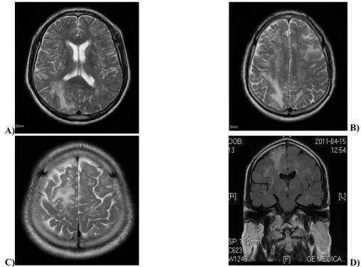
This scanning showed cortico-subcortical temporopolar on both sides, parieto-occipital and posterior parietal both sidedly more dominant on the right, pachy, unsharply bor-dered, partly confluent lesions hyperintense in T2W and FLAIR sequence with mild compressive effect on occipitial horn of the right lateral lobe, without postcontrastive en-hancment of signal intensity (Figure 2).
A) B)
C) D)
Fig. 2 – A) T2W axial tomogram showing the zones of vasogenic edema of cortical/subcortical localization in the parietal lobes, more pronounced on the right; B) T2W axial tomogram showing the asymmetric changes frontally on the left, parietally bilaterally, more pronounced on the right; C) T2W axial tomogram showing the changes frontally on the right;
There were no signs of diffusion restriction. In DWI se-quence occipitoparietally on the right there were no changes in signal intensity, but on ADC map hypointensity of the signal was observed, implicating the existence of vasogenic edema.
Based on the CT and MR scanning results the patient was referred to MR spectroscopy. Using 2D multivoxel spectroscopy the obtained spectres showed normal or slightly higher values of choline for the area in question (Cho/Cr), and NAA was slightly lower. By single voxel spectroscopy spectres of low apsolute concentrations were obtained, ratio Cho/Cr did not significantly deviate from normal values for the area in question. NAA was lower. Resonance lines of lip-ides and lactates were not noticed. Diffusion Tensor Imaging (DTI) reveled that in the area of lesion occipitoparietally on the right fractional anizotrophy was lower.
After diagnosing the patient was regularly treated with antihypertensive, antilypemic and rehydration therapy and was discharged two weeks later with arterial tension 140/80 mmHg and normalized neurological findings.
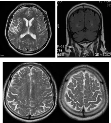
Control MR scanning, two months after the initial one, showed complete regression of T2W and FLAIR hyperin-tense changes which had previously affected cortex and sub-cortical white matter (Figure 3).
Discussion
PRES has been reported in patients aged 4 to 90 years, although most cases occur in young to middle-aged adults. Mechanical ventilation is required in 35% to 40% of patients 12, 13
. The average hospital stay is 20 days, and the mortality rate up to 15% 12, 13 . Numerous conditions may lead to the
syndrome: acute hypertension, renal function disorder, im-munosuppressive therapies being some of them 1. Other pos-sible causes are eclampsia, transplantation, chemotherapy 15, systemic infection, shock 16 and insect bites 17, 18.
Until recently it was thought that PRES tipically affects the white matter, simetrically, predominantly in occipital and posterior parietal areas. Sporadically, changes were de-scribed in frontal and temporal lobes, basal ganglia, brain stem and cerebellum, as well as in cortical grey matter 6.
Recent cohort studies showed that changes are asym-metric in 3–15% of cases, that occipital lobes are affected in 99%, and parietal lobes in 67% to 99% of cases. Changes are less often detected in frontal (68–89%) and temporal (40– 83%) 13, 16, 19, 20 lobes of cerebral parenchyma. The brainstem is affected in 13–58%, cerebellum in 30–58%, and basal gan-glia in 12–34% of cases 16, 19, 20. Till now changes are least often detected in the grey matter, i.e. in 10–44 % of cases 11, 16, 19, 20
. In the presented case the changes were asymmetrical and affecting frontal, temporal, parietal and occipital lobes, cortical grey matter, as well as subcortical and deep cerebel-lar white matter.
The main pathophysiological mechanism which leads to this syndrome is cerebral vasogenic edema. Some authors think that the occurence of edema is the consequence of a
disorder in cerebral autoregulation of blood flow through the brain. Other authors think that it is caused by endothelium dysfunction with cerebral hypoperfusion 7.
Fig. 3 – Control magnetic resonance imaging (MRI) after two month. T2W axial tomogram and coronal T2W tomogram showed almost complete regression of the lesion.
CT scan is often normal or non-specific, as in our case. Topographic regionssuggest the diagnosis of PRES 20.
intensity signal. Changes in PRES are best seen in FLAIR sequence, as hyperintense zones cortically and/or subcorti-cally 8 and such changes are more often frontally local-ized compared to the posterior presentation using this technique21.
Signal intensity on the DWI sequence is normal, but it is higher on ADC 22.
Fractional isotrophy shows zones of decrease, which indicates a mild damage of brain paths which can be re-versible and it is in accordance with a mild decrease of the values of N-acetylaspartate (NAA) obtained by MR spec-troscopy 23, 24.
MR spectroscopy is not superior to conventional MR sequences, but it helps us to rule out other etiology of changes. Signal intensity increase after application of con-trast agents is seen in about one half of cases 19.
In recent years susceptibility-weighted imaging (SWI) is used for detecting microhemorrhagies. This examination shows a higher occurrence rate of microhemorrhage in this syndrome which is associated with vasculopathy 25.
All those characteristics of lesions were also seen in our case.
Many conditions may resemble PRES. The differential diagnosis of findings obtained by the examination of the brain using MR imaging in patients with abnormalities of cerebellar white matter includes the following 5, 26: acute dis-seminated encephalomyelitis (ADEM) in which, unlike the PRES, lesions in T1 can be hypo- to isointense, in DWI the signal is without changes, and on ADC map an increase of signal intensity is seen, changes being contrast uptaken 27; in progressive multifocal leukoencephalopathy (PML) in DWI new and active lesions show hyperintensity of signal, the old ones hypointensity, while in ADC map new and active le-sions are hypointense and the old ones are hyperintense 28; in cerebral autosomal dominant arteriopathy with subcortical infarcts and leukoencephalopathy (CADASIL) in DWI and ADC sequences the restriction of diffusion always occurs,
and the changes after iv application of contrast do not show signs of signal hyperintensity, changes simetrically affect basal ganglia and white matter widening the perivascular space 29; in acute ischemia a restriction of diffusion does exist (signal hyperintensity in DWI, signal hypointensity in ADC map) without contrast increase of signal intensity 30; in mitochon-drial encephalomyopathy, lactic acidosis, and stroke-like (MELAS) episodes signal hyperintensity occurs in T1 W se-quence, in T1 W and T2 FLAIR it is hypointense, and in DWI iso- or hypointense. In ADC it is iso or hyperintense. The in-crease in signal intensity may occur post-contrastivelly 31; in CNS vasculitis lesions show a decrease in signal intensity in T1 W, an increase in signal intensity in T1 W and FLAIR, and in DWI and ADC. Signal hyperintensity occurs post-contrastivelly 32; in creutzfeldt – jakob disease the changes are isointense in T1 W, hyperintense in T2 W, T2 FLAIR and DWI, and in ADC they are hypointense 33.
Conclusion
Posterior reversible encephalopathy syndrome has no specific clinical presentation and mortality rate is up to 15% of cases due to acute hemorrhage and ischemia. Studies show that magnetic resonance scanning is crucial for diagnosing, monitoring the course and assessing the treatment effective-ness of this syndrome.
Although the name of the syndrome implicates that pos-terior cerebral circulation is affected, the changes are often localized in frontal and temporal lobes, as noted in our case study, as well as in the structures of brain stem and cerebel-lum.
Because of the complications (hemorrhage, ischemia), as well as the lethal outcome, the acronym denoting this clinicoradiological entity has been challenged.
We consider that a suggestion to change the term into potentially reversible encephalopathy syndrome should be accepted.
R E F E R E N C E S
1. Hinchey J, Chaves C, Appignani B, Breen J, Pao L, Wang A, et al. A
reversible posterior leukoencephalopathy syndrome. N Engl J Med 1996; 334(8): 494−500.
2. Fugate JE, Claassen DO, Cloft HJ, Kallmes DF, Kozak OS,
Rabin-stein AA. Posterior reversible encephalopathy syndrome:
asso-ciated clinical and radiologic findings. Mayo Clin Proc 2010; 85(5): 427−32.
3. Bartynski WS. Posterior reversible encephalopathy syndrome,
part 1: fundamental imaging and clinical features. AJNR Am J Neuroradiol 2008; 29(6): 1036−42.
4. Bartynski WS. Posterior reversible encephalopathy syndrome,
part 2: controversies surrounding pathophysiology of vasog-enic edema. AJNR Am J Neuroradiol 2008; 29(6): 1043−9.
5. Legriel S, Pico F, Azoulay E. Understand ing posterior reversible
encephalopathy syndrome. In: Vincent JL, editor. Annual Up-date in Intensive Care and Emergency Medicine. Berlin: Springer: 2011. p. 631−53.
6. Schwartz RB, Jones KM, Kalina P, Bajakian RL, Mantello MT,
Ga-rada B, et al. Hypertensive encephalopathy: findings on CT,
MR imaging, and SPECT imaging in 14 cases. AJR Am J Ro-entgenol 1992; 159(2): 379−83.
7. Schwartz RB, Bravo SM, Klufas RA, Hsu L, Barnes PD, Robson
CD, et al. Cyclosporine neurotoxicity and its relationship to hypertensive encephalopathy: CT and MR findings in 16 cases. AJR Am J Roentgenol 1995; 165(3): 627−31.
8. Casey SO, Sampaio RC, Michel E, Truwit CL. Posterior reversible
encephalopathy syndrome: utility of fluid-attenuated inversion recovery MR imaging in the detection of cortical and subcorti-cal lesions. AJNR Am J Neuroradiol 2000; 21(7): 1199−206.
9. Servillo G, Striano P, Striano S, Tortora F, Boccella P, De RE, et al.
Posterior reversible encephalopathy syndrome (PRES) in criti-cally ill obstetric patients. Intensive Care Med 2003; 29(12): 2323−6.
10.Kozak OS, Wijdicks EF, Manno EM, Miley JT, Rabinstein AA.
Status epilepticus as initial manifestation of posterior reversible encephalopathy syndrome. Neurology 2007; 69(9): 894−7.
11.Yoon SD, Cho BM, Oh SM, Park SH, Jang IB, Lee JY. Clinical
encephalopa-thy syndrome. J Cerebrovasc Endovasc Neurosurg. 2013; 15(3): 206−13.
12.Lee VH, Wijdicks EF, Manno EM, Rabinstein AA. Clinical
spec-trum of reversible posterior leukoencephalopathy syndrome. Arch Neurol 2008; 65(2): 205−10.
13.Burnett MM, Hess CP, Roberts JP, Bass NM, Douglas VC,
Joseph-son SA. Presentation of reversible posterior
leukoencephalopa-thy syndrome in patients on calcineurin inhibitors. Clin Neurol Neurosurg 2010; 112(10): 886−91.
14.Legriel S, Schraub O, Azoulay E, Hantson P, Magalhaes E, Coquet I,
et al. Determinants of recovery from severe posterior reversi-ble encephalopathy syndrome. PLoS One 2012; 7(9): e44534.
15.MarroneLC, Marrone BF, de la Puerta RJ, Gadonski G, da Costa
JC. Gemcitabine monotherapy associated with posterior re-versible encephalopathy syndrome. Case Rep Oncol 2011; 4(1): 82−7.
16.Bartynski WS, Boardman JF, Zeigler ZR, Shadduck RK, Lister J.
Posterior reversible encephalopathy syndrome in infection, sepsis, and shock. AJNR Am J Neuroradiol 2006; 27(10): 2179−90.
17.Loh HH, Tan CH. Acute renal failure and posterior reversible
encephalopathy syndrome following multiple wasp stings: a case report. Med J Malaysia 2012; 67(1): 133−5.
18.Luiz C, Porcello M. Posterior Reversible Encephalopathy
Syn-drome Following a Scorpion Sting. J Neuroimaging 2013; 23: 535–6.
19.Bartynski WS, Boardman JF, Zeigler ZR, Shadduck RK, Lister J.
Posterior reversible encephalopathy syndrome in infection, sepsis, and shock. AJNR Am J Neuroradiol 2006; 27(10): 2179−90.
20.McKinney AM, Short J, Truwit CL, McKinney ZJ, Kozak OS,
San-taCruz KS, et al. Posterior reversible encephalopathy
syn-drome: incidence of atypical regions of involvement and imag-ing findimag-ings. AJR Am J Roentgenol 2007; 189(4): 904−12.
21.Covarrubias DJ, Luetmer PH, Campeau NG. Posterior reversible
encephalopathy syndrome: prognostic utility of quantitative diffusion-weighted MR images. AJNR Am J Neuroradiol 2002; 23(6): 1038−48.
22.Kastrup O, Schlamann M, Moenninghoff C, Forsting M, Goericke S.
Posterior Reversible Encephalopathy Syndrome: The Spec-trum of MR Imaging Patterns. Clin Neuroradiol 2014; (In Press)
23.Lee S, Kim SH, Lee SH, Baek HJ, Shon HS, Kim SS. Serial MR
spectroscopy in relapsing reversible posterior leukoencephalo-pathy syndrome. Neurologist 2009; 15(6): 338−41.
24.Alexander AL, Lee JE, Lazar M, Field AS. Diffusion tensor
im-aging of the brain. Neurotherapeutics 2007; 4(3): 316−29.
25.Sonneville R, Klein IF, Wolff M. Update on investigation and
management of postinfectious encephalitis. Curr Opin Neurol 2010; 23(3): 300−4.
26.Thurnher MM, Post MJ, Rieger A, Kleibl-Popov C, Loewe C,
Schin-dler E. Initial and follow-up MR imaging findings in
AIDS-related progressive multifocal leukoencephalopathy treated with highly active antiretroviral therapy. AJNR Am J Neurora-diol 2001; 22(5): 977−84.
27.McKinney AM, Sarikaya B, Gustafson C, Truwit CL. Detection of
microhemorrhage in posterior reversible encephalopathy syn-drome using susceptibility-weighted imaging. AJNR Am J Neuroradiol 2012; 33(5): 896−903.
28.O'Sullivan M. Leukoaraiosis. Pract Neurol 2008; 8(1): 26−38.
29.Hugonnet E, Da Ines D, Boby H, Claise B, Petitcolin V, Lannareix
V, Garcier JM. Posterior reversible encephalopathy syndrome
(PRES): features on CT and MR imaging. Diagn Interv Imag-ing 2013; 94(1): 45−52.
30.Culebras A, Kase CS, Masdeu JC, Fox AJ, Bryan RN, Grossman
CB, et al. Practice guidelines for the use of imaging in transient ischemic attacks and acute stroke. A report of the Stroke Council, American Heart Association. Stroke 1997; 28(7): 1480−97.
31.Matthews PM, Tampieri D, Berkovic SF, Andermann F, Silver K,
Chityat D, et al. Magnetic resonance imaging shows specific
abnormalities in the MELAS syndrome. Neurology 1991; 41(7): 1043−6.
32.Pomper MG, Miller TJ, Stone JH, Tidmore WC, Hellmann DB. CNS
vasculitis in autoimmune disease: MR imaging findings and correlation with angiography. AJNR Am J Neuroradiol 1999; 20(1): 75−85.
33.Kallenberg K, Schulz-Schaeffer WJ, Jastrow U, Poser S, Meissner B,
Tschampa HJ, et al. Creutzfeldt-Jakob disease: comparative
analysis of MR imaging sequences. AJNR Am J Neuroradiol 2006; 27(7): 1459−62.