Article
ISSN 0102-695X http://dx.doi.org/10.1590/S0102-695X2011005000183
Received 25 Jan 2011 Accepted 17 Mar 2011 Available online 7 Oct 2011
medicinal plants on burn wounds in
alloxan-induced diabetic rats
A Ghasemi Pirbalouti,
*,1S Azizi,
2A Koohpayeh
11Shahrekord Branch, Islamic Azad University, Department of Medicinal Plants,
Researches Centre of Medicinal Plants & Ethno-veterinary, Shahrekord, Iran, 2Shahrekord Branch, Islamic Azad University, Department of Pathology, Veterinary
Medicine Faculty, Shahrekord, Iran
Abstract: Malva sylvestris, Punica granatum, Amygdalus communis, Arnebia euchroma and Scrophularia deserti are important medicinal plants in Iranian traditional medicine (Unani) whose have been used as remedy against edema, burn, and wound and for their carminative, antimicrobial and anti-inflammatory activities. The ethanol extracts of M. sylvestris and P. granatum flowers, A. communis leaves, A. euchroma roots and S. deserti stems were used to evaluate the burn healing activity in alloxan-induced diabetic rats. Burns were induced in Wistar rats divided into nine groups as following; Group-I: normal rats were treated with simple ointment base (control), Group-II: diabetic rats were treated with simple ointment base (control), Groups-III and –VII: diabetic rats were treated with simple ointment base containing of extracts (diabetic animals), Groups VIII: diabetic rats were treated with simple ointment base containing of mixed extracts, Group-IX: diabetic rats received the standard drug (Silver Sulfadiazine). The efficacy of treatments was evaluated based on wound area, epithelialization time and histopathological characteristics. Wound contraction showed that there is high significant difference between the different groups (p<0.001). At the 18th day, A. euchroma, S. deserti, A. communis
and mixed extract ointment treated groups healed 80-90%. At the 9th and 18th days
the experiment, the best results were obtained with A. communis and standard drug, when compared to the other groups as well as to the controls. It may be concluded that almond leaves (sweet and bitter) formulated in the simple ointment base is effective in the treatment of burns and thus supports its traditional use.
Keywords: Amygdalus communis
Burn wound healing diabetic Iranian traditional medicine
Introduction
Burn injury is a global public health issue especially for the developing and undeveloped countries, which lack adequate medical facilities. Burn injury may lead to complications such as long-term disability, prolonged hospitalization, loss of body extremities and even death. The skin is maintained by a discrete architecture of cells and extracellular matrix which, serves as the principle barrier to environmental and infectious agents. Tissue injuries resulting from burns, frost-bite, gunshots etc. disrupt this barrier, triggering a healing process (Arturson, 1995). Wound healing is a body’s natural process of regenerating dermal and epidermal tissue. The sequence of events that repairs the damage is categorized into three overlapping phases' viz. inl ammation, proliferation and tissue remodeling (Singer & Clark, 1999). However, burn is characterized by a hypermetabolic state which compromises the immune
Diabetes mellitus is a condition which is known to be associated with a variety of connective tissue abnormalities. The collagen content of the skin is decreased as a result of reduced biosynthesis and/or accelerated degradation of newly synthesized collagen. These abnormalities contribute to the impaired wound healing observed in diabetes (Goodson & Hunt, 1977). Diabetic patients are a special group of patients, known to have an increased risk of wound complications, such as infection and delayed healing. In burn patients, diabetes may have implications for length of hospitalization, hospital course, number of surgical procedures, and burn outcome. A retrospective study was designed in order to identify burn characteristics in diabetic patients admitted to our burn unit, and the impact of diabetes on their hospital course and outcome (Shalom et al., 2005).
Many of the synthetic drugs pose problems such as allergy, drug resistance, etc., forcing scientists to seek alternative drugs (Shanmuga Priya et al., 2002). More than 80% of the world’s population depends upon traditional medicines for various skin diseases (Annan & Houghton, 2008). Recently, the traditional use of plants for wound healing has received attention by the scientiic community (Annan & Houghton, 2008; Houghton et al., 2005). Approximately one-third of all traditional medicine in use are for the treatment of wounds and skin disorders, compared to only 1-3% of modern drugs (Mantle et al., 2001).
Several plants used as traditional healing remedies have been reported to treat skin disorders, including burn and cut wounds. In Iran, a survey of the ethnobotanical studies indicated the use of several of plant species by the inhabitants of the area, especially by those habiting the rural areas for burns healing purpose (Ghasemi Pirbalouti, 2009a; Ghasemi Pirbalouti et al., 2009a; Ghorbani, 2003; Zargari, 1989-1992). For example, tribal (Chaharmahal va Bakhtiari) in South West Iran, roots of Arnebia euchroma (Ghasemi Pirbalouti et al., 2009a), stems of Scrophularia deserti (Ghasemi Pirbalouti et al., 2010a) and leaves of sweet and bitter almond (Amygdalus communis) used as remedy burn wound (Ghasemi Pirbalouti, 2009b).
Many traditional remedies are based on systematic observations and methodologies and have been time-tested but for many of them, scientiic evidence is lacking. There are only few prospective randomized controlled trials that have proved the clinical eficacy of these traditional burns healing agents. The present study was designed to test the in vivo burn wound healing activity of the ethanol extracts of ive selected medicinal plants, namely; Malva sylvestris
L. (Malvaceae), Punica granatum L. (Punicaceae),
Amygdalus communis L. (Rosaceae), Arnebia euchroma
Rolye. (Johnst) (Boraginaceae) and Scrophularia deserti
Del. (Scrophulariaceae).
Material and Methods
Plant material
The Malva sylvestris, Punica granatum,
Amygdalus communis and Arnebia euchroma were
collected from mountain areas of Zagross, Chaharmahal va Bakhtiari, South-West Iran, and Scrophularia deserti was collected from Ilam, West Iran, during May-June, 2009. Provisional identifications of specimens were made with the help of “Flora of Iran” (Ghahreman, 1987–1989), “Flora of Ilam” (Mozaffarian, 2008), “Encyclopedia of Iranian Plants” (Mozaffarian, 1996), "Flora Iranica” (Rechinger, 1963-1998) etc. In addition Mr Shirmardi and Mr Prirani, Research Centre of Agriculture and Natural Resources, Ministry of Agriculture, Iran, authenticated the plants.
Extract preparation
About 100 g of powdered flowers of M.
sylvestris and P. granatum, leaves of A. communis (sweet
and bitter), roots of A. euchroma and stems of S. deserti
were extracted with absolute ethanol (Merk, Germany) using Soxhlet apparatus for 12 h. The concentrated extract were filtered using Whatman No. 1 filter paper and then lyophilized gave a green residue with yield 5.8% for M. sylvestris, a red residue with 10.5 % w/w
for P. granatum, a dark red residue with 12 % w/w for
A. euchroma and green residue with 8.9 % and 6.5 %
w/w for A. communis and S. deserti, respectively. The extract samples were stored in universal bottles and refrigerated at 4 °C prior to use.
Experimental animals
Male Wister rats (150-180 g) of two months were used. The animals were housed in standard environmental conditions of temperature (22±3 ºC), humidity (60±5%) and a 12-h light/dark cycle. During experimental time Wistar rats were given standard pellet diet (Pastor Institute, Iran) and water ad libitum. The rats were used for the experiment after one week of acclimatization period. All the procedures were approved by the Medical Ethics Committee of Shahrekord University of Medical Sciences.
Diabetic animals
Burns were made on the rats showing elevated blood glucose (>250 mg/dL).
Burn induction
Animals were anesthetized with 1.5 mg/kg
i.p. of Ketamin and Xylazine and their dorsal surface was shaved with a sterile blade. The shaved area was disinfected with 70% (v/v) ethanol. The burn wounds were created using the method described by Shanmuga Priya et al. (2002) with some modiications. A cylindrical metal rod (15 mm diameter) was heated to 80-90 ºC and pressed to the shaved and disinfected surface for 20 s in rat under Ketamin and Xylazine anesthesia. The animals were randomly divided into nine groups each containing six animals.
Grouping of animals
Burns were induced in Wistar rats divided into nine groups as following; Group-I: normal rats were treated with simple ointment base, Group-II: diabetic rats were treated with simple ointment base (control), Groups-III and -VII: diabetic rats were treated with simple ointment base containing of extracts (diabetic animals), Groups VIII: diabetic rats were treated with simple ointment base containing of mixed extracts (1:1), Group-IX: diabetic rats received the standard drug (Silver Sulfadiazine-Najo 1%) at 200 mg/kg/day dose in all groups.
Measurement of wound area
The progressive changes in wound area were measured in cm2 by tracing the wound boundaries on
a transparent paper on every 3-day interval. The burn wound area was calculated using Auto CAD RL 14 software.
Evaluation of histopathology
At the 9th and 18th days the experiment was
terminated and the wound area was removed from the surviving animals for histological examination. The excision skin biopsies were ixed in 10% formaldehyde solution 48 h during the experimentation period and were embedded in parafin wax. A 6 µm thickness sections were stained with hematoxylin–eosin stain and observed for the histopathological changes under light microscope (Olympus BX51). Inlammatory cell (neutrophil), re-epithelisation, angiogenesis, ibroblasts, vascularisation, eExtracellular matrix, vascularization and organization of the collagen were qualitatively evaluated by grading as (-), (+), (++), (+++).
Evaluation of microbiological status
The microbiological status of the wounds was determined by taking sterile swabs of each burn on the 9th and 18th days. These swabs were streaked onto sterile
nutrient agar plates and incubated for 24 h at 37 °C. Colony counts were recorded and Gram stains performed on representative colonies.
Analysis of data
Results were expressed as mean±SEM. The differences between experimental groups were compared using one-way Analysis of Variance (ANOVA) and signiicant means were separated using Duncan’s multiple range test (DMRT). Differences were considered signiicant at p<0.001. All data processing was performed with SPSS software Version 11.5.
Results and Discussion
Wound contraction
Wound area was traced manually and was photographed in each three days interval and healed area calculated by subtracting from the original wound area. The percentage wound contraction was determined using the following formula:
Percent wound contraction=Healed area/Total wound area×100
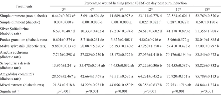
To apply this equation, the wound margins were traced and measured to calculate the non-healed area which was then subtracted from the original wound area to obtain the healed area. Wound contraction on different days is shown in Table 1. Statistically, the percentage of wound contraction showed that there is high signiicant difference between the different groups (p<0.001). The wound healing potential for A. euchroma, S. deserti, A.
communis and mixed extracts was evident on the 18th
day (Table 1). No healing effect was observed with P.
granatum (Table 1).
Epithelialization time
The Epithelialization time was found be high signiicantly (p<0.001) reduced in groups as depicted in Table 2. A better healing pattern with complete wound closure was observed in treated within 21 days while it was about 34 days in non-diabetic control rats. At the 18th day, A. euchroma, S. deserti, A. communis and mixed
Table 2. Effect of the treatments on epithelialization time (day).
Groups epithelialization time (day)
Simple ointment (no diabetic) 34±0.075 b
Silver Sulfadiazine (diabetic rats) 35±0.930 b
Punica granatum (diabetic rats)
-Malva sylvestris (diabetic rats) 24±0.290 a
Arnebia euchroma (diabetic rats) 21±0.186 a
Scrophularia deserti (diabetic rats) 21±0.109 a
Amygdalus communis (diabetic rats) 22±0.250 a
Mixed extracts (diabetic rats) 22±0.492 a Each value represents mean+SEM; -no detected.
Histological evaluation
At the 9th and 18th days the experiment,
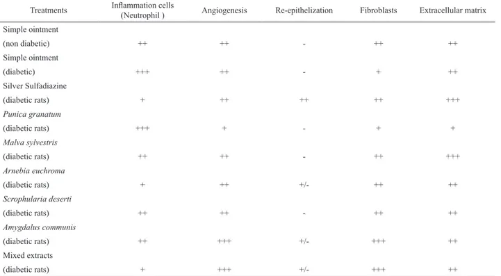
histological evaluation was carried out for the treated and untreated samples. Comparison between controls and some treated animals is shown in Tables 3 and 4. The best results were obtained with A. communis and standard drug, when compared to the other groups as well as to the controls. On the 18th day, groups of A. communis
extract and standard drug showed complete healing as in collagenation, ibroblasts cells and angiogenesis in Table 4. The control groups (diabetic and non-diabetic rats) and some of the groups (P. granatum) presented edema, monocyte cells and area with cellular necrosis (Table 4).
Evaluation of microbiological status
At the 9th and 18th days the experiment,
microbiological evaluation was carried out for groups. At the 9th and 18th days, lowest colonies per swab of Bacillus
and Staphylococcus species were detected in group of
mixed extracts (Table 5), highest colonies were detected in groups of diabetic control and P. granatum extract.
Despite the traditional uses M. sylvestris and P.
granatum, A. communis (sweet and bitter), A. euchroma
and S. deserti in burn wound healing process in Iran,
there are no reported data available in the literature. These species widely distributed plants of Iran are used for the infectious, inlammatory, anti-microbial, skin disease and for wound and burn healing properties according to several ethnobotanical surveys (Ghasemi Pirbalouti, 2009a; Ghasemi Pirbalouti et al., 2009; Ghasemi Pirbalouti et al., 2010b, Ghorbani, 2003; Zargari, 1989-1992). The present study tested the burn wound-healing properties of the ethanol extracts of M. sylvestris, P. granatum, A. communis, A. euchroma, S.
deserti and mixed extracts were used to evaluate the burn
healing activity in alloxan-induced diabetic rats.
On the different days, the results of morphological evaluation showed that A. communis,
A. euchroma, S. deserti and mixed extracts high
significantly increased the percentage of wound contraction (Table 1). At the 18th day the experiment,
A. communis extract and standard drug (Silver
Sulfadiazine) showed increased collagen turnover (Tables 2-4). Collagen, the major component which strengthens and supports extracellular tissue, is composed of the amino acid, hydroxyproline, which has been used as a biochemical marker for tissue collagen (Philips et al., 1991).
Wound healing is a process by which damaged tissue is restored as closely as possible to its normal state and wound contraction is the process of shrinkage of the area of the wound (Nayak et al., 2007). It is mainly Table 1. Effect of the treatments on burn wound expressed as percentage of wound contraction in diabetic rats.
Treatments Percentage wound healing (mean±SEM) on day post burn induction
3th 6th 9th 12th 15th 18th
Simple ointment (non diabetic) 0.449±0.203 e* 5.091±0.504 de 11.689±0.975 e 23.111±0.778 d 33.564±0.621 f 52.769±0.570 c
Simple ointment (diabetic) 0.00±0.000 e 0.00±0.000 e 0.00±0.000 g 0.022±0.022 f 0.287±0.022 h 0.507±0.180 e
Silver Sulfadiazine
(diabetic rats) 6.620±0.487 d 10.333±0.402 d 17.216±0.394 d 24.618±0.602 d 41.178±0.890 e 51.356±1.908 c
Punica granatum (diabetic rats) 0.601±0.374 e 3.710±0.261 de 3.622±0.408 f 4.862±0.916 e 5.966±0.572 g 38.040±1.885 d
Malva sylvestris (diabetic rats) 9.880±0.013 cd 20.087±5.870 c 35.393±0.140 c 47.250±1.350 c 57.410±0.423 d 77.003±0.797 b
Arnebia euchroma
(diabetic rats) 7.742±0.298 d 27.889±0.250 b 43.173±0.522 b 57.056±1.410 b 70.176±0.194 bc 83.549±0.672 a
Scrophularia deserti
(diabetic rats) 13.956±1.241 c 35.478±0.503 ab 44.653±0.032 ab 57.229±0.306 b 67.453±0.587 c 88.829±0.332 a
Amygdalus communis
(diabetic rats) 28.667±2.467 a 42.664±1.467 a 47.511±0.535 a 64.231±0.452 a 75.920±0.151 a 85.789±0.113 a
Mixed extracts (diabetic rats) 21.84±0.518 b 34.229±0.931 b 44.056±0.650 b 58.356±0.637 b 72.753±1.716 ab 84.844±1.374 a
Signiicant † p<0.001 p<0.001 p<0.001 p<0.001 p<0.001 p<0.001
N=6 animals; Each value represents mean+SEM; †One-way Analysis of Variance (ANOVA); *Letters a, b, and etc means in the same column not
a process that occurs throughout the healing process, commencing in the ibroblastic stage whereby the area of the wound undergoes shrinkage. In the maturational phase, the inal phase of wound healing the wound dependent upon the type and extent of damage, the general
state of health and the ability of the tissue to repair. The aims in these processes are to regenerate and reconstruct the disrupted anatomical continuity and functional status of the skin (Philips et al., 1991). Wound contracture is
Table 3. Effect of the treatments on the evolution of wounds in rats after nine days of topical application. Treatments Inlammation cells
(Neutrophil ) Angiogenesis Re-epithelization Fibroblasts Extracellular matrix
Simple ointment
(non diabetic) ++ ++ - ++ ++
Simple ointment
(diabetic) +++ ++ - + ++
Silver Sulfadiazine
(diabetic rats) + ++ ++ ++ +++
Punica granatum
(diabetic rats) +++ + - + +
Malva sylvestris
(diabetic rats) ++ ++ - ++ +++
Arnebia euchroma
(diabetic rats) + ++ +/- ++ ++
Scrophularia deserti
(diabetic rats) ++ ++ - ++ ++
Amygdalus communis
(diabetic rats) ++ +++ +/- +++ ++
Mixed extracts
(diabetic rats) + +++ +/- +++ ++
+: slight, ++: moderate, +++: extensive, -: absent.
Table 4. Effect of the treatments on the evolution of wounds in rats after eighteen days of topical application. Treatments Inlammation cells
(Neutrophil )
Collagen
maturation Re-epithelization
Organization of
the collagen Vascularization
Simple ointment
(non diabetic) ++ ++ + ++ ++
Simple ointment
(diabetic) +++ + - + +++
Silver Sulfadiazine
(diabetic rats) - ++ ++++ +++ +
Punica granatum
(diabetic rats) ++ ++ + + +++
Malva sylvestris
(diabetic rats) - ++ +++ ++ ++
Arnebia euchroma
(diabetic rats) ++ ++ + + +++
Scrophularia deserti
(diabetic rats) - ++ ++ + +++
Amygdalus communis
(diabetic rats) - +++ ++++ +++ +
Mixed extracts
undergoes contraction resulting in a smaller amount of apparent scar tissue. Granulation tissue formed in the inal part of the proliferative phase is primarily composed of ibroblasts, collagen, edema, and new small blood vessels. The increase in dry granulation tissue weight in the test treated animals suggests higher protein content (Philips et al., 1991).
The results of study showed that the extract ointment of M. sylvestris, P. granatum, A. communis, S.
deserti and mixed extracts effectively stimulates burn
wound contraction as compared to controls group. These inding could justify the inclusion of these plants in the management of wound healing. The result of the present study offers pharmacological evidence on the folkloric uses of M. sylvestris lowers, P. granatum lowers, A.
communis leaves and S. deserti stems for healing burn
wound. Hence, the results support the traditional uses of
M. sylvestris, P. granatum, A. communis and S. deserti to
treat skin disorders including burns.
Acknowledgements
The authors are thankful to Mr. Farid, Ph.D (Surgical) and Mr Hamedi for their technical support.
References
Annan K, Houghton PJ 2007. Antibacterial, antioxidant and
ibroblast growth stimulation of aqueous extracts of
Ficus asperifolia Miq. and Gossypium arboreum L., wound-healing plants of Ghana. J Ethnopharmacol 119: 141-144.
Arturson G 1995. Pathophysiology of the burn wound and pharmacological treatment. The Rudi Hermans Lecture.
Burns 22: 255-274.
Diatewa M, Samba CB, Assah TCH, Abena AA 2004. Hypoglycemic and antihyperglycemic effects of diethyl ether fraction isolated from the aqueous extract of the leaves of Cogniauxia podoleana Baillon in normal and alloxan-induced diabetic rats. J Ethnopharmacol 92: 229-232.
Ghahreman A 1987-1989. Flora of Iran. Department of Botany, Institute of and Rangelands Press, Tehran (in Farsi). Ghasemi Pirbalouti A 2009a. Medicinal plants used in
Chaharmahal and Bakhtyari districts, Iran. Herba Pol 55: 34-38.
Ghasemi Pirbalouti A 2009b. Iranian medicinal and aromatic plants (2nd edition). Islamic Azad University Publishers, Shahrekord, Iran (in Farsi).
Ghasemi Pirbalouti A, Yousei M, Nazari H, Karimi I,
Koohpayeh A 2009a. Evaluation of burn healing properties of Arnebia euchroma and Malva sylvestris. E J Bio 5: 62-66.
Ghasemi Pirbalouti A, Koohpayeh A, Karimi I 2009b. Effect of natural remedies on dead space wound healing in Wistar
rats. Pharmacogn Mag 5: 433-436.
Ghasemi Pirbalouti A, Momeni M, Bahmani M 2010a. Studies on pharmaceutical ethnobotany in Ilam, Western Iran. VIII International Ethnobotany Symposium, October 3rd–8th, 2010, Lisbon, Portugal.
Ghasemi Pirbalouti A, Azizi S, Koohpayeh A, Hamedi B 2010b. Wound healing activity of Malva sylvestris and Punica granatum in alloxan-induced diabetic rats. Acta Pol Pharm 67: 511-516.
Ghorbani A 2005. Studies on pharmaceutical ethnobotany in the region of Turkman Sahra, north of Iran. J Ethnopharmacol 102: 58-68.
Goodson WH, Hunt TK 1977. Studies of wound healing in experimental diabetes mellitus. J Surg Res 22: 221-27. Houghton PJ, Hylands PJ, Mensah AY, Hensel A, Deters
AM 2005. In vitro tests and ethnopharmacological investigations: Wound healing as an example. J Ethnopharmacol 100: 100-107.
Mantle D, Gok MA, Lennard TWJ 2001. Adverse and beneicial
effects of plant extracts on skin and skin disorders.
Adverse Drug React T 20: 89-103.
Mokaddas E, Rotimi VO, Sanyal SC 1998. In vitro activity of piperacillin / tazobactam versus other broad antibiotics against nosocomial gram negative pathogens isolated from burn patients. J Chemotherapy 10: 208-214. Mozaffarian V 1996. Encyclopedia of Iranian Plants. Farhang
Moaser Publication, Tehran, Iran, p. 671 (in Farsi). Mozaffarian V 2008. Flora of Ilam. Farhang Moaser Publication,
Tehran, Iran (in Farsi).
Nayak BS, Godwin I, Davis EM, Pillai GK 2007. The Evidence based Wound Healing Activity of Lawsonia inermis
Linn. Phytother Res 21: 827- 831.
Philips GD, Whitehe RA, Kinghton DR 1991. Initiation and pattern of angiogenesis in wound healing in the rat. Am J Anat 192: 257-262.
Raghow R 1994. The Role of extracellular matrix
post-inlammatory wound healing and ibrosis. FASEB J 8:
823-831.
Rechinger KH 1963-1998. Flora Iranica (Vol. 1-173). Austria, Graz: Akademische Druck und Verlagsanstalt.
Shanmuga Priya KS, Gnanamani A, Radhakrishnan N, Babu M 2002. Healing potential of Datura alba on burn wounds in albino rats. J Ethnopharmacol 83: 193-199. Shalom A, Friedman T, Wong L 2005. Burns and diabetes.
Annals of Burns and Fire Disasters - vol. XVIII - n. 1 - March 2005.
Singer AJ, Clark RA 1999. Cutaneous wound healing. New Engl J Med 341: 738-746.
Zargari A 1989-1992. Medicinal Plants (Vol. 1-6). University Publication, Tehran, Iran (in Farsi).
*Correspondence
A. Ghasemi Pirbalouti
Medicinal Plants, Researches Centre of Medicinal Plants & Ethno-veterinary, POBox: 166, Shahrekord, Iran
ghasemi@iaushk.ac.ir

