272
Radiol Bras. 2017 Jul/Ago;50(4):266–276 Letters to the EditorHemangiomas are benign tumor formations of capillaries and blood vessels, common in various organs; they are extremely rare in the urinary bladder, accounting for only 0.6% of all uri -nary bladder tumors(1,2). There have been fewer than 100 re -ported cases of histologically proven hemangioma of the urinary bladder(1).
Most urinary bladder hemangiomas are solitary and smaller than 3 cm in diameter, affecting the dome, posterior wall, or tri -gone of the bladder. Although hemangiomas can occur in indi -viduals of any age, they are seen most often in indi-viduals under 30 years of age and are slightly more common among males(2). A hemangioma usually presents as an incidental inding during the investigation of hematuria. The most common symptom is gross hematuria, which can be accompanied by irritative urinary symptoms and abdominal pain. Urinary bladder hemangiomas occasionally coexist with cutaneous hemangioma or are asso -ciated with one of two conditions(3–5): Sturge-Weber syndrome and Klippel-Trenaunay-Weber syndrome.
In young patients, endoscopic indings of a bluish, ses -sile mass and gross hematuria are highly suggestive of heman-gioma(1). The main differential diagnoses for pigmented lesions seen on endoscopy include endometriosis, melanoma, and sar-coma(6). Imaging tests, such as ultrasound, computed tomogra -phy, and magnetic resonance imaging, are useful in deining the location and extent of a hemangioma(2).
For individuals with hemangioma, the treatment is contro-versial. Although there are many options available, partial cys -tectomy is currently the most widely used treatment for heman-gioma of the urinary bladder(3,6,7). Although hemangioma has a benign course, follow-up is mandatory in order to detect recur-rence or residual disease(3,7,8). The purpose of this case report
was to underscore the importance of early diagnosis of heman-gioma of the urinary bladder and of differentiating it from ma-lignant neoplasms, which would affect the therapeutic strategy and patient survival.
REFERENCES
1. Cheng L, Nascimento AG, Neumann RM, et al. Hemangioma of the uri -nary bladder. Cancer. 1999;86:498–504.
2. Stimac G, Dimanovski J, Katusic J, et al. A large cavernous hemangioma of the urinary bladder: imaging of possible spontaneous regression. Eur J Radiol Extra. 2007;61:61–3.
3. Jibhkate S, Sanklecha V, Valand A. Urinary bladder hemangioma – a rare urinary bladder tumor in a child. APSP J Case Rep. 2015;6:6.
4. Kim YY, Kim MJ, Lee MJ, et al. Multiple hemangiomas of the urinary bladder in a child with gross hematuria. Ultrasonography. 2015;34:231–4. 5. Ikeda T, Shimamoto K, Tanji N, et al. Cavernous hemangioma of the uri
-nary bladder in an 8-year-old child. Int J Urol. 2004;11:429–31. 6. Wong-You-Cheong JJ, Woodward PJ, Manning MA, et al. From the Ar
-chives of the AFIP: Neoplasms of the urinary bladder: radiologic-patho -logic correlation. Radiographics. 2006;26:553–80.
7. Lahyani M, Slaoui A, Jakhlal N, et al. Cavernous hemangioma of the blad -der: an additional case managed by partial cystectomy and augmentation cystoplasty. Pan Afr Med J. 2015;22:131.
8. Castillo OA, Foneron A, Sepúlveda F, et al. Bladder hemangioma: case report. Arch Esp Urol. 2012;65:623–5.
Camila Soares Moreira de Sousa1, Ivo Lima Viana1, Carla Lorena
Vasques Mendes de Miranda1, Breno Braga Bastos2, Ilan Lopes
Leite Mendes1
1. Medimagem, Teresina, PI, Brazil. 2. UDI 24 horas, Teresina, PI, Brazil. Mailing address: Dra. Camila Soares Moreira de Sousa. Medimagem – Ra-diologia. Rua Paissandu, 1862, Centro. Teresina, PI, Brazil, 64001-120. E-mail: [email protected].
http://dx.doi.org/10.1590/0100-3984.2015.0231
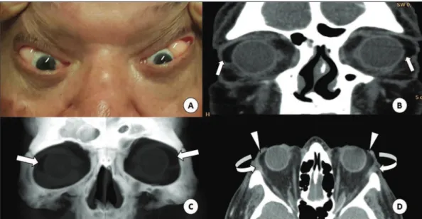
Figure 1. A: Fatty mass in the lateral corner of the orbits, best characterized by the retropul-sion of the globes. CT of the orbits in the coronal plane (B), with volumetric reconstructions in the coronal plane (C) and axial plane (D), showing the masses in the lateral corners (straight arrows), contiguous with the intraconal fat (arrow-heads) and pushing aside the lacrimal glands (curved arrows). Subconjunctival fat prolapse: a disease little known to radiologists
Dear Editor,
A 69-year-old male patient sought outpatient treatment with a 10-year history of fatty masses in the lateral corners of his eyes, best characterized as retropulsion of the globes. He underwent computed tomography (CT) of the orbits, which re -vealed intraconal fat proliferation in the lateral corners of the
eyes, from the orbits to the epibulbar region (Figure 1). Given the clinical presentation and imaging indings, a diagnosis of subconjunctival fat prolapse was made.
273
Radiol Bras. 2017 Jul/Ago;50(4):266–276Letters to the Editor
manifests clinically as a yellowish mass in the lateral corner of the eye, which becomes more evident with retropulsion of the globe(2).
The imaging tests that can facilitate the diagnosis of sub -conjunctival fat prolapse are CT and magnetic resonance imag -ing (MRI) of the orbits, the most important radiological ind-ing being that of a mass with fat density or fat-like signal intensity, respectively, located in the temporal aspect of the orbits, con-tiguous with intraconal fat.
The treatment consists of transconjunctival excision, a simple, safe and effective surgical procedure. The rate of recur -rence after transconjunctival excision is reported to be approxi -mately 9%(3).
Making a clinical diagnosis of subconjunctival fat prolapse is relatively easy. However, due to its rarity, it can be misdiag -nosed as conjunctival dermolipoma, lymphoma, epidermoid cyst, or lacrimal gland prolapse(4). The main differential diag -nosis is conjunctival dermolipoma, which consists of a benign lesion, usually present at birth(5), that affects young women, the mean age of such patients being 22 years(6). Although the clinical presentation of conjunctival dermolipoma is similar to that of the subconjunctival fat prolapse, the former is typically unilateral and fairly immobile. On CT and MRI, conjunctival dermolipoma presents as a crescent-shaped fatty mass in the temporal aspect of the orbit, not in communication with the intraconal fat(1).
In conjunctival dermolipoma, surgical resection is indicated mainly for aesthetic purposes and tends to be more conservative(1). Although resection of a conjunctival dermolipoma is a simple pro -cedure, there can be severe complications, including blepharop-tosis, diplopia, and keratoconjunctivitis sicca. Therefore, a num -ber of different surgical techniques aimed at a lowering the rate of complications and improving the aesthetic results have been described, including resection with conjunctival lap rotation(7).
Cynthia Ramos Tejo1, Péricles Almeida da Costa1, Rafaella Martins
Batista1, Yuri Raoni Ramalho Rocha1, Marcelle Alves Borba1
1. Universidade Federal do Rio Grande do Norte (UFRN), Natal, RN, Brazil. Mail-ing address: Dra. Cynthia Ramos Tejo. Hospital Universitário Onofre Lopes. Avenida Nilo Peçanha, 620, Petrópolis. Natal, RN, Brazil, 59012-300. E-mail: [email protected].
http://dx.doi.org/10.1590/0100-3984.2015.0229
Subconjunctival fat prolapse and dermolipoma present clinically as a fatty epibulbar masses in the lateral corners of the orbits, and in some cases their differentiation by clinical as-pects can be dificult. The subject is little known among radiolo -gists, and there have been few reports of related cases. There -fore, given the difference between these two entities in terms of treatment, it is necessary that radiologists be familiar with both, in order to recognize them promptly and make the differential diagnosis through the use of imaging tests.
REFERENCES
1. Kim E, Kim HJ, Kim YD, et al. Subconjunctival fat prolapse and dermo -lipoma of the orbit: differentiation on CT and MR imaging. AJNR Am J Neuroradiol. 2011;32:465–7.
2. Schmack I, Patel RM, Folpe AL, et al. Subconjunctival herniated orbital fat: a benign adipocytic lesion that may mimic pleomorphic lipoma and atypical lipomatous tumor. Am J Surg Pathol. 2007;31:193–8.
3. Siban M, Weijtens O, van den Bosch W, et al. Eficacy of transconjunc -tival excision of orbital fat prolapse: a long-term follow-up study. Acta Ophthalmol. 2014;92:291–3.
4. Wang X, Yan J. Subconjunctival orbital fat prolapse: an unsuspecting rare lesion. J Craniofac Surg. 2015;26:e92–4.
5. Ferraz LCB, Schellini AS, Wludarski SCL, et al. Dermolipoma e pro -lapso de gordura orbital – duas entidades distintas. Arq Bras Oftalmol. 2002;65:327–31.
6. McNab AA, Wright JE, Caswell AG. Clinical features and surgical man -agement of dermolipomas. Aust N Z J Ophthalmol. 1990;18:159–62. 7. Sa HS, Kim HK, Shin JH, et al. Dermolipoma surgery with rotational
conjunctival laps. Acta Ophtalmol. 2012;90:86–90.
Ogilvie syndrome after use of vincristine: tomographic indings
Dear Editor,
A 33-year-old female patient with diffuse large B-cell non-Hodgkin lymphoma was evaluated two days after the end of the irst cycle of chemotherapy. The chemotherapy regimen com -prised a ive-day cycle, including rituximab, cyclophosphamide, doxorubicin, vincristine, and prednisone on the irst day, where -as prednisone alone w-as administered on the four remaining days. She reported left pleuritic pain and latus with evacuation. She was afebrile. The abdomen was laccid and peristaltic, with
-out painful decompression. Because she had neutropenia, she was hospitalized, after which she evolved to having no bowel movements, with the smell of feces on her breath and painful abdominal decompression. Computed tomography (CT) of the chest and abdomen showed left pleural effusion, intestinal ob-struction in the descending colon adjacent to the splenic lex -ure, that segment being of normal caliber, without occlusive lesions, although the transverse ascending colon and cecum were dilated, the latter being 14 cm in diameter (Figures 1 and 2). There was gas in the rectal ampulla. These indings were suggestive of acute colonic pseudo-obstruction. Colonoscopic
Figure 1. CT scan of the abdo-men, in axial sections, obtained 60 s after injection of iodinated anionic contrast. Note the intes-tinal obstruction at the level of the proximal descending colon, adjacent to the splenic lexure. Distension of the transverse co-lon, ascending coco-lon, and cecum, with the presence of fecal matter. The transitional zone can be seen at the level of the splenic lexure (arrow), with no evident obstruc-tive material.