1
Radiol Bras. 2018 Jan/Fev;51(1):1–7
Extrinsic functional connectivity of the default mode network
in crack-cocaine users
A conectividade funcional extrínseca da rede de modo padrão em usuários de crack-cocaína
Diego Lima Nava Martins1, Talles Destefani de Souza Valiatti2, Júlia D’Ávila2, Lucas Freire Ferreira2, Edson Kruger Batista3, Paulo Rodrigo Bazán4, Rodrigo Stênio Moll de Souza5, Ester Miyuki Nakamura-Palacios6
Martins DLN, Valiatti TDS, D’Ávila J, Ferreira LF, Batista EK, Bazán PR, Souza RSM, Nakamura-Palacios EM. Extrinsic functional connectivity of the default mode network in crack-cocaine users. Radiol Bras. 2018 Jan/Fev;51(1):1–7.
Abstract
Resumo
Objective: This study aimed to explore the functional connectivity of the default mode network (DMN) in crack-cocaine users, in comparison with that observed in age-matched non-drug-using controls.
Materials and Methods: Inpatient crack-cocaine users who had been abstinent for at least four weeks and age-matched non-drug-using controls underwent resting state functional magnetic resonance imaging. Images were acquired while the subjects rested with
their eyes closed. After data preprocessing, DMNs were deined by spatial independent component analysis and seed-based cor -relation analysis, by chosen regions of interest centered in the ventral anterior cingulate cortex and in the posterior cingulate cortex.
Results: The functional connectivity of the DMN determined by independent component analysis did not differ between the crack-cocaine users and the controls. However, the seed-based correlation analysis seeking a single metric of functional connectivity
between speciic brain regions showed that the negative connectivity between the ventral anterior cingulate cortex and the left superior parietal lobule was signiicantly greater in the crack-cocaine users than in the controls.
Conclusion: The results suggest that selective extrinsic network connectivity of the DMN related to motor and executive function is impaired during crack-cocaine addiction.
Keywords: Substance-related disorders; Magnetic resonance imaging/methods; Brain/physiopathology; Image interpretation, com-puter-assisted; Functional neuroimaging.
Objetivo: Este estudo teve como objetivo explorar a conectividade funcional da rede de modo padrão em usuários de crack-cocaína comparando-a a controles não usuários da mesma faixa etária.
Materiais e Métodos: Usuários de crack-cocaína internados em abstinência por pelo menos quatro semanas e controles não usuá-rios pareados por idade foram submetidos a ressonância magnética funcional em estado de repouso, enquanto descansavam com
os olhos fechados. Depois do pré-processamento de dados, a rede de modo padrão foi deinida por análise espacial de componen -tes independen-tes e análise de correlação baseada em semen-tes, por regiões de interesse centradas no córtex cingulado anterior ventral e no córtex cingulado posterior.
Resultados: A conectividade funcional analisada por componentes independentes não foi diferente entre os usuários de crack- cocaína e os controles pareados por idade. No entanto, a análise de correlação baseada em sementes à procura de uma
conecti-vidade funcional métrica única entre regiões especíicas do cérebro mostrou uma negaticonecti-vidade signiicativamente maior da conec -tividade entre o córtex cingulado anterior ventral e o lóbulo parietal superior esquerdo, quando comparada a controles pareados por idade.
Conclusão: Os resultados sugerem que conectividades extrínsecas seletivas da rede de modo padrão relacionadas a funções mo-toras e executivas podem estar comprometidas na dependência de crack-cocaína.
Unitermos: Transtornos relacionados ao uso de substâncias; Ressonância magnética/métodos; Encéfalo/isiopatologia; Interpreta -ção de imagem assistida por computador; Neuroimagem funcional.
Study conducted under the auspices of the Graduate Program in Physiologi-cal Sciences, Federal University of Espírito Santo (UFES), Vitória, ES, Brazil. This work was partially supported by the Conselho Nacional de Desenvolvimento Cien-tíico e Tecnológico (CNPq)—Grant Nos. 475232/2013-5, 443824/2014-2, and 466650/2014-0 to E.M.N-P.). E.M.N-P. is the recipient of a research fellowship grant from CNPq (Grant No. 304374/2014-8).
1. MD, Department of Internal Medicine, Health Sciences Center, Graduate Pro-gram in Medicine, Federal University of Espírito Santo (UFES), Vitória, ES, Brazil.
2. Department of Electric Engineering, Technology Center, Brazilian Research Group on Brain and Cognitive Engineering (BRAEN), Federal University of Espírito Santo (UFES), Vitória, ES, Brazil.
3. MD, MSc, Laboratory of Cognitive Sciences and Neuropsychopharmacology, Graduate Program in Physiological Sciences, Federal University of Espírito Santo (UFES), Vitória, ES, Brazil.
4. Laboratory for Medical Research 44 (LIM-44—Laboratório de Investigação Médica 44), Department of Radiology, University of São Paulo (USP), São Paulo, SP, Brazil.
5. MD, MSc, Department of Internal Medicine, Health Sciences Center, Brazilian Research Group on Brain and Cognitive Engineering (BRAEN), Federal University of Espírito Santo (UFES), Vitória, ES, Brazil.
6. MD, PhD, Graduate Program in Medicine, Laboratory of Cognitive Sciences and Neuropsychopharmacology, Brazilian Research Group on Brain and Cognitive Engineering (BRAEN), Federal University of Espírito Santo (UFES), Vitória, ES, Brazil.
Mailing address: Dra. Ester M. Nakamura-Palacios. Programa de Pós-Graduação em Ciências Fisiológicas, Centro de Ciências da Saúde, Universidade Federal do Es-pírito Santo. Avenida Marechal Campos, 1468, Maruípe. Vitória, ES, Brazil, 29043-900. E-mail: [email protected].
INTRODUCTION
Crack-cocaine has emerged as a substance that readily supplies the active principle (cocaine) through the inhala-tion of smoke from burning rocks of the substance,
typi-cally by means of a pipe(1). How it affects the nervous
sys-tem remains unknown. However, certain neural circuits are supposedly involved in the establishment and mainte-nance of drug addiction(2,3). According to Volkow et al.(4), there are four interrelated circuits that are involved in or affected by drug addiction: the reward circuit—involving several nuclei in the basal ganglia, notably the nucleus ac-cumbens of the ventral striatum, and relaying information to the ventral pallidum; the motivation/drive circuit—in-volving the orbitofrontal and subcallosal cortices, dorsal striatum, and motor cortex; the learning/memory circuit— involving the amygdala and the hippocampus; and the control circuit—which controls cognitive lexibility and planning, involving the dorsolateral prefrontal cortex, an-terior cingulate cortex, and inferior frontal cortex. Many of these brain structures are associated with the default
mode network (DMN)(5–7), one of the brain networks seen
in resting state functional magnetic resonance imaging
(rs-fMRI) studies(8). The DMN is composed of brain
re-gions that are typically deactivated during task demands but exhibit synchronous low frequency oscillation in the
resting state(6,7,9). It is believed that the DMN is related
to attention and self-monitoring(10), or, more precisely, the
general monitoring of sensory information associated with the posterior cingulate and adjacent precuneus, as well as the evaluation of the salience of this information together
with the medial and orbitofrontal cortices(9). This provides
a new perspective on brain function by addressing the im-portance of ongoing or intrinsic activity(7).
Previous rs-fMRI studies have identiied changes in functional connectivity in individuals who abuse or are ad-dicted to drugs such as cocaine, heroin, morphine, nico-tine, alcohol, and caffeine. However, to our knowledge, there have been no such studies involving individuals ad-dicted to crack-cocaine. We hypothesized that the DMN would be affected in crack-cocaine users. Therefore, this study aimed to explore the functional connectivity of the DMN by using independent component analysis (ICA) and seed-based correlation analysis of rs-fMRI data col-lected from crack-cocaine users and age-matched non-drug-using controls.
MATERIALS AND METHODS
For this study, we interviewed 38 individuals with crack-cocaine addiction, as deined in the Diagnostic and Statistical Manual of Mental Disorders, fourth edition (DSM-IV), recruited from among those under treatment at a mental health clinic for drug dependence treatment in the state of Espírito Santo, Brazil. Of those 38 indi-viduals, 10 met the inclusion criteria. At the mental health
clinic, the subjects were evaluated in a highly restricted and controlled environment. Urine samples were collected for drug testing, and all of the subjects tested negative. We acquired rs-fMRI scans in a 1.5 T scanner at the Cassiano Antônio de Moraes University Hospital, operated by the Federal University of Espírito Santo. Because of technical problems during the acquisition of the images, data for two of the subjects were excluded from the analysis. There-fore, data for eight crack-cocaine users were included in all analyses.
As a control group, healthy, non-drug-using, aged-matched male subjects were recruited from among the employees of the University Hospital and of the private clinic, as well as from among students at the Federal Uni-versity of Espírito Santo. The rs-fMRI scans of the control group subjects were acquired in the same scanner as were those of the study group.
The inclusion criteria for the crack-cocaine user group were as follows: being male; being over 18 years of age; meeting the criteria for a clinical diagnosis of crack-cocaine dependence, as deined in the symptom checklist for mental disorders of the International Classiication of Diseases, 10th revision, and in the DSM-IV; being in sta-ble clinical condition (not requiring hospitalization); being able to read, write, and speak Portuguese; and presenting with no signs or symptoms of severe withdrawal at base-line.
Subjects who were intoxicated with or in withdrawal from a substance other than crack-cocaine were excluded, as were those who had been diagnosed with a psychiatric or physical disorder, including substance abuse disorder and addiction to any substance other than crack-cocaine, nicotine, or caffeine. We also excluded subjects who had been diagnosed with epilepsy or convulsive disorder; those who had experienced delirium tremens during abstinence from crack-cocaine use; those with a history of drug hy-persensitivity or adverse reactions to diazepam, other ben-zodiazepines, or haloperidol; those presenting with any contraindication for MRI, such as electronic implants, metal implants, and claustrophobia, as well as permanent makeup or tattoo received within the last three months; and those with vascular, traumatic, inlammatory, or neo -plastic processes detectable by MRI. It should be stated that medications were given to the crack-cocaine users to relieve the symptoms of anxiety and depression that can arise during abstinence.
We assessed ten inpatients diagnosed with crack-co-caine dependence and eight non-drug-using controls. To do so, we used a brief structured interview designed to obtain sociodemographic data and information regarding the characteristics of drug use.
MRI
The rs-fMRI scans were obtained while subjects rested quietly, with their eyes closed but awake. The images were acquired in 1.5 T MRI scanners (Achieva; Philips Medical Systems, Best, the Netherlands), with a speciic 8-channel sensitivity encoding head coil (SENSE; Philips Medical Systems), at the radiology center of the Cassiano Antônio de Moraes University Hospital.
Functional images were acquired using a gradient echo echo-planar imaging sequence—repetition time/ echo time (TR/TE): 3000/50 ms; lip angle = 90°; 35 axial slices; matrix: 64 × 64; ield of view = 230 mm; and voxel size: 3.59 × 3.59 × 4.0 mm—with an echo planar imaging factor of 39 and an acquisition time of 10 min 9 s per sub-ject. For registration purposes, a T1-weighted anatomical image was also acquired: TR/TE: 8.8/4.05 ms; lip angle = 80°; matrix: 240 × 240; voxel size: 1 × 1 × 1 mm; 180 axial slices; ield of view = 240 mm; and acquisition time = 1 min 28 s.
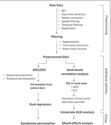
Functional images were pre- and post-processed with the Functional Magnetic Resonance Imaging of the Brain software library (FSL, version 5.0.8; FMRIB Analysis Group, Oxford University, UK). In brief, traditional pre-processing steps were performed: head motion, by realign-ing each volume to the middle volume; slice-timrealign-ing cor-rection; non-brain material extraction; spatial smoothing, using a 6 mm (full width at half maximum) kernel; high-pass temporal iltering (0.01 Hz); and normalization to the Montreal Neurological Institute standard-space template (2-mm resolution). In addition, to reduce physiological noise, tissues were segmented by using the structural T1-weighted images from each subject. The average white matter and cerebrospinal luid signals were iltered using linear regression, the residuals of that analysis being used in the subsequent steps (Figure 1).
ICA and dual regression
Images were spatially and temporally decomposed in a four-dimensional (time × voxels) data matrix into a set of time courses and associated spatial maps with the multi-variate exploratory linear decomposition into independent components interface of the FSL (Figure 1). This instru-ment was used as a primary approach to objectively iden-tify resting state networks, especially the DMN for this
study purpose(11,12). We performed two separate group
ICA runs concatenating the time series of the individuals in each group.
For dual regression analysis (Figure 1), the set of spatial maps from the control group ICA was used as the
template, as described by Rytty et al.(13), to generate
sub-ject-speciic versions of the spatial maps, and associated
time series(11–14) were used in order to test for voxel-wise
group differences. We adopted a nonparametric test, using the “randomize” permutation-testing tool of the FSL and calculating the maximum of 5,000 permutations, with a
threshold of p = 0.05 corrected for multiple comparisons
throughout the brain.
Seed-based correlation analysis
The seed-based correlation analysis employed the
same input data employed in the ICA(14,15). The difference
was that the seed-based correlation analysis was based on regions of interest (ROIs), as shown in Figure 1. The cho-sen ROIs and their Talairach coordinates were based on
those that Greicius et al.(5) deined as the main regions
constituting the DMN. Thus, two ROIs (each 8 mm in diameter) were chosen, one centered in the posterior cin-gulate cortex (Talairach coordinates: x = 2, y = −51, z = 27) and the other centered in the ventral anterior cingu-late cortex (Talairach coordinates: x = 2, y = 38, z = −2). For each participant, the signal extracted from those seed regions were input into two separate whole-brain analy-ses (one for each seed), allowing the positive and negative correlations to be evaluated. Correlation scores were
con-verted to z-scores using Fisher’s z-transformation. We
per-formed group-level analyses using the mixed-effects model Figure 1. Flow chart of the data analysis process.
implemented in the fMRI Expert Analysis Tool of the FSL,
adopting a voxel threshold of z > 2.3 and correcting for
multiple comparisons at the cluster level using Gaussian random ield theory with a cluster signiicance threshold of p = 0.05.
RESULTS
Baseline sociodemographic characteristics and pat-terns of drug use are presented in Table 1. All of the crack-cocaine users were young (mean age, 29.1 ± 10.6 years), were male, typically had a low level of education (fewer than three years of schooling), were mostly unemployed, and were single. In addition, most of them were cigarette smokers. With the exceptions of age and marital status, the sociodemographic characteristics (including level of edu-cation, employment status, and tobacco use) differed be-tween the crack-cocaine user group and the control group. Such differences were expected considering the impover-ishment due to crack-cocaine addiction. On average, the subjects in the crack-cocaine user group had started using crack-cocaine at 22.6 ± 8.9 years of age, consumed 14.8 ± 16.2 rocks per day, and had been abstinent for at least four weeks prior to the beginning of the experimental protocol (Table 1). All of the urine samples collected during the study period tested negative.
rs-fMRI
ICA
The ICA applied to the rs-fMRI data satisfactorily determined the component representing the DMN indi-vidually for subjects in the control and crack-cocaine user groups, accurately providing the mean for each group.
Neither the dual regression analyses considering the con-trol group as the template nor the randomized permuta-tion analyses identiied any differences between the two groups.
Seed-based correlation analysis
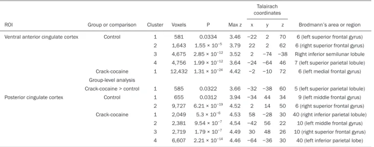
Activities in the ventral anterior cingulate cortex (ROI centered at x = 2, y = 38, z = −2) and in the posterior cin -gulate cortex (ROI centered at x = 2, y = −51, z = 27), as depicted in Figure 2, were found to be positively and nega-tively related to the structures listed in Tables 2 and 3, respectively. No differences were found between the con-trol and crack-cocaine user groups regarding the positive correlations. However, the Figure 3 shows the negative correlation between the ventral anterior cingulate cortex and the region corresponding to the left superior parietal lobule (Brodmann’s area 5). As can be seen in Table 3, that correlation was greater in the crack-cocaine user group than in the control group (p < 0.0322).
DISCUSSION
In this study, we used ICA and seed-based correla-tion analysis to evaluate the funccorrela-tional connectivity of the DMN in crack-cocaine addicts. The seed-based cortion analysis showed that the negative or antiphasic rela-tionship between the medial frontal site of the DMN and the superior parietal lobule was stronger in crack-cocaine addicts who had abstinent for at least four weeks than it was in age-matched non-drug-using control subjects.
Exploring the DMN and its connectivity in drug ad-diction has been of great interest to help improve the
un-derstanding of this complex disease(16). Studies employing
Table 1—Sociodemographic characteristics and patterns of crack-cocaine use in a sample of male drug users, abstinent for at least four weeks, who underwent
rs-fMRI in a 1.5 T scanner (n = 8), in comparison with age-matched non-drug-using male control subjects who also underwent rs-fMRI (n = 8).
Characteristic
Age, in years, mean (SD) Years of schooling, mean (SD) Employment situation, n (%)
Formal job Informal job Unemployed Self-employed On disability Not reported Marital status, n (%)
Single Married Divorced Tobacco use, n (%)
Yes No
Crack-cocaine use
Age, in years, at onset, mean (SD) Amount consumed (rocks/day), mean (SD)
Non-drug-using controls
31.4 (7.0) 12.7 (3.3)
6 (75%) 1 (12.5%)
0 (0%) 0 (0%) 0 (0%) 1 (12.5%)
5 (62.5%) 3 (37.5%) 0 (0%)
0 (0%) 8 (100%)
Crack-cocaine users
29.1 (10.6) 1.9 (0.8)
0 (0%) 0 (0%) 4 (50%) 2 (25%) 1 (12.5%) 1 (12.5%)
6 (75%) 1 (12.5%) 1 (12.5%)
5 (62.5%) 3 (37.5%)
22.6 (8.9) 14.8 (16.2)
Statistic
t(14) = 0.50 t(13) = 9.14
χ2 = 14
χ2 = 2.09
Fisher’s
P
0.62 < 0.0001
0.016
0.35
rs-fMRI to investigate the effects or consequences of cocaine dependence have produced mixed results, from
hyperconnectivity of the anterior cingulate cortex(17) to
decreases in connectivity between the DMN and other networks(18–21).
The precise brain function supported by the DMN
remains unknown. According to Raichle(6), the DMN
sup-ports processes related to emotional processing (involving the ventral medial prefrontal cortex), self-referential men-tal activity (involving the dorsal medial prefronmen-tal cortex), and the recollection of prior experiences (involving pos-terior elements of the DMN). The DMN is a functioning
network that is never inactive; its level of activity seems
to vary only according to the level of consciousness(22,23).
It remains active during mild sedation(24), anesthesia(25),
and even during a vegetative state(26), although with some
variations of its connectivity(23), and is completely silent
only in brain death(26). Therefore, it seems reasonable
to consider that the DMN would be affected only under extremely life-threatening conditions, which fortunately might not be the case for crack users. In that sense, it is heartening to have found that the overall DMN connec-tivity in crack users is not yet affected in comparison with that observed in age-matched non-drug-using controls. Table 2—Regions positively related to ROIs (8 mm in diameter) centered in the ventral anterior cingulate cortex (Talairach coordinates: x = 2, y = 38, z = –2) and
posterior cingulate cortex (Talairach coordinates: x = 2, y = –51, z = 27) in crack-cocaine users (n = 8) and age-matched non-drug-using controls (n = 8).
Brodmann’s area
21 (left middle temporal gyrus) 10 (left medial frontal gyrus) 20 (left inferior temporal gyrus) 32 (right dorsal anterior cingulate)
21 (right middle temporal gyrus) 10 (left superior frontal gyrus)
6 (right middle frontal gyrus) 31 (left dorsal posterior cingulate)
21 (right middle temporal gyrus) 10 (left medial frontal gyrus)
39 (left angular gyrus) 8 (left superior frontal gyrus) 31 (left dorsal posterior cingulate) Talairach
coordinates
z
−16 −8 −26
−4 −22
6 58 28 −10
4 34 52 24 y
−12 40 −2 44 0 66 28 −52
−2 64 −70
22 −52 x
−56 −6 −48
2 56 −8 30 −4 68 0 −46 −36 −2 Max. z
3.89 5.61 3.87 5.97 4.39 4.27 4.39 5.95 4.26 4.77 5.61 4.11 6.26 P
0.00152 1.64 × 10⁻¹⁶
0.0012 7.68 × 10⁻¹⁹
0.0268 0.000885 1.19 × 10⁻⁷ 1.63 × 10⁻²³ 0.00197 9.45 × 10⁻⁵ 4.92 × 10⁻⁵ 1.8 × 10⁻⁵ 4.92 × 10⁻²⁶ Voxels
966 7,058
998 8,499
675 1,166 2,781 13,250
1,043 1,532 1,645 1,824 15,337 Cluster
1 2 1 2 1 2 3 4 1 2 3 4 5 Group
Control
Crack-cocaine
Control
Crack-cocaine ROI
Ventral anterior cingulate cortex
Posterior cingulate cortex
Figure 2. Functional connectivity determined by seed-based correla-tion analysis centering on ROIs in an 8-mm diameter circle (depicted in green). In A the ventral anterior cingulate cortex (vACC: x = 2, y = 38, z = −2) and in B the posterior cingulate cortex (PCC: x = 2, y = −51, z = 27), considering the co -ordinates used by Greicius et al.(5),
Consequently, although the use of crack is highly detri-mental to our young patients in many ways, there is hope that there will be a good recovery, given that intrinsic brain functioning is preserved or re-established after abstinence.
Compulsive cocaine use has been associated with a balance between increased striatal-anterior prefrontal/ orbitofrontal connectivity and decreased striatal-dorsal
an-terior cingulate connectivity(27). In the present study, the
seed-based correlation analysis showed that negative con-nectivity between the ventral anterior cingulate cortex and the left superior parietal lobule was greater in crack-co-caine users. This brain area, corresponding to Brodmann’s area 5, is situated immediately posterior to the primary somatosensory areas (postcentral gyrus), anterior and to the right of Brodmann’s area 7. The superior parietal lobule has been proven to be necessary for the executive
rearrangement of information in working memory(28),
im-pairment of the connectivity between the medial frontal region and the superior parietal lobule observed here in crack-cocaine users, thus potentially being strictly related to the severe executive dysfunction typically seen in
crack-cocaine dependent subjects(29), a condition that aggravates
and maintains the drug addiction(2,3,30). Albeit intriguing,
this result needs to be interpreted with great caution and conirmed in a larger sample, especially considering that the veracity of the “negativity” between network
relation-ships is highly uncertain and is still a matter of debate(31).
This study has certain limitations. We included data from a small number of subjects. Although data from a larger sample were collected, many of the subjects had to be excluded because of technical issues such as artifacts and large head movements. This study was focused only Table 3—Regions negatively related to ROIs (8 mm in diameter) centered in the ventral anterior cingulate cortex (Talairach coordinates: x = 2, y = 38, z = −2) and posterior cingulate cortex (Talairach coordinates: x = 2, y = −51, z = 27) in crack-cocaine users (n = 8) and age-matched non-drug-using controls (n = 8).
Brodmann’s area or region
6 (left superior frontal gyrus) 6 (right superior frontal gyrus) Right inferior semilunar lobule 7 (left superior parietal lobule) 6 (left medial frontal gyrus)
5 (left superior parietal lobule) 9 (left middle frontal gyrus) 6 (right superior frontal gyrus) 40 (right inferior parietal lobule)
10 (left middle frontal gyrus) 10 (right superior frontal gyrus)
40 (left inferior parietal lobe) Talairach
coordinates
z
70 62 −38
46 72
60 34 50 30 22 26 30 y
2 2 −74 −64 −10
−38 44 14 −28
56 48 −36 x
−22 22
2 −24
−2
−32 −34 2 58 −42
30 −64 Max z
3.46 3.79 3.52 3.64 4.42
3.66 3.94 4.52 4.53 4.54 4.49 4.46 P
0.0334 1.55 × 10⁻⁵ 2.85 × 10⁻¹² 1.99 × 10⁻¹² 1.31 × 10⁻²⁴
0.0322 0.0312 6.21 × 10⁻¹⁹
5.3 × 10⁻⁶ 9.54 × 10⁻⁷ 1.79 × 10⁻⁷ 2.21 × 10⁻¹⁴ Voxels
581 1,643 4,675 4,756 12,432
585 655 9,727 2,049 2,381 2,719 6,607 Cluster
1 2 3 4 1
1 1 2 1 2 3 4 Group or comparison
Control
Crack-cocaine Group-level analysis Crack-cocaine > control
Control
Crack-cocaine ROI
Ventral anterior cingulate cortex
Posterior cingulate cortex
Figure 3. Group-level comparison of negative correlations between crack-cocaine users and age-matched non-drug-using controls determined by seed-based correlation analysis of the ROI centered in the ventral an-terior cingulate cortex (x = 2, y = 38, z = −2) and the region (x = −32, y = −38, z = 60) corresponding to the left superior parietal lobule (Brodmann’s area 5). The negativity was greater (p
on the analysis of the DMN as the principal resting state network of interest. Therefore, in this exploratory study, the total DMN functional connectivity determined by ICA was found to be preserved in crack-cocaine dependent subjects who had been abstinent for at least four weeks in comparison with that observed for age-matched non-drug-using controls. However, the negative connectivity be-tween the superior parietal lobule and the ventral anterior cingulate cortex in crack-cocaine users was greatest when we performed seed-based correlation analysis with a single metric of functional connectivity. Our results suggest that, in crack-cocaine addiction, the DMN is intrinsically unaf-fected, although there might be restricted functional con-nectivity with a region extrinsically related to the DMN.
Acknowledgments
We would like to thank Dr. Luis Henrique Casagrande, Elton Francisco Pavan Batista, and José Luiz Aranda.
REFERENCES
1. Duailibi LB, Ribeiro M, Laranjeira R. Proile of cocaine and crack
users in Brazil. Cad Saude Publica. 2008;24 Suppl 4:s545–57. 2. Koob GF, Volkow ND. Neurocircuitry of addiction.
Neuropsycho-pharmacology. 2010;35:217–38.
3. Volkow ND, Wang GJ, Fowler JS, et al. Addiction circuitry in the human brain. Annu Rev Pharmacol Toxicol. 2012;52:321–36. 4. Volkow ND, Wang GJ, Fowler JS, et al. Addiction: decreased reward
sensitivity and increased expectation sensitivity conspire to over-whelm the brain’s control circuit. Bioessays. 2010;32:748–55. 5. Greicius MD, Krasnow B, Reiss AL, et al. Functional connectivity
in the resting brain: a network analysis of the default mode hypoth-esis. Proc Natl Acad Sci U S A. 2003;100:253-–8.
6. Raichle ME. The brain’s default mode network. Annu Rev Neuro-sci. 2015;38:433–47.
7. Raichle ME, Snyder AZ. A default mode of brain function: a brief history of an evolving idea. Neuroimage. 2007;37:1083–90; discus-sion 1097–9.
8. Rosazza C, Minati L. Resting-state brain networks: literature review and clinical applications. Neurol Sci. 2011;32:773–85.
9. Raichle ME, MacLeod AM, Snyder AZ, et al. A default mode of brain function. Proc Natl Acad Sci U S A. 2001;98:676–82. 10. Ma N, Liu Y, Fu XM, et al. Abnormal brain default-mode network
functional connectivity in drug addicts. PLoS One. 2011;6:e16560. 11. Beckmann CF. Modelling with independent components.
Neuroim-age. 2012;62:891–901.
12. Beckmann CF, Smith SM. Probabilistic independent component analysis for functional magnetic resonance imaging. IEEE Trans Med Imaging. 2004;23:137–52.
13. Rytty R, Nikkinen J, Paavola L, et al. GroupICA dual regression analysis of resting state networks in a behavioral variant of fronto-temporal dementia. Front Hum Neurosci. 2013;7:461.
14. Smith DV, Utevsky AV, Bland AR, et al. Characterizing individual
differences in functional connectivity using dual-regression and seed-based approaches. Neuroimage. 2014;95:1–12.
15. Joel SE, Caffo BS, van Zijl PC, et al. On the relationship between seed-based and ICA-based measures of functional connectivity. Magn Reson Med. 2011;66:644–57.
16. Sutherland MT, McHugh MJ, Pariyadath V, et al. Resting state functional connectivity in addiction: lessons learned and a road ahead. Neuroimage. 2012;62:2281–95.
17. Camchong J, MacDonald AW 3rd, Nelson B, et al. Frontal hyper-connectivity related to discounting and reversal learning in cocaine subjects. Biol Psychiatry. 2011;69:1117–23.
18. Gu H, Salmeron BJ, Ross TJ, et al. Mesocorticolimbic circuits are impaired in chronic cocaine users as demonstrated by resting-state functional connectivity. Neuroimage. 2010;53:593–601.
19. Liang X, He Y, Salmeron BJ, et al. Interactions between the salience and default-mode networks are disrupted in cocaine addiction. J Neurosci. 2015;35:8081–90.
20. Kelly C, Zuo XN, Gotimer K, et al. Reduced interhemispheric rest-ing state functional connectivity in cocaine addiction. Biol Psychia-try. 2011;69:684–92.
21. Ding X, Lee SW. Cocaine addiction related reproducible brain regions of abnormal default-mode network functional connectiv-ity: a group ICA study with different model orders. Neurosci Lett. 2013;548:110–4.
22. Boveroux P, Bonhomme V, Boly M, et al. Brain function in physi-ologically, pharmacphysi-ologically, and pathologically altered states of consciousness. Int Anesthesiol Clin. 2008;46:131–46.
23. Vanhaudenhuyse A, Noirhomme Q, Tshibanda LJ, et al. Default
network connectivity relects the level of consciousness in
non-communicative brain-damaged patients. Brain. 2010;133:161–71. 24. Greicius MD, Kiviniemi V, Tervonen O, et al. Persistent
default-mode network connectivity during light sedation. Hum Brain Mapp. 2008;29:839–47.
25. Buckner RL, Vincent JL. Unrest at rest: default activity and sponta-neous network correlations. Neuroimage. 2007;37:1091–9. 26. Boly M, Tshibanda L, Vanhaudenhuyse A, et al. Functional
con-nectivity in the default network during resting state is preserved in a vegetative but not in a brain dead patient. Hum Brain Mapp. 2009;30:2393–400.
27. Hu Y, Salmeron BJ, Gu H, et al. Impaired functional connectivity within and between frontostriatal circuits and its association with compulsive drug use and trait impulsivity in cocaine addiction. JAMA Psychiatry. 2015;72:584–92.
28. Koenigs M, Barbey AK, Postle BR, et al. Superior parietal cortex is critical for the manipulation of information in working memory. J Neurosci. 2009;29:14980–6.
29. Fein G, Di Sclafani V, Meyerhoff DJ. Prefrontal cortical volume
reduction associated with frontal cortex function deicit in 6-week
abstinent crack-cocaine dependent men. Drug Alcohol Depend. 2002;68:87–93.
30. Goldstein RZ, Volkow ND. Dysfunction of the prefrontal cortex in
addiction: neuroimaging indings and clinical implications. Nat Rev
Neurosci. 2011;12:652–69.
31. Cole DM, Smith SM, Beckmann CF. Advances and pitfalls in the analysis and interpretation of resting-state FMRI data. Front Syst Neurosci. 2010;4:8.