original article
RBCDH
1 Miguel Hernandez University of Elche. Sports Research Centre. Elche (Alicante), Spain.
Received: 18 September 2013 Accepted: 22 October 2013
Trunk stabilization exercises for healthy
individuals
Exercícios de estabilização do tronco para indivíduos
saudáveis
Francisco J. Vera-Garcia1
David Barbado1
Manuel Moya1
Abstract– he aim of this study was to analyze the trunk muscular response during diferent variations of some of the most popular stabilization exercises: front-bridge, back-bridge, side-bridge, and bird-dog. Surface electromyography was bilaterally re-corded from rectus abdominis, external and internal oblique and erector spinae during 25 variations of the aforementioned exercises. Compared to the conventional form of the front- and side-bridge, performing these exercises kneeling on a bench or with elbows extended reduced the muscular challenge. Conversely, performing the back-bridge with elbows extended elicited higher muscular activation than the conventional exercise. While bridge exercises with double leg support produced the highest activation levels in those muscles that counteracted gravity, single leg support while bridging increased the activation of the trunk rotators, especially internal oblique. he highest activation levels were found in three exercises: sagittal walkout in a front-bridge position, rolling from right side-bridge into front-bridge position, and side-bridge with single leg support on a BOSUTM balance trainer. Although the exercises performed on unstable surfaces usu-ally enhanced the muscle activation, performing the exercises on the BOSUTM balance trainer did not always increase the trunk muscle activity. Overall, this information may be useful to guide itness instructors and clinicians when establishing stabilization exercise progressions for the trunk musculature.
Key words: Electromyography; Exercise; Postural balance.
Resumo– O objetivo do presente estudo foi analisar a resposta muscular durante a realização de diferentes variações de alguns dos exercícios mais populares para estabilização do tronco: front bridge, back bridge, side bridge e bird-dog. Registou-se bilateralmente a electromiograia dos músculos recto abdominal, oblíquo externo, oblíquo interno e eretor da espinha durante 25 variações desses exercícios. Comparado com a forma tradicional do front bridge, o side bridge reduziu a ativação muscular na execução dos exercícios com os joelhos apoiados ou com os cotovelos estendidos. Contrariamente, a execução do back bridge com os cotovelos estendidos produziu maior ativação comparativamente com o exercício tradicional, enquanto a realização dos exercícios bridge com as duas pernas apoiadas produziu níveis mais altos de ativação dos músculos antigravitacionais. Os exercícios bridge realizados com apoio mono-podal incrementaram a ativação dos rotadores do tronco, especialmente, do oblíquo interno. O maior nível de ativação encontrou-se em três exercícios: sagittal walkout na posição de front bridge, rolling desde right side bridge para front bridge, e side bridge com uma perna apoiada sobre uma superfície instável (BOSUTM balance trainer). Embora os exercícios sobre
superfícies instáveis normalmente aumentem a ativação muscular, a utilização do BOSUTM
INTRODUCTION
he concept of spine stability has become a major focus in the scientiic and clinical literature. From a biomechanical point of view, spine stabil-ity has been understood as the abilstabil-ity of a loaded spine to maintain static equilibrium even when subjected to small luctuations around this position1.
Passive and active trunk structures under the control of the neural system participate in spine stabilization2. In this respect, several biomechanical
studies have shown that the coordinated activation of the trunk musculature provides a stifening mechanism to the spine and thus enhances stability3-5.
Many trunk exercises have been suggested as stabilization exercises in clinical, recreational and sport settings. According to Professor Stuart McGill’s research group6,7, a stabilization exercise is any exercise that
challenges the spine stability while grooving trunk co-activation patterns that ensure a stable spine. From a practical point of view, these exercises usually consist of holding the spine in a “neutral” position with minimal associated movement, while the trunk is loaded using diferent strategies, for example: a) moving the upper and/or lower limbs in several positions, such as quadruped (e.g.: “bird-dog”) or lying positions (e.g.: “dead-bug”)8-12;
b) maintaining the pelvis lited of the loor against gravity in supine, prone or lateral positions (e.g.: “bridging”)8,10,11,13,14; c) using diferent devices such
as itball9,14-17, BOSUTM balance trainer14, cable pulley machines18,19 or
oscil-lation poles20,21, and d) combining any of the above strategies.
In the present study, the trunk muscle electromyography (EMG) of a healthy individual was recorded and analyzed during a batch of stabiliza-tion exercises which were performed based on all the aforemenstabiliza-tioned strate-gies. he aim was to obtain information to help establish a progression for four of the most popular stabilization exercises: front-bridge, back-bridge, side-bridge and bird-dog.
PROCEDURES METHODS
Participant
he participant in this study was an asymptomatic 31-year-old man, with a body weight of 59.7 kg and a height of 165.1 cm. He was healthy without current back, hip or shoulder pain or past pathology in these regions. In addition, he was recreationally trained and familiar with trunk stabilization exercises. Written informed consent was obtained from this participant prior to testing. he experimental procedures used in this study were in accordance with the Declaration of Helsinki.
Trunk stabilization exercises
he participant was instructed to perform several variations of the con-ventional front-bridge, back-bridge, side-bridge and bird-dog exercises:
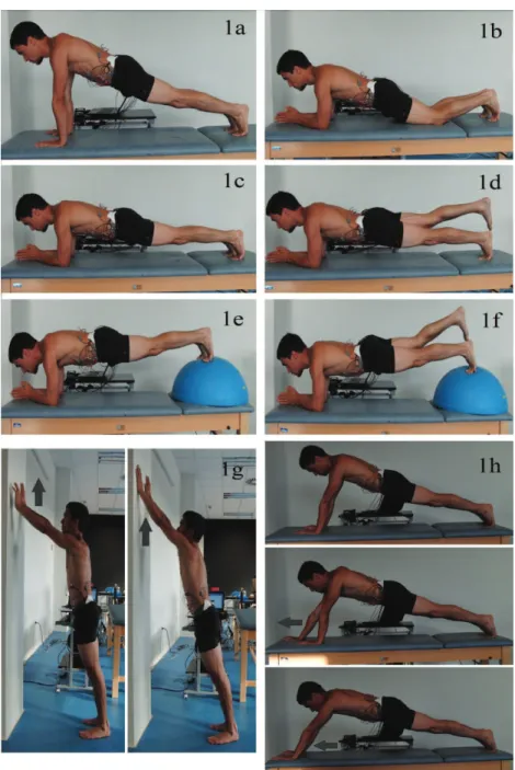
variations of this exercise were performed: with elbows extended (Figure 1a); kneeling on a bench (Figure 1b); conventional front-bridge (Figure 1c); with elevated right leg (Figure 1d); with feet resting on the BOSUTM balance
trainer (Figure 1e); with let foot resting on the BOSUTM balance trainer
and elevated right leg (Figure 1f); starting from the front-bridge position on the wall, with elbows extended, walkout moving the hands up 30 cm (Figure 1g); starting from the front-bridge position on the bench, with elbows extended, walkout moving the hands forward 30 cm (Figure 1h).
Figure 1. Images of the subject performing diferent forms of the front-bridge exercise: 1a) elbows extended; 1b) kneeling on the bench; 1c) conventional front-bridge; 1d) elevated right leg; 1e) feet resting on the BOSUTM
balance trainer; 1f) left foot on the BOSUTM balance trainer and elevated right leg; 1g) sagittal walkout on the
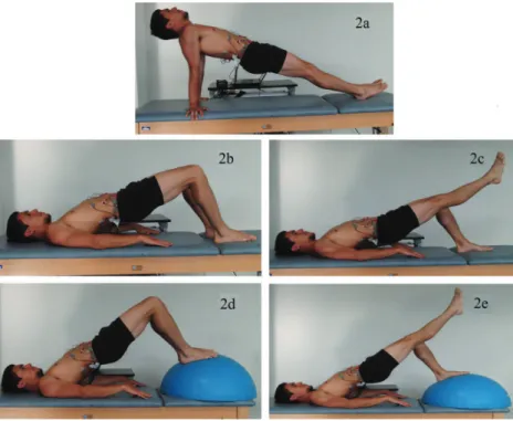
Back-bridges (Figure 2): the subject held the pelvis lited of the bench in a supine position, with the knees bent and the trunk aligned with the thighs. Five variations of this exercise were performed: with elbows extended (Fig-ure 2a), conventional back-bridge (Fig(Fig-ure 2b), with elevated right leg (Fig(Fig-ure 2c), with feet resting on BOSUTM balance trainer (Figure 2d), with let foot
resting on the BOSUTM balance trainer and elevated right leg (Figure 2e).
Figure 2. Images of the subject performing diferent forms of the back-bridge exercise: 2a) elbows extended; 2b) conventional back-bridge; 2c) elevated right leg; 2d) feet resting on the BOSUTM balance trainer; 2e) left foot
on BOSUTM balance trainer and elevated right leg.
Right side-bridges (Figure 3): the subject maintained the pelvis lited of the bench in a right lateral position, with the trunk fully aligned with the thighs. Eight variations of this exercise were performed: with right elbow extended (Figure 3a), resting on the right elbow and knee (Figure 3b), conventional right side-bridge (Figure 3c), with let hip lexion (Figure 3d), with feet resting on BOSUTM balance trainer (Figure 3e), with right
foot resting on the BOSUTM balance trainer and let hip lexion (Figure
3f), rolling on the wall from the right side-bridge into the front-bridge position (Figure 3g), rolling on the bench from the right side-bridge into the front-bridge position (Figure 3h).
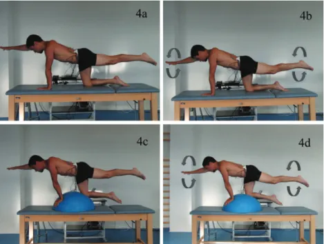
Bird-dogs (Figure 4): the subject maintained a 2-point kneeling position, with a contralateral arm and leg raise. Four variations of this exercise were performed: with elevated right arm and let leg (Figure 4a); with elevated right arm and let leg while drawing circles in the air with the raised limbs (Figure 4b); with elevated right arm and let leg on the BOSUTM balance
trainer (Figure 4c); with elevated right arm and let leg on the BOSUTM
Figure 3. Images of the subject performing diferent forms of the right side-bridge exercise: 3a) right elbow extended; 3b) right knee on the bench; 3c) conventional right side-bridge; 3d) elevated left leg with hip lexion; 3e) feet resting on BOSUTM balance trainer; 3f) right foot on the BOSUTM balance trainer and left hip lexion; 3g)
rolling on the wall; 3h) rolling on the bench.
Figure 4. Images of the subject performing diferent forms of the bird-dog exercise: 4a) elevated right arm and left leg; 4b) elevated right arm and left leg while drawing circles with the raised limbs; 4c) elevated right arm and left leg on the BOSUTM balance trainer; 4d) elevated right arm and left leg on the BOSUTM balance trainer
while drawing circles with the raised limbs.
Data collection
Surface EMG signals were collected bilaterally (R = right, L = let) from the following trunk muscles and locations: rectus abdominis (RA), approximately 3 cm lateral to the umbilicus; external oblique (EO), approximately 15 cm lateral to the umbilicus; internal oblique (IO), in the geometric center of the triangle formed by the inguinal ligament, the outer edge of the rectus sheath and the imaginary line between the anterior superior iliac spine and the umbilicus; and erector spinae (ES), 3 cm lateral to L3 spinous process. he Muscle Tester ME6000® (Mega Electronics Ltd., Finland) was used for the EMG recordings. his is a portable microcomputer with an 8-channel A/D conversion (14 bit resolution), a CMRR of 110 dB and a band-pass ilter of 8-500 Hz. Sampling frequency was programmed at 1000 Hz. he EMG signals were transferred with an optical cable to a compatible laptop where they were monitored by the Megawin® 2.5 program (Mega Electronics Ltd., Finland). Before data collection, pre-gelled disposable bipolar Ag-AgCl disc surface electrodes were placed in bipolar coniguration on the aforemen-tioned sites, previously cleaned with alcohol. he center-to-center electrode distance was 3 cm. Care was taken to ensure precise electrode placement to guarantee reproducibility of the measure. he subject was asked to contract his muscles to check the detection of an appropriate signal.
EMG signals. he MVC protocol has been described elsewhere in detail22.
Data reduction
In order to remove possible artifacts, the raw EMG signals were visually reviewed. hen they were full wave rectiied, averaged every 0.1 s and nor-malized to maximum EMG values obtained during the MVCs. In order to rank the exercises by level of muscular activation, the center 3 s window of the normalized EMG signal was averaged for each exercise and muscle.
RESULTS
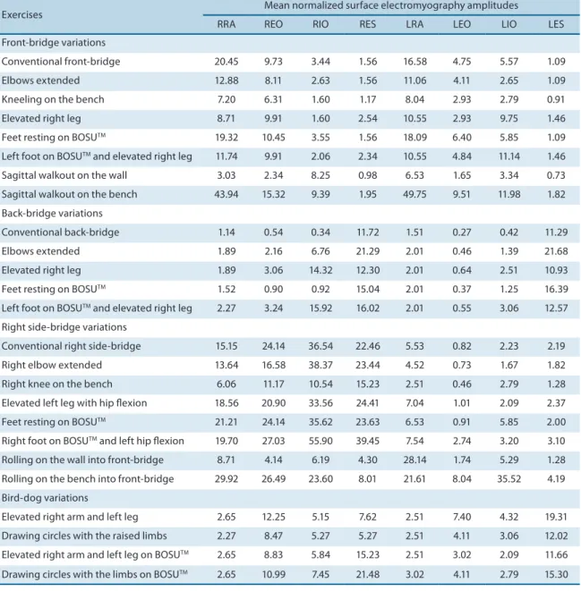
Table 1 shows the mean activation level of the recorded trunk muscles during diferent variations of the conventional front-bridge, back-bridge, side-bridge and bird-dog exercises.
he mean levels of trunk muscle activation were low to moderate. RA was mainly activated during the front-bridge and side-bridge variations, with maximum RA activity occurring in the sagittal walkout on the bench (RRA: 43.94% MVC; LRA: 49.75% MVC). For EO, the higher levels of mus-cular activation were recorded during the side-bridge variations, especially when performing the rolling on the bench into the front-bridge position (right EO: 26.49% MVC). Similarly, the higher levels of IO activation were found during the side-bridge variations: 55.90% MVC for RIOin the right side-bridge on the BOSUTM balance trainer with let hip lexion, and 35,52%
MVC for LIO in the rolling on the bench into the front-bridge position. he obliques were also activated during the diferent forms of the front-bridge, mainly when they were required to prevent twisting trunk motion in asymmetric postures or limb motions. Finally, ES was activated during the back-bridge, the side-bridge and the bird-dog variations. In particular, the right side-bridge on the BOSUTM balance trainer with let hip lexion
and the back-bridge with elbows extended produced the maximum acti-vation level for RES (39.45% MVC) and LES (21.68% MVC), respectively. he conventional front-bridge mainly activated the RA, followed by the obliques. he activation of the abdominal muscles was reduced when the front-bridge was performed kneeling on the bench or with elbows extended. Similarly, the RA and EO normalized EMG was very low dur-ing the sagittal walkout on the wall (e.g.: RRA: 3.03% MVC; REO: 2.34% MVC). When the front-bridge exercises were carried out with the right leg elevated, the LIO activation increased and the RA activation decreased. he sagittal walkout on the bench was the only front-bridge variation that increased the activation of all abdominal muscles. Interestingly, perform-ing the front-bridges on the BOSUTM balance trainer did not produce any
efect on the trunk muscle EMG.
he conventional back-bridge isolated the activation of the ES. he activa-tion of this muscle increased when the back-bridge was performed with elbows extended or with feet resting on the BOSUTM balance trainer. In addition,
he conventional right side-bridge activated the right side trunk muscles. he activation of these muscles was reduced when the right side-bridge was performed kneeling on the bench or when rolling on the wall into the front-bridge. In addition, the exercise with right elbow extended reduced the REO activation. In contrast, the rest of the side-bridge forms increased trunk muscle activity, especially the side-bridge with right foot on the BOSUTM balance
trainer and let hip lexion, and the rolling on the bench into the front-bridge. Interestingly, in the latter exercise, which involves an important rotational torque, the REO and LIO were co-activated to prevent twisting.
he bird-dog variations mainly activated the ES, followed by the obliques. Drawing circles with the raised limbs and performing these exercises on the
Table 1. Mean normalized surface electromyography amplitudes (% MVC) for each muscle tested during diferent variations of the conventional front-bridge, back-front-bridge, side-bridge and bird-dog exercises
Exercises Mean normalized surface electromyography amplitudes
RRA REO RIO RES LRA LEO LIO LES
Front-bridge variations
Conventional front-bridge 20.45 9.73 3.44 1.56 16.58 4.75 5.57 1.09
Elbows extended 12.88 8.11 2.63 1.56 11.06 4.11 2.65 1.09
Kneeling on the bench 7.20 6.31 1.60 1.17 8.04 2.93 2.79 0.91
Elevated right leg 8.71 9.91 1.60 2.54 10.55 2.93 9.75 1.46
Feet resting on BOSUTM 19.32 10.45 3.55 1.56 18.09 6.40 5.85 1.09
Left foot on BOSUTM and elevated right leg 11.74 9.91 2.06 2.34 10.55 4.84 11.14 1.46
Sagittal walkout on the wall 3.03 2.34 8.25 0.98 6.53 1.65 3.34 0.73
Sagittal walkout on the bench 43.94 15.32 9.39 1.95 49.75 9.51 11.98 1.82
Back-bridge variations
Conventional back-bridge 1.14 0.54 0.34 11.72 1.51 0.27 0.42 11.29
Elbows extended 1.89 2.16 6.76 21.29 2.01 0.46 1.39 21.68
Elevated right leg 1.89 3.06 14.32 12.30 2.01 0.64 2.51 10.93
Feet resting on BOSUTM 1.52 0.90 0.92 15.04 2.01 0.37 1.25 16.39
Left foot on BOSUTM and elevated right leg 2.27 3.24 15.92 16.02 2.01 0.55 3.06 12.57
Right side-bridge variations
Conventional right side-bridge 15.15 24.14 36.54 22.46 5.53 0.82 2.23 2.19
Right elbow extended 13.64 16.58 38.37 23.44 4.52 0.73 1.67 1.82
Right knee on the bench 6.06 11.17 10.54 15.23 2.51 0.46 2.79 1.28
Elevated left leg with hip lexion 18.56 20.90 33.56 24.41 7.04 1.01 2.09 2.37
Feet resting on BOSUTM 21.21 24.14 35.62 23.63 6.53 0.91 5.85 2.00
Right foot on BOSUTM and left hip lexion 19.70 27.03 55.90 39.45 7.54 2.74 3.20 3.10
Rolling on the wall into front-bridge 8.71 4.14 6.19 4.30 28.14 1.74 5.29 1.28
Rolling on the bench into front-bridge 29.92 26.49 23.60 8.01 21.61 8.04 35.52 4.19
Bird-dog variations
Elevated right arm and left leg 2.65 12.25 5.15 7.62 2.51 7.40 4.32 19.31
Drawing circles with the raised limbs 2.27 8.47 5.27 5.27 2.51 4.11 3.06 12.02
Elevated right arm and left leg on BOSUTM 2.65 8.83 5.84 15.23 2.51 3.02 2.09 11.66
Drawing circles with the limbs on BOSUTM 2.65 10.99 7.45 21.48 3.02 4.11 2.79 15.30
BOSUTM balance trainer did not have a clear efect on the trunk muscular
recruitment. It seems that the activation migrated from LES to RES.
DISCUSSION
Conventional bridge and bird-dog exercises challenge the trunk muscu-lature while applying relatively small loads to the lumbar spine6,23. In the
same way as observed in the present study, many EMG studies have shown that back-bridges mainly activate the trunk extensor muscles8,10,13,14,16,
front-bridges the sagittal lexor muscles10,11,13-15, side-bridges the lateral bend
muscles10,11,13-15, and bird-dogs the trunk extensor and rotator muscles6,10-12.
In this study, several forms of these exercises were analyzed in order to guide itness instructors and clinicians when prescribing stabilization exercises: starting level, progression, dosage, etc.
Bridge exercises kneeling on the bench
Compared with the conventional forms, performing the front- and side-bridges kneeling on the bench reduced the weight lited of the bench and its lever arm, and therefore the muscular challenge (Table 1). hese “short” bridges seem to be a good option for beginners or for people who are concerned with safety, as they impose minimal compressive penalty on the spine24.
Bridge exercises with elbows extended
Similarly, when the front- and side-bridges were performed with elbows extended, the lever arm of the weight lited of the bench was shorter than during the conventional forms, and consequently the muscular activa-tion levels were lower (Table 1). his reducactiva-tion in muscular activaactiva-tion was smaller for the side-bridge with elbows extended, maybe because the mus-cles were recruited in some way to ensure body balance (since during this side-bridge variation the height of the center of gravity was higher and the base of support smaller). Regarding the back-bridge variations, the weight lited of the bench and its lever arm were higher during the exercise with elbows extended. herefore, unlike the front- and side-bridges, the back-bridge with elbows extended produce higher muscular activation levels than the conventional form (Table 1). We have reviewed the literature in order to compare these results with previous data; however, despite our best eforts, we did not ind studies that analyzed the efect of modifying the elbow position during bridge exercises on trunk muscular recruitment.
Bridge exercises with single leg support (an elevated leg)
he results of the present study conirm those obtained recently by our research group10. While bridge exercises with double leg support produced
back-bridges, performing these exercises with elevated right leg increased the activation of LIO and RIO, respectively (Table 1). he increase of the IO activation was needed to combat the rotational torque and the more unstable position caused by the unilateral leg support. Although hold-ing asymmetric postures while bridghold-ing elicits a major muscular chal-lenge6,8,10,13,16, it also increases the load on the spine6.
Stabilization exercises with limb/shoulder motion
Before moving the limbs in diferent positions, trunk muscle co-activation is needed to control spine stability25. Unilateral limb motions while
per-forming a stabilization exercise challenge the motor system in order to stabilize the trunk and to control body balance. In this way, as determined by McGill and Karpowicz11, drawing squares in the air with the elevated
limbs during a conventional bird-dog exercise increases the activation of diferent muscles. However, a reduction in muscular activation was ob-served in our study when drawing circles in the air during the bird-dog (Table 1). It was the irst time the participant performed this exercise and he was not able to hold the posture with minimal associated movement. Possibly, he paid more attention to moving the arm and leg simultaneously than to controlling the trunk posture and the neutral spine position.
he sagittal walkout can be classiied as a front-bridge exercise with limb motion. he walkout on the bench elicited higher muscular activation levels than walkout on the wall (Table 1), since the lever arm of the weight supported during the former exercise was higher, data which support the results obtained by McGill et al.18. Similarly, rolling on the bench from the
right side-bridge into the front-bridge produced higher activation levels than rolling on the wall (Table 1). Rolling and walkout on the bench gen-erated the highest activation levels for most muscles. hese exercises are not for beginners, as it is diicult to control the rotational torque applied to the trunk when the shoulder moves. In this respect, when rolling on the bench from the right side-bridge into the front-bridge position, REO and LIO were co-activated (Table 1) to produce rotational torque and to prevent twisting between thorax and pelvis. his oblique co-activation on opposite trunk sides to generate twisting torque was also observed in previous studies10,26,27.
Stabilization exercises on the BOSU
TMbalance trainer
Labile surfaces (e.g.: BOSUTM balance trainer or itball) are frequently
used during trunk exercises to challenge spine stability. As compared to exercises on stable surfaces, many studies have demonstrated that ex-ercises performed on unstable surfaces usually enhanced trunk muscle activation14,17. However, as shown in Table 1, the addition of an unstable
surface to trunk exercises does not always have an efect on the muscular response9,14,15, and may even reduce the muscular challenge9. Possibly,
Limitations of the study
In order to establish safe trunk stabilization exercise progressions, further biomechanical studies should assess both muscular recruitment and spine loading. In addition, interpretation of the results of this study is limited because our subject was relatively young, healthy and physically it. In order to provide a major insight on trunk muscle response during stabilization exercises, future studies should analyze the trunk muscular recruitment of older and unit people, and/or of patients with diferent spinal conditions.
CONCLUSION
he EMG data reported in this study may be used to support decision-making when prescribing trunk stabilization exercise progressions. In this respect, the following key points should be taken into consideration: i) conventional front-, back- and side-bridges mainly activate the trunk lexor, extensor and lateral bend muscles, respectively; ii) performing front- and side-bridges with elbows extended, and especially kneeling on the bench, reduces muscle activation; iii) performing back-bridges with elbows extended increases muscle activation; iv) single leg support and/or limb motions while performing stabilization exercises increase the activa-tion of the trunk rotators, especially the internal oblique; v) performing stabilization exercises on unstable surfaces can increase the muscular challenge, but this depends on the way these devices are used.
Acknowledgments
Research supported by Ministerio de Ciencia e Innovación of Spain (DEP2010-16493).
REFERENCES
1. Bergmark A. Stability of the lumbar spine: a study in mechanical engineering. Acta Orthop Scand Suppl 1989;230:1-54.
2. Panjabi MM. he stabilizing system of the spine. Part I. Function, dysfunction, adaptation, and enhancement. J Spinal Disord Tech 1992;5:383-9.
3. Brown SHM, Vera-García FJ, McGill SM. Efects of abdominal muscle coactivation on the externally pre-loaded trunk: variations in motor control and its efect on spine stability. Spine 2006;31:387-93.
4. Vera-Garcia FJ, Brown SHM, Gray JR, McGill SM. Efects of diferent levels of torso coactivation on trunk muscular and kinematic responses to posteriorly applied sudden loads. Clin Biomech 2006;21:443-55.
5. Vera-Garcia FJ, Elvira JLL, Brown SHM, McGill SM. Efects of abdominal stabiliza-tion maneuvers on the control of spine mostabiliza-tion and stability against sudden trunk perturbations. J Electromyogr Kinesiol 2007;17:556-67.
6. Kavcic N, Grenier S, McGill SM. Quantifying tissue loads and spine stability while performing commonly prescribed low back stabilization exercises. Spine 2004;29:2319-29.
7. McGill SM, Grenier S, Kavcic N, Cholewicki J. Coordination of muscle activity to assure stability of the lumbar spine. J Electromyogr Kinesiol 2003;13(4):353-9. 8. Bjerkefors A, Ekblom MM, Josefsson K, horstensson A. Deep and supericial
Corresponding author
Francisco Jose Vera-Garcia Centro de Investigación del Deporte. Universidad Miguel Hernández de Elche.
Avda. de la Universidad s/n. 03202 - Elche (Alicante). Spain.
E-mail: [email protected]
9. Drake JD, Fischer SL, Brown SH, Callaghan JP. Do exercise balls provide a training advantage for trunk extensor exercises? A biomechanical evaluation. J Manipulative Physiol her 2006;29(5):354-62.
10. García-Vaquero MP, Moreside JM, Brontons-Gil E, Peco-González N, Vera-Garcia FJ. Trunk muscle activation during stabilization exercises with single and double leg support. J Electromyogr Kinesiol 2012;22(3):398-406.
11. McGill SM, Karpowicz A. Exercises for spine stabilization: motion/motor pat-terns, stability progressions, and clinical technique. Arch Phys Med Rehabil 2009;90:118-26.
12. Stevens VK, Vleeming A, Bouche KG, Mahieu NN, Vanderstraeten GG, Danneels LA. Electromyographic activity of trunk and hip muscles during stabilization exercises in four-point kneeling in healthy volunteers. Eur Spine J 2007;16:711-8. 13. Ekstrom RA, Donatelli RA, Carp KC. Electromyographic analysis of core trunk,
hip, and thigh muscles during 9 rehabilitation exercises. J Orthop Sport Phys 2007;37:754-62.
14. Imai A, Kaneoka K, Okubo Y, Shiina I, Tatsumura M, Izumi S, et al. Trunk muscle activity during lumbar stabilization exercises on both a stable and unstable surface. J Orthop Sport Phys 2010;40:369-75.
15. Lehman GJ, Hoda W, Oliver S. Trunk muscle activity during bridging exercises on and of a Swiss ball. Chiropr Osteopat 2005;13:14.
16. Stevens VK, Bouche KG, Mahieu NN, Coorevits PL, Vanderstraeten GG, Dan-neels LA. Trunk muscle activity in healthy subjects during bridging stabilization exercises. BMC Musculoskel Dis 2006;7:75.
17. Vera-Garcia FJ, Grenier SG, McGill S. Abdominal response during curl-ups on both stable and labile surfaces. Physical her 2000;80:564-9.
18. McGill SM, Karpowicz A, Fenwick CM, Brown SH. Exercises for the torso per-formed in a standing posture: spine and hip motion and motor patterns and spine load. J Strength Cond Res 2009;23(2):455-64.
19. Santana JC, Vera-Garcia FJ, McGill SM. A kinetic and electromyographic comparison of the standing cable press and bench press. J Strength Cond Res 2007;21(4):1271-7.
20. Moreside JM, Vera-Garcia FJ, McGill SM. Trunk muscle activation patterns, lumbar compressive forces, and spine stability when using the bodyblade. Physical her 2007;87:153-63.
21. Sánchez-Zuriaga D, Vera-Garcia FJ, Moreside JM, McGill SM. Trunk muscle acti-vation patterns and spine kinematics when using an oscillating blade: inluence of diferent postures and blade orientations. Arch Phys Med Rehabil 2009;90:1055-60. 22. Vera-Garcia FJ, Moreside JM, McGill SM. MVC techniques to normalize trunk
muscle EMG in healthy women. J Electromyogr Kinesiol 2010;20:10-6.
23. Axler CT, McGill, SM. Low back loads over a variety of abdominal exercises: searching for the safest abdominal challenge. Med Sci Sports Exerc 1997;29:804-11. 24. McGill SM. Low back exercises: evidence for improving exercise regimens. Phys
her 1998;78(7):754-65.
25. Hodges PW, Richardson C. Contraction of the abdominal muscles associated with movement of the lower limb. Physical her 1997;77(2):132-44.
26. McGill SM. Electromyographic activity of the abdominal and low back musculature during the generation of isometric and dynamic axial trunk torque: implications for lumbar mechanics. J Orthop Res 1991;9:91-103.