Introduction
Contemporary lifestyle is often characterized by sedentary behav-ior and an excess intake of a high-fat diet (HFD), increasing the chances of developing of obesity1 and several chronic diseases, including arterial hypertension2 and autonomic alterations3. Conlicting results have emerged over the nature of autonomic activity in obesity4. These investigators showed that obese subjects presented lower sympathetic activity but no change in parasympathetic modulation. On the contrary, an increase in body weight (BW) is associated with sympathetic overactivity and attenuation in parasympathetic activity5. Likewise, obese subjects have shown an imbalance in autonomic nervous system (ANS) activity, which is characterized by an increase in para-sympathetic nervous system (PNS) tonus and/or a decrease in the sympathetic nervous system (SNS) tonus6.
Obesity was found to be associated with decreased sympathetic activity in obesity7. Others researcher’s, however, also reports a sympathetic hyperactivity2. In animal models, HFD-induced obesity increases sympathetic activity and renin-angiotensin-aldosterone system, consequently increasing blood pressure (BP)8. Obesity not only shifts SNS, but it also changes cardiac PNS drive. The HFD has been found to reduce parasympathetic activity in rats9. Body mass index (BMI), waist circumference and sum of skinfolds are associated with a reduced cardiac vagal tonus and indicators of central obesity as compared to total indicators of obesity10. Increase of BW is associated with a decline in PNS, accompanied by a rise in heart rate (HR), and conversely, HR declines during weight reduction This is a point important because increment in HR has been shown as a marker associated with increased mortality11.
Exercise training ameliorates BP levels in overweight and lean subjects, as well as was effective in reducing sympathetic and parasympathetic nervous activities in obesity12. Chronic exercise may prevent obesity development and metabolic alteration13. Despite much study in this ield, the mechanisms through which excessive fat accumulation results in an increased incidence of cardiovascular autonomic dysfunction and the possible ef-fects of ET in these parameters are still a matter of debate. The mechanisms underlying this relationship are multifactorial, and for some time there are controversy as to whether the SNS and PNS were activated or inhibited in obesity and the effects of ET. Therefore, we determined the alteration of baseline pressoric regulation, autonomic nerve activity by HR and BP variability, cardiac autonomic tonus and barorelex sensitivity in HFD-fed rats after swimming exercise training. This study aimed to clarify the therapeutic effect of exercise training on sympathetic acti-vation and parasympathetic attenuation as well as bradycardic and tachycardic barorelex responses in diet-induced obese rats.
Materials and methods
Animal model
Adult male Wistar rats at 10-12 weeks of age and BW around 380-400g were procured from Physiology Laboratory. The ani-mals were kept at a constant temperature of 22 ± 1°C, relative humidity of 50–60% and light/dark cycle (12/12 h). They were fed with different diets as described below and drinking water ad libitum. The rats were randomly assigned to 4 experimental Original Article (short paper)
Exercise training reverse autonomic dysfunction and hypertension in rats fed
with high-fat diet
Carla Cristina de Sordi1, Marina de Paiva Lemos1, Moacir Marocolo2, Rosangela Soares Chriguer3, Edmar Lacerda Mendes1, Gustavo Ribeiro da Mota1, Octávio Barbosa Neto1
1Universidade Federal do Triângulo Mineiro, UFTM, Uberada, MG, Brazil; 2Universidade Federal de Juiz de
Fora, UFJF, Juiz de Fora, MG, Brazil; 3Universidade Federal de São Paulo, UNIFESP, Santos, SP, Brazil.
Abstract — Aims: We evaluated whether exercise training ameliorates cardiovascular autonomic dysfunction in obesity-induced by high-fat diet (HFD) in rats. Methods: Wistar male rats were assigned in normal diet, sedentary (ND-S) and trained (ND-T) and HFD, sedentary (HFD-S) and trained (HFD-T). Blood pressure (BP), heart rate (HR), HR variability (HRV), BP variability (BPV), cardiac barorelex and cardiac autonomic tonus were assessed. Results: HFD-S showed higher bodyweight increase (19.4%) compared to all other groups (HFD-T: 13.2%, ND-S: 14% and ND-T: 12.4%). Relative epididymal, retroperitoneal and visceral fat was also greater in HFD-S compared to all other groups. Resting bradycardia in ND-T (339.5±10.6 bpm) and HFD-T (341.0 ± 9.4 bpm) was more pronounced than ND-S (438.4 ± 6.3 bpm; p<0.05) and HFD-S (448.5 ± 18.7 bpm; p<0.05). The HFD-T group showed lower systolic (125.3 ± 1.9 mmHg), diastolic (88.5 ± 2.0 mmHg) and mean BP (100.3 ± 2.5 mmHg) in comparison with HFD-S (153.8 ± 3.7; 103.5 ± 2.6 and 120.5 ± 3.7 mmHg; p<0.05, respectively). Lower variance of HRV and higher variance of diastolic BPV was observed in HFD-S compared to other groups while sympathetic modulation of HRV and BPV was higher in HFD-S, as well as the LF/HF ratio. HFD-T showed a decrease in sympathetic tonus compared to HFD-S. ND-T and HFD-T showed higher cardiac vagal tonus than respective sedentary groups. Analysis showed an association between visceral fat, sympathetic activity and BP. Conclusions: HFD induces hypertension and is associated with autonomic imbalance in rats, while exercise training may reverse these harmful changes.
groups that received either a normal diet plus sedentary activity (ND-S, n=8), a high-fat diet plus sedentary activity (HFD-S, n=8), a normal diet plus exercise training activity (ND-T, n=8) and a high-fat diet plus exercise training activity (HFD-T, n=8). All experimental procedures were performed in accordance with the Ethical Principles in Animal Research of the Brazilian College of Animal Experimentation and were approved by the local Ethical Committee for Animal Research (Protocol 167/2010).
Diet and exercise training
A 9-week dietary intervention was followed by a same period of exercise intervention. The both ND groups received a standard diet, normocaloric (commercial Nuvilab® CR1 rat feeding) containing (% weight) 19% protein, 56% carbohydrate, 3.5% lipids, 4.5% cellulose and 5% vitamins and minerals, while the HFD groups received a diet consisted of a mixture of ground and calorie foods that were offered in the form of pellets. They were fed with (% weight) 15g of standard diet (Nuvilab® CR1), 10g roasted peanuts, 10g milk chocolate and 5g of cornstarch cookie. This type of diet had in its composition 20% protein, 48% carbohydrate, 20% as fat, 4% cellulose and 5% of vitamins and minerals14.The swimming protocol was performed in a glass tank and ambient water temperature was kept at 30º ± 1ºC. The trained animals received a 20 min adaptation period on the irst day and were increased 10-min each day until reaching 1 h on the ifty day15. Following, these rats trained 5 days/week with a gradual progression toward a 2 h session. This protocol is deined as an aerobic endurance training and low-intensity, as the animals swam without additional work load, this method corresponds the intensity below the anaerobic threshold in rats16. Sedentary animals were placed in the swimming apparatus for 10 min twice a week to mimic the water stress associated with the experimental protocol. BW was measured in a conscious state at weekly intervals.
Experimental procedures and hemodynamic recording protocol
One day after the last exercise session, acute experiments were performed under anesthesia induced by intraperitoneal injection of sodium pentobarbital (40 mg/kg). Arterial and venous catheters (PE-10 attached to PE-50) illed with heparinized saline were inserted in the femoral artery and vein to direct recordings of pulsatile BP and drug administration, respectively. Followed, the catheters were tunneled subcutaneously and exteriorized at the scapular region. Twenty-four hours after catheter surgery comple-tion, BP and HR were monitored in conscious freely moving rats by connecting the arterial catheter to a pressure transducer (P23Db, Gould-Statham) connected to ampliier (ML224 Quad Bridge Amp, ADInstruments, NSW, Australia) and a microcomputer equipped with an analog-to-digital converter board (CODAS, 4-kHz sampling frequency, Di220 Dataq Instruments, Inc., Akron, OH, USA). After animal acclimation to the experimental room, the hemodynamic parameters were recorded for 30 min. Mean
(MBP), systolic (SBP), and diastolic BP (DBP) were analyzed off-line. Beat-by-beat pulse interval (PI) values were generated off-line from the pulsatile arterial BP signal by measuring the time interval between two systolic peaks.
Linear analysis of HR and BP variability
For the cardiovascular variability study, the signals of BP were processed to generate beat-to-beat time series of PI, DBP, and SBP. The variance of these values in each period was considered a variability index in the time-domain assessed by the standard deviation of the time series. The PI, DBP and SBP luctuations were assessed in the frequency domain by means of autoregres-sive spectral estimation method as described elsewhere17. Briely, beat by beat time series of PI, DBP, and SBP were divided into serial segments of 300 beats with a 50% overlap on the previ-ous segments (Welch’s method). The spectra of each segment were estimated via the Levinson-Durbin recursion, and the order of the model chosen according to Akaike’s criterion18. The oscillatory components were labeled as very low (VLF, 0.01 to 0.20 Hz), low (LF, 0.20 to 0.75 Hz) or high frequency (HF, 0.75 to 3.00 Hz). The normalization procedure, applied only to the variability of the PI, was performed by dividing the power of the LF or HF component by total spectral power, which is subtracted from the power of the VLF band and multiplying the result by 10019. The normalization procedure tends to minimize the effect of the changes in total power on the absolute values of LF and HF components of HRV17.
Barorelex sensitivity and cardiac autonomic tonus
Removal of white adipose tissue depot.
At the completion of the experiment, rats were euthanized with an overdose of pentobarbital sodium (300 mg/kg). The epi-didymal, retroperitoneal (surrounding the kidney) and visceral (surrounding the stomach and intestines) white adipose tissue were rapidly removed, rinsed in deionized water to remove excess blood and weighed.
Statistical analysis
The normality of the data was checked with the Kolmogorov-Smirnov test and Levene’s F test was used to assess the ho-mogeneity. Signiicance of differences of continuous variables between normal-fed and high fat-diet animals and exercise training was tested using 2-way ANOVA with Tukey post-test (normally distributed variables). Non-normally distributed
variables were tested by non-parametric Friedman test followed by Mann–Whitney U-tests. Pearson correlation coeficient was used to test the correlation between white adipose tissue with sympathetic activity and blood pressure levels. All data are represented as mean ± SEM. Probability values of p<0.05 were considered statistically signiicant. Analyses were performed using SigmaStat® SPSS, Chicago, IL.
Results
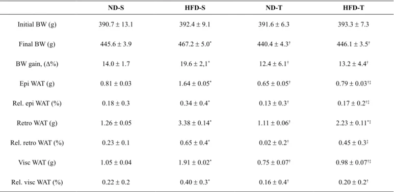
HFD intake induced obesity in sedentary rats as evidenced by the increased inal BW, BW gain and adiposity (masses of epididymal, retroperitoneal and visceral adipose depots) in comparison to the other groups (p<0.05). The ET was able to prevent the increases in the above abnormal anatomic data in HFD-induced obese rats (p<0.05), similarly to sedentary and trained animals with normal diet (Table 1).
Table 1. Descriptive data for inal BW mass, BW gain (relative D%), absolute and relative weights of epididymal, retroperitoneal and visceral adi -pose tissue weights from sedentary normal diet (ND-S, n=8), sedentary high-fat diet (HFD-S, n=8), trained normal diet (ND-T, n=8) and trained high-fat diet (HFD-T, n=8) groups for 9 weeks.
ND-S HFD-S ND-T HFD-T
Initial BW (g) 390.7 ± 13.1 392.4 ± 9.1 391.6 ± 6.3 393.3 ± 7.3
Final BW (g) 445.6 ± 3.9 467.2 ± 5.0* 440.4
± 4.3† 446.1
± 3.5†
BW gain, (D%) 14.0 ± 1.7 19.6 ± 2,1* 12.4 ± 6.1† 13.2 ± 4.4†
Epi WAT (g) 0.81 ± 0.03 1.64 ± 0.05* 0.65 ± 0.05† 0.79 ± 0.03†‡
Rel. epi WAT (%) 0.18 ± 0.3 0.34 ± 0.4* 0.13
± 0.3† 0.17
± 0.2†‡
Retro WAT (g) 1.26 ± 0.05 3.38 ± 0.14* 1.11
± 0.06† 2.23
± 0.11*‡
Rel. retro WAT (%) 0.23 ± 0.1 0.65 ± 0.4* 0.02 ± 0.2† 0.45 ± 0.3‡
Visc WAT (g) 1.05 ± 0.04 1.91 ± 0.02* 0.75 ± 0.07† 0.98 ± 0.07†‡
Rel. visc WAT (%) 0.22 ± 0.2 0.40 ± 0.3* 0.16
± 0.4† 0.20
± 0.2†
Values are mean ± SEM. Abbreviations: BW; body weight, WAT; white adipose tissue, Epi; epididymal, Retro; retroperitoneal, Visc; visceral. Signiicances are
*p<0.05 vs. ND-S; †p<0.05 vs. HFD-S and ‡p<0.05 vs. ND-T. (Statistical differences were determined by 2-way ANOVA followed by Tukey post-test).
Under baseline conditions, resting bradycardia in both ND-T (339.5 ± 10.6 bpm) and HFD-T (341.0 ± 9.4 bpm) male rats has been more closely pronounced when compared with their respective sedentary groups (438.4 ± 6.3 bpm in ND-S and 448.5 ± 18.7 bpm in HFD-S, p<0.05; Fig. 1A). The sedentary rats fed a HFD exhibited a higher systolic BP (153.8 ± 3.7 mmHg) when compared with HFD-T (125.3 ± 1.9 mmHg; p<0.001), ND-S
ND-T (98.4 ± 4.1 mmHg; p<0.001), respectively (Fig. 1D). No differences were observed in BP between sedentary and trained rats fed a normal diet.
The variance of HRV was lower in HFD-S when compared with ND-S, ND-T and HFD-T animals (p<0.05). On the other hand, the variance of DBPV increased in comparison to the same groups
(p<0.05). The LF component of HRV, SBPV and DBPV were higher in HFD-S than in the other groups (p<0.05). The HF power of HRV was lower in sedentary rats submitted to HFD compared to ND-S, ND-T and HFD-T (p<0.05). Considered a hallmark of sympatho-vagal balance, the LF/HF relation was higher in HFD-S rats in comparison to the other groups (p<0.05) (Table 2).
Figure 1. Values of (a) resting heart rate (HR, bpm), (b) systolic blood pressure (SBP, mmHg), (c) diastolic blood pressure (DBP, mmHg) and (d) mean blood pressure (MBP, mmHg) in non-anesthetized Wistar rats submitted to normal diet plus sedentary activity (ND-S, n=8), high-fat diet plus sedentary activity (HFD-S, n=8), normal diet plus exercise training activity (ND-T, n=8) and high-fat diet plus exercise training activity (HFD-T, n=8) treatment during 9 weeks. Bars represent mean ± SEM. *p<0.05 vs. ND-S, †p<0.05 vs. HFD-S, ‡p< 0.05 vs. ND-T. (Statistical differences were determined by 2-way ANOVA followed by Tukey post-test).
*
*
†
*
†
*
†
†
†
†
*
†
†
500
400
300
200
100
0 a)
HR (bpm)
120
80
40
0 c)
DBP
(mmHg)
b) 200
150
100
50
0
SBP
(mmHg)
0 50 100 150 d)
MBP
(mmHg)
ND-S HFD-S ND-T HFD-T ND-S HFD-S ND-T HFD-T
Table 2. Mean values (± SEM) of variance and VLF, LF, and HF spectral components of systolic and diastolic blood pressure variability and pulse interval variability in non-anesthetized Wistar rats submitted to normal diet plus sedentary activity (ND-S, n=8), high-fat diet plus seden-tary activity (HFD-S, n=8), normal diet plus exercise training activity (ND-T, n=8) and high-fat diet plus exercise training activity (HFD-T, n=8) treatment during 9 weeks.
ND-S HFD-S ND-T HFD-T
Spectral parameters
SBP
Variance, mmHg2 11.7 ± 1.8 12.7 ± 1.7 12.9 ± 1.5 14.5 ± 2.4
LF, mmHg2 5.7
± 0.9 12.9 ± 1.8* 4.2
± 1.0† 7.5
± 1.7†
HF, mmHg2 7.3± 0.4 5.9±3.2 8.6±1.5 7.9±0.6
DBP
Variance, mmHg2 11.2 ± 0.7 17.0 ± 1.5* 11.3 ± 0.9† 12.8 ± 1.4†
LF, mmHg2 3.4
± 0.4 9.6 ± 0.5* 3.5
± 0.4† 4.7
± 0.5†
PI
Variance, ms2 27.7 ± 0.9 19.9 ± 0.6* 30.6 ± 1.3† 24.3 ± 5.4
VLF, ms2 8.3
± 1.2 11.4 ± 1.7 7.4 ± 1.8 8.9 ± 1.6
LF, ms2 4.1 ± 1.0 10.2 ± 1.6* 4.2 ± 1.1† 4.1 ±1.8†
LF, nu 13.7 ± 2.5 19.5 ± 3.9 12.7 ± 1.5 11.8 ± 1.6
HF, ms2 14.7 ± 0.7 7.7 ± 0.9* 20.9 ± 2.2† 18.3 ± 1.2†
HF, nu 74.7 ± 2.4 70.3 ± 6.3 77.1 ± 6.2 83.3 ± 1.7
LF/HF 0.27 ± 0.0 1.58 ± 0.3* 0.26 ± 0.0† 0.23 ± 0.0†
Abbreviations: LF; low-frequency component, HF; high-frequency component, nu; indicates normalized units. Signiicances are *p<0.05 vs. ND-S; †p£0.05 vs.
HFD-S and ‡p<0.05 vs. ND-T. (The parametric variables were tested using 2-way ANOVA with Tukey post-test and non-parametric data were determined by Friedman test followed by Mann–Whitney U-tests.)
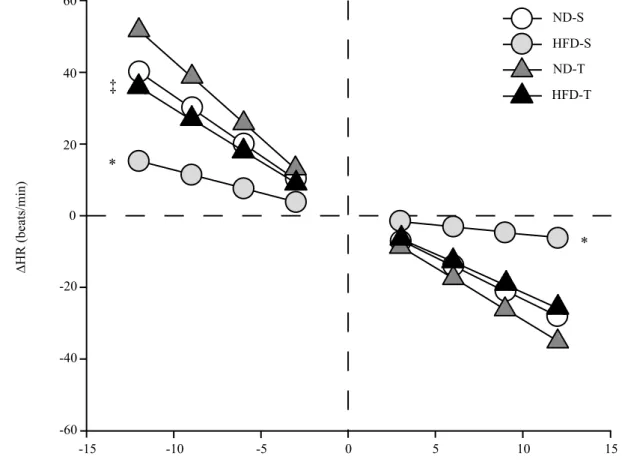
Figure 2 shows that the linear regression slope between changes in HR and MBP (bpm/mmHg) corresponding to relex bradycardia and relex tachycardia was greatly attenuated by high-fat diet in sedentary animals (-0.52 ± 0.1 and 1.65 ± 0.3 bpm/ mmHg; respectively). The exercise training improved barorelex control of HR in HFD-induced obese rats. Consequently, at the end of the 9-week intervention period, the relex bradycardic
sensitivity in HFD-T (-2.15 ± 0.2 bpm/mmHg) exceeder that observed in their respective sedentary group (p<0.001). The relex tachycardic sensitivity was also higher in HFD-T (2.83 ± 0.2 bpm/mmHg) than HFD-S rats (p<0.001) and was similar to that observed in ND-S group (3.04 ± 0.1 bpm/mmHg). The relex tachycardia in trained rats fed a normal diet was more pronounced in relation to trained rats fed a high-fat diet (p<0.001).
Figure 2. Effects of chronic exercise training on the relex control of the heart rate in non-anesthetized Wistar rats that were assigned to sedentary normal diet (ND-S, n=8), sedentary high-fat diet (HFD-S, n=8), trained normal diet (ND-T, n=8) and trained high-fat diet (HFD-T, n=8). Baro-relex bradycardic gain and baroBaro-relex tachycardic gain, respectively, induced by phenylephrine and sodium nitroprusside injections. BaroBaro-relex gains were obtained by DHR/DMAP index (bpm/mmHg). Data are mean ± SEM. *p<0.05 vs. all groups and ‡p<0.05 vs. ND-T. (Statistical dif-ferences were determined by 2-way ANOVA followed by Tukey post-test).
60
40
20
0
-20
-40
-60
-15 -10 -5 0 5 10 15
∆MBP (mmHg)
* *
‡
∆HR (beats/min)
ND-S
HFD-S
ND-T
Compared with all the groups, the HFD-S presented higher values of sympathetic tonus (p<0.05). The aerobic exercise train-ing could decreased sympathetic tonus in HFD in comparison to
their respective sedentary group (p<0.05). In contrast, both the ND-T and HFD-T groups shows a higher cardiac vagal tonus in comparison to ND-S and HFD-S (p<0.05) (Fig. 3).
Figure 3. Bar graphs showing sympathetic (ST) and vagal (VT) tone, basal heart rate (HR), intrinsic HR (IHR) and changes (%) in HR (DHR) after atropine (A) and propranolol (P) injections in conscious Wistar rats that were divided in four treatment groups, respectively, sedentary normal diet (ND-S, n=8), sedentary high-fat diet (HFD-S, n=8), trained normal diet (ND-T, n=8) and trained high-fat diet (HFD-T, n=8). The VT was estimated by the difference between the IHR (double blockade by atropine and propranolol) and the maximal bradycardia achieved after sympathetic blockade with propranolol. The ST was evaluated by the difference between the IHR the highest tachycardia observed after vagal blockade with atropine. Data presented as mean ± SEM. (Statistical differences were determined by 2-way ANOVA followed by Tukey post-test).
p < 0.05
p < 0.05
p < 0.05 p < 0.05
p < 0.05
ND-S HFD-S ND-T HFD-T
500
450
400
350
300
250
200
IHR HR
IHR
HR HR
IHR A
A
A
A
P P
P P
Sympathetic tonus Vagal tonus
Heart Rate (bpm)
IHR HR
Further analysis showed a signiicant association between visceral fat, sympathetic nervous activity and BP levels (Fig. 4), demonstrated by correlation of visceral white adipose tissue and LF component of HRV (r=0.44, p<0.05; Fig. 4a), white adipose tissue and LF component of SBPV (r=0.56, p<0.001; Fig. 4b), white adipose tissue and LF component of DBPV (r=0.45,
p<0.05; Fig. 4c). The Figure 4d show a correlation between visceral white adipose tissue and MBP (r=0.39, p<0.05). In addition, a strong positive association was also found between sympathetic modulation represented by the LF component of SBPV and MBP (r=0.81, p<0.001; Fig. 4e) and LF component of DBPV and MBP (r=0.76, p<0.001; Fig. 4f).
Discussion
One important inding of our research was that HFD-induced obesity in rats is correlated with autonomic imbalance represented by a sympathetic overactivity, parasympathetic attenuation and reduced response of barorelex sensitivity, as well as an associa -tion between elevated sympathetic activity and higher levels of BP. In addition, conducting a simple aerobic exercise training protocol is effective to reverse these changes.
Previous studies with animals have shown that obesity increases the risk of developing hypertension and other cardio-vascular diseases, and excessive sympathetic outlow contributes to the progression of obesity20. Feeding rat with a HFD is known to closely mimic many characteristics of human obesity21. Corroborating these studies, here we have demonstrated that HFD intake for 9 weeks induced obesity, as evidenced by the increased body weight and adiposity. The above variables in-dicated the rat model of diet induced obesity was successfully established and are suitable for investigating mechanistic links between obesity and hypertension.
The mechanisms underlying the development of obesity-induced hypertension are multifactorial and complex. Several researches demonstrated that, among neurohumoral, renal, and vascular factors, hyperactivity of the sympathetic nervous system contributes to the etiology of hypertension in obese humans and animal models2. In this sense, our data indicate that HFD-induced hypertension is associated with an increase in the LF component of pulse interval, systolic and diastolic BP, as well as an elevated sympathetic tonus to the heart, which are indexes of cardiac sympathovagal balance and vascular sympathetic outlow, respectively22.
Strengthening the involvement of sympathetic overactivity in obesity-induced hypertension are the previous indings that: irstly, drugs that block the sympathetic activity lowers BP to a greater extent in obese compared with lean subjects23; secondly, chronic treatment with clonidine (centrally acting a-agonist hypotensive agent) or a- and b-adrenergic receptor antagonists reduces SNS activity and prevents the increase in BP in animals fed with HFD and in hypertensive obese patients2.
The mechanisms involved in the increased high BP and concomitant sympathetic hyperactivity in high HFD-induced obesity were not addressed in the present study. However, some potential mechanisms may explain these effects observed. Among several neuromodulators involved in activation of the sympa-thetic modulation, one of the most studied in obesity is leptin, an adipokine secreted by adipocytes proportionally to the degree of adiposity. High levels of leptin could explain, at least in part, the obesity-induced hypertension and sympathetic hyperactiv-ity of humans24 and animals models25. Thus, leptin would be a link between the excess of adiposity and sympatho excitation26.
We showed that exercise training by swimming was able in attenuated the high BP and provoked fat loss, this indicates the therapeutic effect of chronic exercise on the symptoms of obesity. Exercise training has been shown to modulate sympa-thetic activity and maintain the balance between parasympasympa-thetic and sympathetic activity in the long term of many diseases27. Based on these studies, the regular physical exercise shows
beneicial effects and may be a non-pharmacological strategy for this particular patient group. As previously reviewed28, sympathetic hyperactivity is accompanied by the elevated sympathetic outlow to organs such as the heart, kidneys, and blood vessels29. Moreover, it can contribute to a further decline of insulin sensitivity, creating a vicious cycle that may promote the development of the metabolic syndrome. Sympathetic ner-vous system plays a key role in the control of energy balance and body weight29, so when the exercise training causes body weight loss and energy expenditure, the excess sympathetic activity is consequentially attenuated.
Another relevant inding of our study was that HFD-induced obesity rat model shown a reduced cardiac vagal tonus, mea-sured directly after cardiac pharmacological parasympathetic blockade. In addition, the evaluation of the heart rate variability (HRV) indices in the present study suggests a decrease in the activity of the parasympathetic autonomic nervous systems in obese animals observed by reduced of the HF component of pulse interval. Previous researches could demonstrate reduced activity in the parasympathetic division of the autonomic nervous system in obese in comparison to lean subjects. Moreover, in other study, have shown the relation between obesity duration and a lowest parasympathetic tone by means of reduced HRV30.
Clinical31 as well as experimental studies32 have shown that individuals with the greatest reduction in parasympathetic tone have the greatest propensity for sudden cardiac death. Several pathophysiological mechanisms may partially explain the strong association between obesity and the development of cardiovas-cular disease, including autonomic dysfunction, which, due to a reduced cardiac vagal tone, can alone be an important risk factor for cardiovascular disease and mortality33.
The mechanisms by which vagal dysfunction is related to obesity are under debate, and it is uncertain whether this dys-function is a consequence of obesity or facilitates their develop-ment. The hypothalamus is a regulatory center of satiety and of the autonomic nervous system. Therefore, abnormalities in the hypothalamus may cause obesity and autonomic dysfunction34. This may explain the alterations observed in the HRV indices.
Our data demonstrate that exercise training may improve the imbalanced parasympathetic activity of HFD-induced obese rats, which was observed to be closely associated with reduc-tion on the fat deposireduc-tion in these obese rats. In fact, regular exercise is eficient in modify the cardiac autonomic balance by increasing the parasympathetic activity35. Exercise training promotes an increase in the capacity of the ANS to meet the demands of the cardiovascular system, thereby increasing HRV. Previous studies have suggested that exercise training promotes adaptations in the cardiac autonomic balance, characterized by a predominance of the parasympathetic autonomic component over the sympathetic component. This statement is based on studies that showed that high frequency oscillations are mostly due to autonomic parasympathetic modulation36.
the conscious exercise trained demonstrates the effectiveness of the exercise training used in the present study.
The arterial baroreceptor relex is designed to buffer beat-to-beat luctuations in BP. Afferent information from arterial baro -receptors is carried to the dorsal medulla and is integrated in the ventrolateral medulla. These areas modulate the cardiovascular function through the ANS39. We analyzed in the current study the arterial baroreceptor relex control in a HFD model in rats. This analysis demonstrated that HFD-induced obese animals had markedly an impairment of arterial barorelex control of BP. Our interpretation for these data is that the arterial baroreceptor relex impairment of sympathetic activity is an early marker of cardiovascular dysfunction related to diet-induced obesity and probably a major mechanism leading to sympathoexcitation related to obesity.
Previously, the literature has reported that the sympathoinhi-bition and sympathoexcitatory effects induced by baroreceptor stimulation and deactivation, respectively, are impaired in differ-ent models of hypertension, including obesity40. Arterial barore-lex activity is also blunted in obese patients with hypertension, insulin resistance, and early onset diabetes suggesting barorelex impairment may play a causal role in the sympathoexcitatory state observed in obesity41. Physical inactivity, more likely in obese patient, may also be one of the reasons for reduction in barorelex sensitivity.
Our data showed that 9 weeks of aerobic training restored arterial baroreceptor relex in HFD-induced obese rats. Whilst exercise training has been shown to improve sympatho inhibition and potentiate barorelex sensitivity there is a strong evidence that weight loss associated with exercise, in particular abdominal fat loss, is the most important determinant of sympathetic neural adaption to improve hemodynamic and metabolic parameters in adults42. It is possible that an improvement in barorelex function may have something to do with the sympathoinhibition after weight loss. Previous studies have demonstrated that a hypo-caloric diet increased the arterial baroreceptor relex control of sympathetic activity in obese individuals. There is large body of evidence, showing that exercise training attenuates sympathetic overactivity, increases parasympathetic heart rate modulation and enhanced barorelex sensitivity in several clinics situations43.
Some potential mechanisms may explain the increased baroreceptor gain sensitivity after exercise training presently observed. According to the mechanoelastic concept, in the pres-ence of increased vascular compliance, the same pulse pressure can result in increased baroreceptor activation. Because exercise training increases intrinsic aortic compliance in rats and arterial compliance44 in humans, we postulate that the improvement in aortic baroreceptor gain sensitivity may be due to an increase in aortic compliance. Although the increase in arterial compliance is an attractive explanation for the enhancement of barorecep-tor gain sensitivity produced by exercise training, it appears to apply to normotensive but not hypertensive rats.
Both the magnitude and frequency of shear stress on the endothelial cells during exercise increase the release of endo-thelial factors and/or the sensitivity of endoendo-thelial cells, which in turn enhances baroreceptor ending activity. In fact, some authors reported that exercise training increases the vasodilatory
response to acetylcholine in hypertensive rats45. The increase in aortic baroreceptor gain sensitivity may be also explained by a reduction nerve sympathetic nerve activity. Exercise training reduces sympathetic activity and the spillover of catecholamine in humans and reduces the renal sympathetic nerve activity in rats. These changes in sympathetic outlow could modify the distensibility of the sinus area and, in consequence, improve afferent baroreceptor discharge46.
Epidemiological and clinical studies have been shown that exercise training is an important approach in preventing of many cardio metabolic diseases and exerts important adapta-tions in skeletal muscle and adipose tissue metabolism47. In this study, we found that regular physical exercise did not change any parameters of body weight, white adipose tissue, BPV and HRV between both sedentary and trained groups that received normal diet. A possible explanation for these results is that administration of the normal diet was not suficient to cause deleterious changes of these parameters in rats. On the other hand, the exercise training was able to decrease basal heart rate in ND-T group compared to ND-S, as well as increased cardiac vagal tonus. Thus, long-term exercise training used in our study had positive effects on cardiovascular autonomic parameters.
Lifestyle changes, including regular physical activity, have been reported to be more effective in preventing diseases than drug therapy47. It is evident that the beneits of exercise training result from a combination of effects on various organs. Previous studies showed that animal models with low aerobic treadmill running capacity (low-intrinsic running capacity) in the absence of exercise training express a number of characteristics com-mon to metabolic disease phenotypes, such as increased body mass and adiposity, hyperinsulinemia, and impaired glucose tolerance48, 49. In contrast, rats of high aerobic treadmill running capacity (high-intrinsic running capacity) live 6–8 month lon-ger50 and present with superior metabolic health characterized by resistance to weight gain and an increased capacity for the uptake and oxidation of glucose and fat48, 49.
Conclusions
In summary, we have demonstrated that daily administration of fat diet in rats leads to obesity and cause hypertension and cardiovascular autonomic dysfunction. The elevated fatness levels are associated with sympathetic activation as well as an association between sympathetic hyperactivity and high blood pressure. Nevertheless, the non-pharmacological treatment through exercise training is effective in reversing these harm-ful changes, and may be considered fundamental to protect of the cardiovascular risk factors acquired with excess body fat.
Reference
2. Hall JE, da Silva AA, do Carmo JM, Dubinion J, Hamza S, Munusamy S, et al. Obesity-induced hypertension: role of sym-pathetic nervous system, leptin, and melanocortins. J Biol Chem. 2010;285(23):17271–6.
3. Hyatt Sachs H, Rohrer H, Zigmond RE. The conditioning lesion effect on sympathetic neurite outgrowth is dependent on gp130 cytokines. Exp. Neurol. 2010;223(2):516–22.
4. Bedi M, Khullar S, Varshney V P. Assessment of Autonomic Function Activity in Obese Children. Vascular Disease Prevention. 2009;6(2):139–41.
5. Rajalakshmi R, VijayaVageesh Y, Nataraj SM, MuraliDhar SC.. Heart rate variability in Indian obese young adults. Pak. J. Physiol. 2012;8(1):39–44.
6. Mitrani P, Srinivasan M, Dodds C, Patel MS. Autonomic in-volvement in the permanent metabolic programming of hyper-insulinemia in the high-carbohydrate rat model. Am. J. Physiol. Endocrinol. Metab. 2007;292(5):1364–77.
7. Laederach-Hofmann K, Mussgay L, Rúddel H. Autonomic cardio-vascular regulation in obesity. J Endocrinol. 2000;164(1):59–66. 8. Sowers JR. Diabetes mellitus and vascular disease. Hypertension.
2013;61(5):943–47.
9. Afonso RA, Lautt WW, Schafer J, Legare DJ, Oliveira AG, Macedo MP. High-fat diet results in postprandial insulin resis-tance that involves parasympathetic dysfunction. Br. J. Nutr. 2010;104(10):1450–59.
10. Chen GY, Hsiao TJ, Lo HM, Kuo CD. Abdominal obesity is as-sociated with autonomic nervous derangement in healthy Asian obese subjects. Clin Nutr. 2008;27(2):212–7.
11. Li Y. Association between resting heart rate and cardiovascular mortality: evidence from a meta-analysis of prospective studies. International Journal of Clinical and Experimental Medicine. 2015;8(9):15329–39.
12. Nagai N, Moritani T. Effect of physical activity on autonomic nervous system function in lean and obese children. Int. J. Obes. Relat. Metab. Disord. 2004;28(1):27–33.
13. Rennie KL, McCarthy N, Yazdgerdi S, Marmot M, Brunner E. Association of the metabolic syndrome with both vigorous and moderate physical activity. Int. J. Epidemiol. 2003;32(4):600–6. 14. Estadella D, Oyama LM, Dâmaso AR, Ribeiro EB, Oller
Do Nascimento CM. Effect of palatable hyperlipidic diet on lipid metabolism of sedentary 39 and exercised rats. Nutrition. 2004;20(2):218–24.
15. Seo TB, Han LS, Yoon JH, Hong KE, Yoon SJ, Namgung UK. Involvement of Cdc2 in axonal regeneration enhanced by exercise training in rats. Med. Sci. Sports Exerc. 2006;38(7):1267–76. 16. Gobatto CA, Mello MA, Sibuya CY, Azevedo JR, Santos LA,
Kokubun E. Maximal lactate steady state in rats submitted to swimming exercise. Comp. Biochem. Physiol. A. Mol. Integr. Physiol. 2001;130(1):21–7.
17. Malliani A, Pagani M, Lombardi F, Cerutti S. Cardiovascular neural regulation explored in the frequency domain. Circulation. 1991;84(2):482–92.
18. Tfescnaspe. Task Force of the European Society of Cardiology and the North American Society of Pacing and Electrophysiology. Heart rate variability: standards of measurement, physiological interpretation and clinical use. Circulation. 1996;93(5):1043–65.
19. Rubini R, Porta A, Baselli G, Cerutti S, Paro M. Power spec-trum analysis of cardiovascular variability monitored by te-lemetry in conscious unrestrained rats. J. Auton. Nerv. Syst. 1993;45(3):181–90.
20. Chaar LJ, Coelho A, Silva NM, Festuccia WL, Antunes VR. High-fat diet-induced hypertension and autonomic imbalance are associated with an upregulation of CART in the dorsomedial hypothalamus of mice. Physiological Reports. 2016;4(11):1–14. 21. Northcott CA, Fink GD, Garver H, Haywood JR, Laimon-Thomson EL, Mcclain JL, et al. The development of hypertension and hyperaldosteronism in a rodent model of life-long obesity. Endocrinology. 2012;153(4):1764–73.
22. Thireau J, Zhang BL, Poisson D, Babuty D. Heart rate vari-ability in mice: a theoretical and practical guide. Exp. Physiol. 2008;93(1):83–94.
23. D’Angelo G, Mintz JD, Tidwell JE, Schreihofer AM, Pollock DM, Stepp DW. Exaggerated cardiovascular stress responses and impaired-adrenergic-mediated pressor recovery in obese Zucker rats. Hypertension. 2006;48(6):1109–15.
24. Eikelis N, Schlaich M, Aggarwal A, Kaye D, Esler M. Interactions between leptin and the human sympathetic nervous system. Hypertension. 2003;41(5):1072–9.
25. Simonds SE, Pryor JT, Ravussin E, Greenway FL, Dileone R, Allen AM, et al. Leptin mediates the increase in blood pressure associated with obesity. Cell. 2014;159(6):1404–16.
26. Rahmouni K, Morgan DA, Morgan GM, Mark AL, Haynes WG. Role of selective leptin resistance in diet-induced obesity hyper-tension. Diabetes. 2005;54(7):2012–8.
27. Broderick TL, Wang D, Jankowski M, Gutkowska J. Unexpected effects of voluntary exercise training on natriuretic peptide and receptor mRNA expression in the ob/ob mouse heart. Regul. Pept. 2014;188(2):52–9.
28. Davy KP, Orr JS. Sympathetic nervous system behavior in human obesity. Neurosci Biobehav Rev. 2009;33(2):116–24.
29. Messina G, De Luca V, Viggiano A, Ascione A, Iannaccone T, Chiefi S, Monda M. Autonomic nervous system in the control of energy balance and body weight: personal contributions. Neurol. Res. Int. 2013;639280.
30. Rabbia F, Silke B, Conterno A, Grosso T, DeVito B, Rabbone I, et al. Assessment of cardiac autonomic modulation during adolescent obesity. Obes Res. 2003;11(4):541–8.
31. Kleiger RE, Miller JP, Bigger JT Jr, Moss AJ. Decreased heart rate variability and its association with increased mortality after acute myocardial infarction. Am. J. Cardiol. 1987;59(4):256–62. 32. Collins MN, Billman GH. Autonomic response to coronary oc-clusion in animals susceptible to ventricular ibrillation. Am. J. Physiol. 1989; 257(6 Pt 2):1886–94.
33. Lauer MS. Autonomic function and prognosis. Cleve Clin. J. Med. 2009;76(1):18–22.
34. Tonhajzerova I, Javorka M, Trunkvalterova Z, Chroma O, Javorkova J, Lazarova Z, et al. Cardio-respiratory interaction and autonomic dysfunction in obesity. J. Physiol. Pharmac. 2008;59(Suppl 6):709–18.
36. Souza HC, De Araujo JE, Pinge MC, Cozza IC, Martins-Dias DP. Nitric oxide synthesis blockade reduced the barorelex sensitivity in trained rats. Auton. Neurosci. 2009;150(1-2):38–44. 37. Sugawara J, Murakami H, Maeda S, Kuno S, Matsuda M.
Changes in post-exercise vagal reactivation with exercise training and detraining in young men. Eur. J. Appl. Physiol. 2001;85(3-4):259–63.
38. Schaefer ME, Allert JA, Adams HR, Laughlin MH. Adrenergic responsiveness and intrinsic sinoatrial automaticity of exercise-trained rats. Med. Sci. Sports Exerc. 1992;24(8):887–94. 39. Dampney RA. Functional organization of central pathways
regulat-ing the cardiovascular system. Physiol. Rev. 1994;74(2):323–64. 40. Huber DA, Schreihofer AM. Attenuated barorelex control of
sympathetic nerve activity in obese Zucker rats by central mecha-nisms. J. Physiol. 2010;588(Pt 9):1515–25.
41. Straznicky NE, Lambert EA, Lambert GW, Masuo K, Esler MD, Nestel PJ. Effects of dietary weight loss on sympathetic activity and cardiac risk factors associated with the metabolic syndrome. J. Clin. Endocrinol. Metab. 2005;90(11):5998–6005.
42. Patel KP, Salgado HC, Liu X, Zheng H. Exercise training nor-malizes the blunted central componente of the barorelex in rats with heart failure: role of the PVN. Am. J. Physiol. Heart. Circ. Physyol. 2013;305(2):173–81.
43. Liu, JL, Kulakofsky J, Zucker IH. Exercise training enhances barorelex control of heart rate by a vagal mechanism in rabbits with heart failure. J. Appl. Physiol. 2002; 92 (6): 2403-2408. 44. Kingwell BA, Cameron JD, Gillies KJ, Jennings GL, Dart
AM. Arterial compliance may inluence barorelex function in athletes and hypertensives. Am. J. Physiol. 1995; 268 (1 Pt 2): H411–H418.
45. Yen MH, Yang JH, Sheu JR, Lee YM, Ding YA. Chronic exercise enhances endothelium-mediated dilation in spontaneously hyper-tensive rats. Life Sci. 1995; 57 (24): 2205–2213.
46. Seagard JL, Hopp FA, Kampine JP. Effect of sympathetic sensi-tization of baroreceptors on renal nerve activity. Am. J. Physiol. 1987; 252 (2 Pt 2): R328–R335.
47. Gordecke JH, Micklesield LK. The effect of exercise on obesity, body fat distribution and risk for type 2 diabetes. Med Sport Sci. 2014; 60, 82-93.
48. Noland RC, Thyfault JP, Henes ST, Whitield BR, Woodlief TL, Evans JR, et al. Artiicial selection for high-capacity endurance running is protective against high-fat diet-induced insulin resis-tance. Am J Physiol Endocrinol Metab. 2007; 293 (1): E31–E41. 49. Stephenson EJ, Stepto NK, Koch LG, Britton SL, Hawley JA.
Divergent skeletal muscle respiratory capacities in rats artiicially selected for high and low running ability: a role for Nor1? J Appl Physiol. 2012; 113 (9): 1403–1402.
50. Koch LG, Kemi OJ, Qi N, Leng SX, Bijma P, Gilligan LJ, et al. Intrinsic aerobic capacity sets a divide for aging and longevity. Circ Res. 2011; 109 (10): 1162–1172.
Acknowledgements
We acknowledge the Federal University of Triângulo Mineiro that made this study possible. We also thank Izabella Cristina Ferreira and Tatiane Mocellin Bolelli for contributions to this study.
Corresponding author
*Octávio Barbosa Neto, Ph.D.
Human Performance and Sport Research Group, Department of Sport Sciences, Federal University of Triangulo Mineiro. Av. Getúlio Guaritá, 159, Postcode (CEP): 38025-440, Uberaba, Brazil
Email: [email protected]
Manuscript received on September 1, 2017
Manuscript accepted on November 4, 2017