INTRODUCTION
The most frequently encountered primary form of progressive motoneuron disease is amyotrophic lateral sclerosis (ALS), a devastating neurological disorder affecting upper and lower motoneurons. The only drug currently used to slow down the progression of ALS, although with only modest effect, is riluzole, a putative blocker of glutamate release (Doble and Kennle, 2000; Meininger et al., 2000). Passive transfer of the disease occurs when immunoglobulins (IgGs) from ALS patients are injected into experimental animals (Appel et al., 1991). Neuronal death due to excitotoxicity has been suggested to contribute to ALS etiopathogen-esis. Excitotoxicity might be produced by abnor-mally high levels of glutamate released by nerve terminals following increase of intracellular free calcium ([Ca2+]
i) content through an action of IgGs
from ALS patients on ligand and/or voltage-gated Ca2+ channels, thus suggesting an immunological
mechanism involved in this disease. In addition, in ALS autoantibodies against ganglioside GM1,
neurofilament proteins, tumor necrosis factor recep-tor member FAS (CD95), and voltage-dependent calcium channels (Smith et al., 1992; Drachman, 2000) have been reported. In rodents treated with ALS IgGs, a [Ca2+]
i increment associated with
degenerative structural alterations in motoneurons and synaptic plasticity during neuromuscular junc-tions were also observed (Engelhardt et al., 1995; Fratantoni et al., 2000;Pullen and Humphreys, 2000; Pullen et al., 2004; Pagani et al., 2006). However, electrophysiological evidence for ALS IgG modulation of voltage-activated Ca2+ currents
of central neurons has provided contrasting results, ranging from depression (Zhainazarov et al., 1994) to potentiation (Llinas et al., 1993).
In our previous studies, the effects of ALS IgGs on spontaneous release of glutamate were tested on hippocampal cells in culture. It was reported that ALS IgGs induce a significant increase in frequency but not in amplitude of spontaneous and miniature glutamatergic currents through a mechanism that is independent of external calcium (Andjus et al., ImmunoglobulIns From AmyotrophIc lAterAl sclerosIs pAtIents enhAnce the Frequency oF glycIne-medIAted spontAneous InhIbItory postsynAptIc
currents In rAt hypoglossAl motoneurons
P. R. ANDjUS
Faculty of Biology, University of Belgrade, 11000 Belgrade, Serbia
International School for Advanced Studies (SISSA), 34014 Trieste, Italy
Abstract — Amyotrophic lateral sclerosis (ALS) is a devastating, still incurable neurological disorder affecting upper and lower motoneurons. Passive transfer of the disease occurs when immunoglobulins from ALS patients are injected into experimental animals. It is suggested that ALS IgGs cause excitotoxicity by acting on voltage-gated Ca2+ channels. We
reported previously that ALS IgGs increase spontaneous release of glutamate in hippocampal neurons. Since these cells are not normally affected in ALS, we here studied the effect of ALS IgGs on hypoglossal motoneurons in rat brain-stem slices. The frequency of spontaneous glycine-mediated inhibitory postsynaptic currents (sIPSCs) was augmented, but not that of miniature ones (mIPSCs), thus pointing to an indirect effect on release.
Key words: Amyotrophic lateral sclerosis, IgG, glycinergic synapses, postsynaptic currents, brain-stem slices, patch-clamp
UDC 612.83:59:612.82
1997). A specificity of the previous study was the use of hippocampal neurons, not normally affected in ALS. However, although survival of such neurons may simply originate from inadequate exposure to ALS IgGs, testing these immunoglobulins on moto-neurons is essential to prove the above hypotheses. We have therefore undertaken a study on a rat brain-stem slice preparation containing hypoglossal motoneurons, which are often one of the first targets in ALS pathology.
MATERIALS AND METHODS
ALS patients with approximately one average year of illness duration provided the sera for the pool of IgGs for experimental applications. Healthy donors of comparable age (around 50 years old) served as control. IgGs were isolated using affin-ity chromatography (protein A-sepharose). Elution was performed with 1 M acetic acid. The first elu-tion peak contained the IgG-free fracelu-tion, while the second peak included IgGs. Samples were dialyzed, lyophilized, resuspended in Hank’s balanced salt solution (Sigma) without Ca2+ and Mg2+ (pH 7.4),
pooled, and frozen until used. Aliquots of diluted IgGs (0.1 mg/ml in standard external solution; see below), kept frozen until used, were applied by pres-sure from a pipette located close to the patched cell.
Experiments were carried out using brain-stem slices obtained from 0- to 9- day-old rats. Thin slices were prepared following a previously published procedure (Viana et al., 1994). The brain-stem was isolated from neonatal rats and immersed in modi-fied, ice-cold Krebs solution (see below). A tissue block containing the lower medulla was then affixed with insect pins to an agar block inside a Vibratome chamber filled with ice-cold Krebs solution (bubbled with O2/CO2 ) to obtain 200-µm-thick slices. Slices were first transferred to an incubation chamber for 1 h at 32oC under continuous oxygenation and
sub-sequently maintained at room temperature for ~1 h before use.
For electrophysiological experiments, brain-stem slices were placed in a small recording chamber, con-tinuously superfused (2–5 ml/min) with Krebs
solu-tion (see below) and viewed with a Zeiss Axioscope microscope (Carl Zeiss AG, Germany) connected to an infrared video camera, in order to identify indi-vidual motoneurons within the hypoglossal nucleus. All cell recordings were obtained with whole cell patch-clamp electrodes (3–5 MΩ resistance) via an L/M PCA patch clamp ampliier (List Medical, Germany). Data acquisition was achieved with a PC using pClamp 7.1 sotware (Axon Instruments). All the recorded currents were iltered at 3 kHz and sampled at 5–10 kHz. Spontaneous inhibitory post-synaptic currents (sIPSCs) were mainly glycinergic (strychnine sensitive) while the residual GABAergic component was eliminated by 10 µM bicuculline (Sigma, Italy) (Donato and Nistri, 2000). In order to obtain glycinergic miniature inhibitory postsyn-aptic currents (mIPSCs), 1 µM tetrodotoxin (TTX, Ainiti Research, UK) was applied in the perfusion.
The solution for slice preparation and mainte-nance was (in mM): 130 NaCl, 3 KCl, 26 NaHCO3, 1.5 Na2HPO4, 1 CaCl2, 5 MgCl2, and 10 glucose (osmolarity 290–310 mOsm). The extracellular solution for electrophysiological recording was (in mM): 130 NaCl, 3 KCl, 26 NaHCO3, 1.5 Na2HPO4, 2 CaCl2, 2 MgCl2, and 10 glucose (osmolarity 290–310 mOsm). The patch pipette solution was (in mM): 110 K-gluconate, 20 KCl, 5 NaCl, 2 MgCl2, 1 CaCl2, 10 HEPES (N-2-hydroxyethylpiperazine-N’-2-eth-ane-sulphonic acid), 10 EGTA, and 2 ATP-Mg (pH 7.2, 260–270 mOsm).
Postsynaptic currents were detected as previ-ously reported (Donato and Nistri, 2000) using AxoGraph 4.6 (Axon Instruments) software, while Sigma Plot (jandel Scientific, USA) and Clampfit (Axon Instruments) software were used for linear regression analysis of experimental data. Data are presented as means ± SE.
RESULTS
Under whole cell patch clamp conditions, hypo-glossal motoneurons in brain-stem slices exhibited spontaneous inhibitory synaptic currents which were Cl--dependent sIPSCs and mainly mediated by
was eliminated with bicuculline. The remaining gly-cine-mediated inhibitory postsynaptic currents were tested for the effect of ALS IgGs. IgGs were applied at a concentration of 0.1 mg/ml by pressure from a pipette located close to the patched cell. Recording
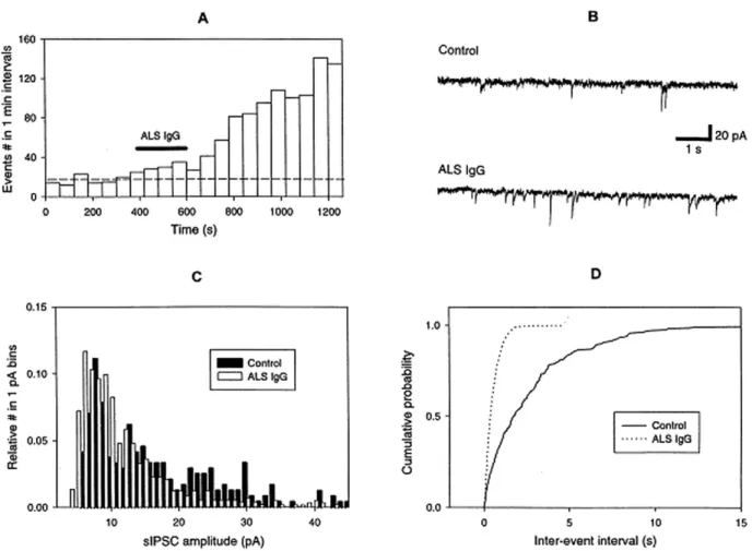
of postsynaptic events usually started 400 s before IgGs application, which lasted for 200 s. After IgGs application, the solution flow was stopped and the synaptic activity recorded continuously for about 10 min. Minutes after ALS IgGs application, a rise in the frequency of sIPSCs was already observed (Fig. 1A and D). On average for the period after applica-tion of ALS IgGs this frequency enhancement was 2.7 ± 1- fold (n=8) as compared to the period prior to their application (Fig. 2). However, the sIPSC
amplitude was not changed (see example in Fig. 1C and average data in Fig. 2). Control IgGs from healthy donors were ineffective in changing either frequency or amplitude of sIPSCs (n= 3; Fig 2). In order to check the effect of ALS IgGs on the release
of glycine, hypoglossal slices were pre-treated with 1 µM TTX (in addition to 10 µM bicuculline to knock down residual GABA-mediated responses). However, ALS IgGs did not have any effect on gly-cinergic miniature currents (mIPSC) recorded in the presence TTX (Fig. 2).
DISCUSSION
he presented work demonstrates that in addi-tion to lower motoneurons of the spinal cord (Appel
et al., 1991; Engelhardt et al., 1995; Pullen and Humphreys, 2000; Pullen et al., 2004) and neu-romuscular junctions (Fratantoni et al., 2000; Pagani et al., 2006), ALS IgGs also afect higher motoneurons in the brain stem. ALS IgGs have been shown to afect transmitter release in moto-neurons (Fratantoni et al., 2000; Pagani et al., 2006; Uchitel et al., 1988), as well as in glutama-tergic central synapses (Andjus et al., 1997). In hypoglossal neurons, the frequency of sIPSCs was afected by ALS IgGs but not the amplitude, thus
pointing to a presynaptic efect. However, although the frequency of sIPCSs was afected, mIPSCs were insensitive to ALS IgGs. A possible explanation may lie in the fact that unlike the glutamatergic synapses and neuromuscular junctions, glycinergic synaps-es are dually regulated by mechanisms of opposite sign triggered by a rise in [Ca2+]
i (Mukhtarov et
al., 2005). hese mechanisms are: (i) a decrease of glycinergic inhibitory postsynaptic currents due to a reduction of presynaptic glycine release, predomi-nantly induced by retrograde action of endogenous cannabinoids; and (ii) a potentiation of postsynaptic glycine receptors. Under normal physiological con-ditions, the postsynaptic efect is masked by pow-erful presynaptic inhibition. hus, although ALS IgGs have a potentiating efect on Ca2+ channels and
Ca2+ signaling as demonstrated in several systems
(Engelhardt et al., 1995; Fratantoni et al., 2000; Pullen et al., 2004; Pagani et al., 2006; Llinas et al., 1993),this may not have had an inluence on gly-cinergic synapses, where the two opposite efects of [Ca2+]
i rise may have cancelled each other.
he facilitation of spontaneous glycine-mediated events observed in the absence of TTX was probably indirect via changes in the basic membrane conduc-tance of glycinergic cells or network-driven through enhanced glutamatergic drive to these neurons. ALS IgGs could have increased the release of glutamate in these neurons, as in the case of hippocampal cells (Andjus et al., 1997). However, further experi-mental evidence is needed to prove that ALS IgGs act speciically on glutamate release and/or speciic membrane conductance in glycinergic cells.
Acknowledgments — We are largely in debt to Dr. Remigius Lape (SISSA) for his experimental expertise and to Profs. Andrea Nistri and Enrico Cherubini for their constant support in obtaining and discussing the results. his work was supported by grant # 823 (heleton Foundation) and grant # 143054B (MS RS).
REFERENCES
Andjus, P. R., Stević-Marinković, Z., and E. Cherubini (1997). Immunoglobulins from motoneurone disease patients enhance glutamate release from rat hippocampal neu-rones in culture. J. Physiol. (London) 504.1, 103-122.
Appel, S. H., Engelhardt, J. I., Garcia, J., and E. Stefani (1991). Immunoglobulins from animal models of motor neuron disease and from human amyotrophic lateral sclerosis pa-tients passively transfer physiological abnormalities to the neuromuscular junction. Proc. Natl. Acad. Sci. USA 88, 647-651.
Doble, A., and P. Kennel (2000). Animal models of amyotrophic lateral sclerosis. Amyotroph. Lateral Scler. Other Motor Neuron Disord. 1, 301–312.
Donato, R., and A. Nistri (2000). Relative contribution by GABA or glycine to Cl--mediated synaptic transmission on rat hypoglossal motoneurons in vitro. J. Neurophysiol. 84, 2715–2724.
Drachman, D. B. (2000). Does autoimmunity play a role in amyotrophic lateral sclerosis? In: Amyotrophic Lateral Sclerosis (Eds. R. H. Brown, V. Meininger, and M. Swash), 327–339. Dunitz, London.
Engelhardt, J. I., Siklos, L., Komuves, L., Smith, R. G., and S. H. Appel (1995). Antibodies to calcium channels from ALS
patients passively transferred to mice selectively increase intracellular calcium and induce ultrastructural changes in motoneurons. Synapse 20, 185–199.
Fratantoni, S. A., Weisz, G., Pardal, A. M., Reisin, R. C., and O. D. Uchitel (2000). Amyotrophic lateral sclerosis IgG-treated neuromuscular junctions develop sensitivity to L-type calcium channel blocker. Muscle Nerve 23, 543–550.
Llinas, R., Sugimori, M., Cherksey, B. D., Glenn Smith, R., Delbono, O., and E. Stefani (1993). IgG from amyotrophic lateral sclerosis patients increases current through P-type calcium channels in mammalian cerebellar Purkinje cells and in isolated channel protein in lipid bilayer. Proc. Natl. Acad. Sci. USA 90, 11743-11747.
Meininger, V., Lacomblez, L., and F. Salachas (2000). What has changed with riluzole? J. Neurol. 247, 19–22.
Mukhtarov, M., Ragozzino, D., and P. Bregestovski (2005) Dual Ca2+ modulation of glycinergic synaptic currents in rodent hypoglossal motoneurons. J. Physiol. (London)
569.3, 817-831.
Pagani, M. R., Reisin, R.C., and O. D. Uchitel (2006). Calcium signaling pathways mediating synaptic potentiation trig-gered by amyotrophic lateral sclerosis IgG in motor nerve terminals. J. Neurosci. 26, 2661–2672.
Pullen, A. H., and P. Humphreys (2000). Ultrastructural analysis of spinal motoneurons from mice treated with IgG from
ALS patients, healthy individuals, or disease controls. J. Neurol. Sci. 180, 35– 45.
Pullen, A. H., Demestre, M., Howard, R. S., and R. W. Orrell (2004). Passive transfer of purified IgG from patients with amyotrophic lateral sclerosis to mice results in degeneration of motor neurons accompanied by Ca2+ enhancement. Acta Neuropathol. (Berlin) 107, 35– 46.
Smith, R. G., Hamilton, S., Hofmann, F., Schneider, T., Nastainczyk, W., Birnbaumer, L., Stefani, E., and S. H. Appel (1992). Serum antibodies to L-type calcium channels in patients with amyotrophic lateral sclerosis. N. Engl. J. Med. 327, 1721–1728.
Uchitel, O. D., Appel, S. H., Crawford, F., and L. Sczcupak (1988). Immunoglobulins from amyotrophic lateral sclerosis patients enhance spontaneous transmitter release from motor-nerve terminals. Proc. Natl. Acad. Sci. USA 85, 7371-7374.
Viana, F., Bayliss, D. A., and A. J. Berger (1994). Postnatal chang-es in rat hypoglossal motoneuron membrane propertichang-es. Neuroscience 59, 131–148.
Zhainazarov, A. B., Annunziata, P., Toneatto, S., Cherubini, E., and A. Nistri (1994). Serum fractions from amyotrophic lateral sclerosis patients depress voltage-activated Ca2+ currents of rat cerebellar granule cells in culture. Neurosci. Lett. 172, 111-114.
ИМУНОГЛОБУЛИНИ Als ПАЦИЈЕНАТА ПОВЕЋАВАЈУ ФРЕКВЕНЦИЈУ ГЛИЦИНОМ ИЗАЗВАНИХ СПОНТАНИХ Ipsc У ХИПОГЛОСАЛНИМ МОТОНЕУРОНИМА ПАЦОВА
П. Р. АНЂУС
Биолошки факултет, Универзитет у Београду, 11000 Београд, Србија
Интернационална школа за више студије (СИССА), 34014 Трст, Италија
Амтрна латрална склрза (ALS) ј разарајћа, јш вк нзлчва нрлшка блст кја пгађа грњ дњ мтнрн. Пасвн транср блња настај прнсм мнглблна ALS пацјната крвтк кс прмнталн жвтњ. Сматра с да ALS IgG зазвај ксцттксчнст дјствм на влта жнзавсн Ca2+ канал. Наша ранја страж вања пказала с да ALS IgG пвћавај спн