Comparing the Primary and Recall Immune
Response Induced by a New EV71 Vaccine
Using Systems Biology Approaches
Jie Shao1☯, Junnan Zhang2☯, Xing Wu2, Qunying Mao2, Pan Chen2, Fengcai Zhu3,
Miao Xu2, Wei Kong1*, Zhenglun Liang2*, Junzhi Wang2*
1School of Life Sciences, Jilin University, Changchun, Jilin, P.R.China,2Division of Hepatitis Virus Vaccines, National Institutes for Food and Drug Control, Beijing, P.R.China,3Jiangsu Provincial Center for Disease Control and Prevention, Nanjing, Jiangsu, P.R.China
☯These authors contributed equally to this work.
*[email protected](WK);[email protected](ZL);[email protected](JW)
Abstract
Three inactivated EV71 whole-virus vaccines have completed Phase III clinical trials in mainland China, with high efficacy, satisfactory safety, and sustained immunogenicity. However, the molecular mechanisms how this new vaccine elicit potent immune response remain poorly understood. To characterize the primary and recall responses to EV71 vac-cines, PBMC from 19 recipients before and after vaccination with EV71 vaccine are col-lected and their gene expression signatures after stimulation with EV71 antigen were compared. The results showed that primary and recall response to EV71 antigen have both activated an IRF7 regulating type I interferon and antiviral immune response network. How-ever, up-regulated genes involved in T cell activation regulated by IRF1, inflammatory response, B-cell activation and humoral immune response were only observed in recall response. The specific secretion of IL-10 in primary response and IL-2,IP-10,CCL14a, CCL21 in recall response was consistent with the activation of immune response process found in genes. Furthermore, the expression of MX1 and secretion of IP-10 in recall response were strongly correlated with NTAb level at 180d after vaccination (r = 0.81 and 0.99). In summary, inflammatory response, adaptive immune response and a stronger anti-viral response were indentified in recall response.
Introduction
Hand foot and mouth disease (HFMD) is a serious public health problem in Western Pacific region countries[1]. From May 2008 to March 2015, 11.96 million cases of HFMD had been reported in mainland China, of which 3,227 were fatal[2]. Based on the epidemiological and clinical etiological data published in recent years, more than 80% of the pathogens isolated from patients died from HFMD were identified as enterovirus 71 (EV71)[3–6]. There are no efficient drugs available for EV71 treatment, sovaccines will be the essential way to control the EV71 outbreak. Research and development of EV71 vaccine was carried out in several Asian OPEN ACCESS
Citation:Shao J, Zhang J, Wu X, Mao Q, Chen P, Zhu F, et al. (2015) Comparing the Primary and Recall Immune Response Induced by a New EV71 Vaccine Using Systems Biology Approaches. PLoS ONE 10(10): e0140515. doi:10.1371/journal. pone.0140515
Editor:Shixia Wang, University of Massachusetts Medical School, UNITED STATES
Received:July 23, 2015
Accepted:September 25, 2015
Published:October 14, 2015
Copyright:© 2015 Shao et al. This is an open access article distributed under the terms of the
Creative Commons Attribution License, which permits unrestricted use, distribution, and reproduction in any medium, provided the original author and source are credited.
Data Availability Statement:We have submitted our dataset in Gene Expression Omnibus, and the accession number is GSE72618.
countries. Currently three inactivated EV71 whole-virus vaccines in mainland China have completed Phase III clinical trials in more than 30,000 infants and children. Results showed that these vaccines were safe and there were over 90% efficacy in preventing EV71-associated HFMD, 80% efficacy in preventing EV71-associated diseases[7–9]. However, a comprehensive understanding of immune responses to this new vaccine is still lacking.
Recently, systems biology approach has been used to predict the development of protective immunity after vaccination by profiling gene expression of PBMC samples from vaccinated individuals. This approach has been pioneered in the studies of yellow fever vaccine[10–11], influenza vaccine[12] and HPV virus-like particles vaccine[13]. In these studies, genes involved in innate immune response pathways were enriched after vaccination, and specific biomarkers were found to predict the immune effect of vaccines with high accuracy[10–13]. Since most of these researches were focused on the primary responses after initial immunization, little is known about the recall response. The recall response is important due to its close relationship with neutralizing antibodies (NTAb) titer and immune persistence[14].
In this study, microarray analysis and cytokine profiling have been performed to compare gene expression patterns between primary and recall immune response induced by EV71 vac-cines. Our results provide a better understanding of the immune response induced by EV71 vaccine.
Materials and Methods
1. Study design
Participants were selected from a randomized, double-blind, placebo-controlled phase III trial of inactivated EV71 whole-virus vaccines, which was conducted in a sample of 10245 healthy children aged 6–35 months (ClinicalTrials.gov, number NCT01508247)[7]. This clinical trial study was approved by the institutional review board of Jiangsu Provincial Center of Disease Control and Prevention, and all guardians of participants provided written informed consent. The immunization schedule consisted of two doses given on day 0 and day 28. Vaccine or pla-cebo was administered intramuscularly to the anterolateral side of the thigh (for participants aged 6–11 months) or the deltoid muscle (those aged 12–35 months). Blood specimens were collected before the initial dose (day 0) and day 56 and 180 after the first vaccination. Plasma and peripheral blood mononuclear cells (PBMCs) were separated on the scene and frozen at -80°C.
Nineteen vaccine recipients whose antibody level is negative before vaccination and sero-conversion on day 56 after vaccination were selected in this study. However, 5 samples pre-vac-cination and 3 samples post-vacpre-vac-cination were excluded from the microarray detection because of poor RNA purity and integrity.Only day 0 and 56 samples were selected for this study because earlier findings indicated that the largest increases in cytokine responses were typically observed at month 2[15]. Thirteen vaccine recipients (5 on day 0 and 8 on day 56) were selected for protein array analysis.
2. Inactivated EV71 whole-virus vaccine and EV71 liquid bulk
Inactivated alum-adjuvanted EV71 whole-virus vaccine (vero cell), containing 320U of antigen and 0.18mg of alum, was developed by Beijing Vigoo Biological with a seed virus of EV71 strain FY7VP5/AH/CHN/2008 (subgenotype C4). Each dose of placebo contains 0.18mg of alum adjuvant and no EV71 antigen.
EV71 liquid bulk for stimulation in vitro contained 4000U/ml of antigen and no alum. Competing Interests:The authors have declared
that no competing interests exist.
3. PBMCs incubation and RNA extraction
Cryopreserved PBMCs were thawed and cultured. PBMCs (10
106) were plated in each well of a 6-well plate (Costar) in RPMI 1640 medium supplemented with penicillin-streptomycin (100g/ml-100U/ml; Invitrogen), L-glutamine (2mM), HEPES buffer (10mM), and 10% heat-inactivated FCS (HyClone). Cells were cultured for 24h at 37°C with EV71 Ag (200u/ml). Media was used as a background measurement for untreated cells. A total of four incubations were set up on the same day for each subject. The order of sample preparation was randomly defined from the list of selected subjects. When harvesting, cultures were centrifuged and cell precipitation were frozen at -80°C.
Total RNA extracts were performed using an RNeasy Total RNA isolation kit (Qiagen). RLT lysis buffer was quickly added to the culture well and then to the cell pellet to include both adherent and suspension cells. RNA purity and integrity were tested by microcapillary electro-phoresis using the Agilent 2100 bioanalyzer (Agilent Technologies). The sample of RIN>6 was
selected for follow-up experiment.
4. Microarray analysis
Microarray gene expression analysis was performed using a human genome 70-mer oligonu-cleotide microarray (CapitalBio Corporation 35K Human Genome Array),that contains 35035 probesets to 25100 characterized human genes. Total RNA preparation and labeled cDNA syn-thesis and hybridization were performed according to the manufacturer’s recommended proto-col (CapitalBio). In short, 1 ug total RNA was used for double-stranded cDNA (ds-cDNA) synthesis with the CbcScript reverse transcriptase kit (Capitalbio) with the T7 Oligo (dT). Kle-now enzyme labeling strategy was adopted after reverse transcription using CbcScriptⅡreverse transcriptase. Labeled cDNA were hybridized performed in a CapitalBio BioMixerTMII Hybridization Station and washed in a Capitalbio SlideWasherTM(CapitalBio) with two conse-cutive solutions. Arrays were scanned with a confocal LuxScanTMscanner and the images obtained were then analyzed using LuxScanTM3.0 software (CapitalBio). After background subtracted in both channels (Cy3 and Cy5),faint spots were extracted for which intensities were exceed 400 units. A space- and intensity-dependent normalization based on a LOWESS program was employed[16]. To determine the significant differentially expressed genes, Signif-icance Analysis of Microarrays (SAM, version 3.02) were performed.
5. Quantitative real-time PCR and confirmation of microarray results
The gene expression levels were measured by RT-PCR. A total of 17 genes in 60 samples were selected for confirmation. The primers are listed in Table A inS1 File. Briefly, total RNA was extracted from the PBMCs as described above. For quantification, a single-tube RT-PCR assay was performed using the TaqMan 1-step RT-PCR Master Mix in a 7500 Fast Real-time RT-PCR system. The following protocol was used for all PCR assays: 5 min at 42°C and 10s at 95°C,followed by 40 cycles at 95°C for 5s and 60°C for 30s.
6. Protein array analysis
four times and then detected by protein array, according to the protocol of RayBio1Human Cytokine Antibody Array G Series 4000.
7. Neutralizing Antibody
EV71-neutralizing Ab titers were determined using classical cytopathic effect (CPE) assay per-formed as previously described[17]. Plasma samples were inactivated at 56°C for 30 minutes, serially diluted two fold from 1:8 and mixed with equal volumes of TCID50 of a EV71 strain. The mixture was dispensed into a 96-well microplate and incubated at 37°C for 2 hours. RD cells (1–2105cells/mL) were added to the mixture. The plates were then placed in a CO
2 incu-bator at 35°C for 7 days. CPE was observed by microscopy. EV71 national standards were included in each test as a control for the reproducibility of the results[18]. Neutralizing anti-body titers of EV71 were defined as the dilution rate showing 50% inhibition of the CPE. Unde-tectable Ab levels were considered as 4. All individuals selected for the microarray analysis had no detectable Abs before vaccination. After vaccination, all individuals developed Ab titers against EV71 in the group of vaccination.
Results
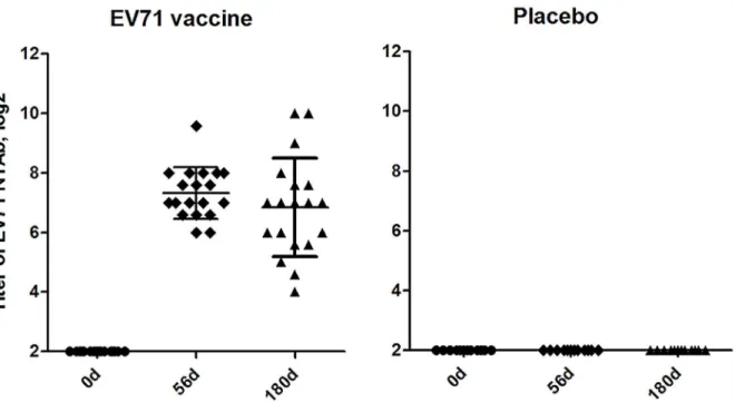
1. The neutralizing antibodies response to EV71 vaccine
As shown inFig 1, NTAb titers were detected on day 56 (56d) and 180 (180d) after initial inoc-ulated with inactivated EV71 vaccine. NTAb seroconversion rates reached 100% at 56d and persisted up to 180d. The geometric mean titer (GMT) of anti-EV71 NTAb elicited by EV71 vaccine was 1:160 at 56d and 1:86 at 180d. NTAb titers in placebo group were both negative before and after vaccination.
2. Comparison of the gene expression patterns between primary and
recall immune response
2.1 Gene expression patterns in primary response. To find changes of gene expression in primary response, we compared gene expression levels in pre-immunization PBMCs stimu-lated by EV71 Ag. After filtration, we observed that overall 220 of 35035 probe sets were signifi-cantly and differentially up-regulated after stimulation(fold change1.5,p<0.05),
Using the DAVID Bioinformatics Database (http://david.abcc.ncifcrf.gov/), we analyzed the Gene Ontology (GO) terms of the 220 genes, which revealed an enrichment of genes related to various immunological responses, such as genes mediating response to virus, including ISG15, EIF2AK2, MX1, IFI27, MX2, PLSCR1, IFI35, TRIM22, IRF9, IFIH1, CCL22; genes involved in transcription, including UBTF, ZNF205, ELL2, IRF9, ZNF432, POLR3G, TMF1, ZNF431, ZNF665, ZNF417; immune response, including OAS1, IFI35, TRIM22, IFITM2, GBP1, CCL22; interspecies interaction between organisms, including ISG15, EIF2AK2, IFIH1, SF3B2; cell-cell signaling, including ISG15, BST2, POMC, CCL22 (Table B inS1 File).
2.2 Gene patterns in recall response. The recall immune responses were investigated by comparing post-vaccination gene expression patterns of PBMCs stimulated by EV71 Ag. After data filtration, we observed that 565 of 35035 probe sets met the fold change criteria (fold change1.5,p<0.05). Among these genes, 487 were up-regulated and 78 were down-regulated.
We analyzed the GO terms of the 565 genes, and results showed 89 of the 487 significantly up-regulated probe sets were identified within the top 5 GO terms including immune response, transcription inflammatory response,signal transduction,response to virus (Table C inS1 File). The down-regulated probe sets were identified within some metabolic pathways, such as lipid metabolism, cell wall catabolism and protein amino acid glycosylation.
Ingenuity Pathways Analysis revealed that differentially expressed genes (DEGs) were mainly involved in Interferon Signaling (9/34), Activation of IRF by Cytosolic Pattern Recogni-tion Receptors (8/64), and Role of RIG1-like Receptors in Antiviral Innate Immunity (5/45). Gene networks analysis revealed that IRF7 also canactivate MDA-5 and LGP2, and played as a node of transcriptional regulation network in recall response. Several antivirus-genes were upregulated by IFR7 such as:MX1, IFI27 and IFIH1, similar with those in primary response. Another node of transcriptional regulation observed in recall immune response was IRF1. It directly regulated the expression of genes related to activation of MHC-I, including: PDIA3, TAP1, HLA-F, HLA-G, and LILRB1. These genes took a part in the activation of cytotoxic T cells (Fig 2B).
Expression level of MX1 mRNA had a high correlation with the NTAb level at 180d after vaccination (r = 0.81).
2.3 Comparison between primary and recall immune response. In order to explore the similarities and differences between primary and recall immune response induced by EV71 Ag, a comparative analysis was carried out between them for the DEGs. By comparing, 124 DEGs were observed both in primary response and recall response; 96 DEGs were observed only in primary response; and 441 DEGs were observed only in recall response.
Fig 1. Titers of neutralizing antibodies responses to EV71 vaccine on days 0, 56 and 180 post vaccination.Each symbol represents an individual donor; median value and standard deviation were indicated. There were 19 subjects in the EV71 vaccine group and 13 in the placebo group. After vaccination (the first vaccination on day 0 and boosted on day 28), seroconversion was 100% in EV71 vaccine group.
We analyzed the GO terms of the 124, 96 and 441 genes respectively. Results showed that the 124 genes both activated in primary and recall response were involved in response to virus, transcription, immune response and signal transduction. However, the fold change of these genes in recall response was higher than that in primary response (Fig 3A). Ninety-six genes observed only in primary response were involved in several metabolic pathways, such as lipid metabolism, fatty acid metabolism and DNA replication, but not immune response. 441 DEGs observed only in recall response involved in inflammatory response and the induction of an adaptive immune response, based on published literatures[19–33], such as TNFSF13B, CDKN1A and BANK1 involved in B Cell activation; IRF1, PVRL2, TNFSF13B and TRAF2 involved in T Cell activation;CCL2,RBP4,TNFSF13B,PVRL2,KDELR3,PSMB10 and PDIA3 involved in humoral immune response (Fig 3B).
Ingenuity Pathways Analysis revealed that the 124 common genes were enriched in Inter-feron Signaling, Activation of IRF by Cytosolic Pattern Recognition Receptors and Role of RIG1-like Receptors in Antiviral Innate Immunity. The visualization of gene networks showed a similar interacting network containing dozens of IFN-I regulated and antiviral genes both in primary response and recall response, and IRF7 played as a node of transcriptional regulation of these genes (Fig 2A & 2B). One of the most significant distinctions between networks of pri-mary and recall immune response was that genes involved in MHC-I activation regulated by IRF1 were only observed in the recall response. This may be associated with the effect of the cytotoxic T cells (Fig 2B).
Fig 2. Molecular signatures of immune reponses to EV71 vaccine.(a) Ingenuity Pathways Analysis of DEGs in primary response. IRF7 was found as a hub of transcriptional regulation. (b) Gene network analysis of recall response. IRF7 was also found as a major node of transcriptional regulation. There was another key node, IRF1, which upregulated the genes related to activation of MHC-I as shown in the solid box.
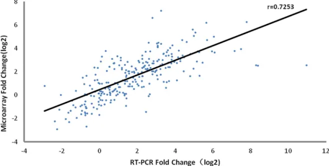
3. Confirmation of a subset of DEGs by quantitative RT-PCR
To evaluate the expression profiles of the genes detected in the microarray assay, RT-PCR veri-fication was performed for 17 genes (defined in Materials and Methods) using samples from microarray analysis of vaccine recipients (n = 60). As shown inFig 4and Table D inS1 File, the expression pattern of the selected genes was consistent with the microarray data. A signifi-cant correlation (r = 0.7253) existed between the microarray data and RT-PCR results, suggest-ing that the microarray assay was reliable.
4. Cytokine secretion profiles in primary and recall immune response
In order to compare the cytokine secretion profiles between primary and recall response, cyto-kines in culture supernatants of PBMC after stimulation with EV71 Ag were tested by Capital-Bio protein array which had a capacity of measuring 274 cytokines.
Our results showed that 7 of 274 cytokines met the fold change criteria in primary response. Of these, 5 were up-regulated, including GDF-15, MMP-13, IL-10, TIMP-2 and Angiopoietin-2; and 2 were down-regulated, including PDGF-R alpha and GRO (Fig 5). Twenty-seven cyto-kines were found to be significantly differentially secreted in recall response. Among these 20 were up-regulated, such as Th1 type cytokines IL-2 and IP-10; competitive inhibitor of Th1 type cytokine receptor IL-13Ralpha2; chemokines CCL14a and CCL21 and so on. Seven were down-regulated,such as CD23, CRP, DPPIV, Bate2M and NAP-2 (Fig 6).
A correlation analysis between the level of cytokine secretion and the NTAb at 180d after vaccination showed that IP-10 had a high correlation coefficient with the antibody titers (r = 0.99).
Fig 3. Heat map of DEGs in primary and recall response.Colors ranging from blue to red corresponded represent the DEGs’average fold change among the subjects (n = 19). (a) Common genes identified in primary and recall response. However, the fold change of these genes in recall response was higher than that in primary response. (b) Pathways that were only observed in recall response,including inflammatory response, antigen processing and presentation, B cell activation, T cell activation and humoral immune response.
doi:10.1371/journal.pone.0140515.g003
Fig 4. Verification of selective genes with RT-PCR.Correlation analysis of gene expressions changes with microarray and RT-PCR (r = 0.7253). 17 genes were selected to confirm within 60 samples. Each point represents a single gene at a given sample.
Discussion
“EV71 vaccine is an invaluable gift for children in the Asia-Pacific regions and worldwide”[34]. Exploring the effective protective immune mechanism will contribute greatly to the develop-ment of novel vaccines and the improvedevelop-ment of existing vaccines. To have a comprehensive understanding of the EV71 vaccines-induced immune response, the recall immune response has been explored as well as the primary immune response in this study.
Our results show that IFN-I and antiviral immune response were activated in both primary and recall response. The activation of IFN-I pathway may be necessary to the protective immune response elicited by EV71 vaccine. Because we have already found IFN-I related genes were gen-erally activated on the early stage after vaccination with 5 different kinds of vaccines by a meta-analysis[35], and IFN-I contributes significantly to the procedure that mature B cells differentiate into antibody-secreting plasma cells[36]. IRFs are master regulators for IFN-I and antiviral genes signalling by Toll-like receptors and other cytosolic pattern-recognition receptors (PRRs)[37]. In our study IRF7 were identified as a node prominently involved in regulating transcription of interferon and antiviral genes in both primary and recall response. IRF7 governs both the sys-temic production of IFN in innate immune response and the local action of IFN from plasmacy-toid dendritic cells in adaptive immunity[38]. Similar regulatory role of IRF7 was also observed in the immune response to yellow fever vaccines[10–11] and seasonal influenza vaccine[12].
More genes are activated in recall response to EV71 vaccine compared with the primary response. A dozen of them are enriched in activation of MHC class I, which is probably associ-ated with the effect of memory CD8+T cells. IRF1, another member of IRFs, is indentified as a master regulator of these genes. IRF1 could promote CD8+T cell maturity and induce a shift of
Fig 5. Cytokines profiling ofin vitroEV71 antigen stimulated PBMCs which were from recipients before vaccination.PBMC were incubated with either EV71 antigen or medium as indicated in Materials and Methods. Five were up-regulated cytokines (GDF-15, MMP-13, IL-10, TIMP-2 and Angiopoietin-2) and 2 were down-regulated cytokines (PDGF-R alpha and GRO). Seven cytokines were significantly induced by EV71 antigen compared with control (P<0.05).
the Th subset balance to a Th1 dominant state during Listeria infection, as well as activating expression of the cytokine Interferon beta like IRF7[39]. The activation of genes involved in inflammatory response was indentified in our study, which might assist in the effect of memory CD8+T cells[40]. Other specific up-regulated genes involved in generating Ab procedure, such as B-Cell activation, antigen processing and presentation and humoral immune response, are important in generating high titer of antibodies after the boost.
The profiles of cytokines secretion were consistent with our finding in genes. IL-10, a specific secreted cytokine in primary response, coincides exactly with the process that naive B cell differ-entiate into the plasma cells in primary response, because it serves as a down-regulator of inflam-matory cytokines and an enhancer of B cell proliferation[41]. In addition, IL-10 is involved in the regulation of the JAK-STAT signaling pathway[42] which was observed activate in the DEGs pathway analysis. An increased secretion of IL-2, IP-10, CCL14a, and CCL21 is observed in recall response. IL-2 and IP-10 promote the differentiation of certain immature T cells into regulatory T cells, effector T cells and memory T cells[43–44], and CCL14a and CCL21 promotes the migra-tion and adhesion of T lymphocytes[45–46]. The increased secretion of IL-2, IP-10, CCL14a, and CCL21 is consistent with the up-regulation of IRF1, inflammatory genes and genes involved in activation of CD8+ T cells observed in microarray analysis.
The mRNA expression level of MX1 and cytokine secretion level of IP-10 in recall response were strongly correlated with NTAb level at 180d after vaccination. MX1 is responsible for a specific antiviral state against dozens of virus infection[47]. IP-10 is regarded as a marker to evaluate the antigen-specific cytotoxic T-lymphocytes induced by EV71 vaccine, and it was sig-nificantly induced by TIV and EV71 infection[12,48]. But further studies are needed to verify if MX1 and IP-10 can be used as markers to predict the immune persistence of EV71 vaccines.
Our study compared the different molecular mechanisms between primary and recall immune response on a new EV71 vaccine. IFN-I and antiviral immune response were actived in both primary and recall response. However, the up-regulation of genes involved in memory CD8+T activation, inflammatory response, B-cell activation and humoral immune response were only found in recall response. Furthermore, MX1 and IP-10 are identified as two potential markers to predict the immune persistence of EV71 vaccines.
There are two limitations in this study. The recall immune response on EV71 vaccine was explored by re-stimulating PBMCs in vitro in this study, which did not completely simulate to the response in vivo. In addition, bio-markers indentified in our study still need be verified in broader vaccination receivers.
Supporting Information
S1 File. RT-PCR primers (Table A).Enrichment of GO terms in primary response (Top 10) (Table B). Enrichment of GO terms in recall response (Top 10) (Table C). Confirmation of microarray results with real-time PCR (n = 60) (Table D).
(DOC)
Acknowledgments
We would like to acknowledge the staff members at the Jiangsu Center for Disease Control and Prevention, for collecting clinical specimens from the vaccines for use in this study.
Fig 6. Cytokines profiling ofin vitroEV71 antigen stimulated PBMCs which were from recipients after vaccination.PBMC were incubated with either EV71 antigen or medium as indicated in Materials and Methods. Up-regulated cytokines are shown in red and down-regulated cytokines in green. Each row represents a probeset and each column represents an individual.
Author Contributions
Conceived and designed the experiments: JS JZ MX WK ZL JW. Performed the experiments: JS JZ XW. Analyzed the data: JS JZ XW QM. Contributed reagents/materials/analysis tools: FZ. Wrote the paper: JS JZ PC.
References
1. Kok CC. Therapeutic and prevention strategies aganist human enterovirus 71 infection. World J Virol. 2015; 4:78–95. doi:10.5501/wjv.v4.i2.78PMID:25964873
2. Chinese center for disease control and prevention. National Notifiable Disease Reported System (HFMD).Available:http://www.nhfpc.gov.cn/zhuzhan/yqxx/lists.shtml. Accessed April 2015. 3. Ho M, Chen ER, Hsu KH, Twu SJ, Chen KT, Tsai SF, et al. An epidemic of enterovirus 71 infection in
Taiwan. Taiwan Enterovirus Epidemic Working Group. N. Engl J Med. 1999; 341: 929–935. PMID:
10498487
4. Lin TY, Chang LY, Hsia SH, Huang YC, Chiu CH, Hsueh C, et al. The 1998 enterovirus 71 outbreak in Taiwan: pathogenesis and management. Clin Infect Dis. 2002; 34 Suppl 2: S52–57. PMID:11938497
5. Chen KT, Chang HL, Wang ST, Cheng YT, Yang JY. Epidemiologic features of hand-foot-mouth dis-ease and herpangina caused by enterovirus 71 in Taiwan, 1998–2005. Pediatrics. 2007; 120: e244–
252. PMID:17671037
6. Lin TY, Twu SJ, Ho MS, Chang LY, Lee CY. Enterovirus 71 outbreaks, Taiwan: occurrence and recog-nition. Emerg Infect Dis. 2003; 9: 291–293. PMID:12643822
7. Zhu FC, Meng FY, Li JX, Li XL, Mao QY, Tao H, et al. Efficacy, safety, and immunology of an inacti-vated alum-adjuvant enterovirus 71 vaccine in children in china: a multicentre, randomised, double-blind, placebo-controlled, phase 3 trial. Lancet. 2013; 381:2024–2032. doi:10.1016/S0140-6736(13) 61049-1PMID:23726161
8. Zhu F, Xu W, Xia J, Liang Z, Liu Y, Zhang X, et al. Efficacy, safety, and immunogenicity of an enterovi-rus 71 vaccine in China. N Engl J Med.2014; 370:818–828. doi:10.1056/NEJMoa1304923PMID:
24571754
9. Hu YM, Wang X, Wang JZ, Wang L, Zhang YJ, Chang L, et al. Immunogenicity, safety, and lot consis-tency of a novel inactivated enterovirus 71 vaccine in chinese children aged 6 to 59 months. Clin Vac-cine Immunol.2013; 20:1805–1811. doi:10.1128/CVI.00491-13PMID:24108780
10. Querec TD, Akondy RS, Lee EK, Cao W, Nakaya HI, Teuwen D, et al. Systems biology approach pre-dicts immunogenicity of the yellow fever vaccine in humans. Nat Immunol.2009; 10:116–125. doi:10. 1038/ni.1688PMID:19029902
11. Gaucher D, Therrien R, Kettaf N, Angermann BR, Boucher G, Filali-Mouhim A, et al. Yellow fever vac-cine induces integrated multilineage and polyfunctional immune responses. J Exp Med.2008; 205:3119–3131. doi:10.1084/jem.20082292PMID:19047440
12. Nakaya HI, Wrammert J, Lee EK, Racioppi L, Marie-Kunze S, Haining WN, et al. Systems biology of vaccination for seasonal influenza in humans. Nat Immunol.2011; 12:786–795. doi:10.1038/ni.2067
PMID:21743478
13. Garcia-Pineres AJ, Hildesheim A, Dodd L, Kemp TJ, Yang J, Fullmer B, et al. Gene expression patterns induced by hpv-16 l1 virus-like particles in leukocytes from vaccine recipients. J Immunol.2009; 182:1706–29. PMID:19155521
14. Church SE, Jensen SM, Twitty CG, Bahjat K, Hu HM, Urba WJ. Multiple Vaccinations: Friend or Foe. Cancer J. 2011; 17:379–396. doi:10.1097/PPO.0b013e3182346320PMID:21952289
15. Pinto LA, Edwards J, Castle PE, Harro CD, Lowy DR, Schiller JT, et al. Cellular immune responses to human papillomavirus (HPV)-16 L1 in healthy volunteers immunized with recombinant HPV-16 L1 virus-like particles. J Infect Dis. 2003; 188:327–338. PMID:12854090
16. Yang YH, Dudoit S, Luu P, Lin DM, Peng V, Ngai J. Normalization for cDNA microarray data: a robust composite method addressing single and multiple slide systematic variation. Nucleic Acids Res.2002; 30: e15. PMID:11842121
17. Mao Q, Cheng T, Zhu F, Li J, Wang Y, Li Y, et al. The cross-neutralizing activity of enterovirus 71 sub-genotype c4 vaccines in healthy chinese infants and children. PLoS One.2013; 8:e79599. doi:10.1371/ journal.pone.0079599PMID:24260259
19. Thibault-Espitia A, Foucher Y, Danger R, Migone T, Pallier A, Castagnet S, et al. BAFF and BAFF-R levels are associated with risk of long-term kidney graft dysfunction and development of donor-specific antibodies. Am J Transplant. 2012; 12: 2754–2762. doi:10.1111/j.1600-6143.2012.04194.xPMID:
22883025
20. Chen H, Li C, Huang J, Cunq T, Seiss K, Beamon J, et al. CD4+ T cells from elite controllers resist HIV-1 infection by selective upregulation of p2HIV-1. J Clin Invest.20HIV-1HIV-1; HIV-12HIV-1:HIV-1549–1560. doi:10.1172/JCI44539
PMID:21403397
21. Bernal-Quirós M, Wu YY, Alarcón-Riquelme ME, Castillejo-López C. BANK1 and BLK act through phospholipase C gamma 2 in B-cell signaling. Plos One. 2013; 8:e59842. doi:10.1371/journal.pone. 0059842PMID:23555801
22. Taki S, Sato T, Oqasawara K, Fukuda T, Sato M, Hida S, et al. Multistage regulation of TH1-type immune responses by the transcription factor IRF-1. Immunity. 1997; 6:673–679. PMID:9208840
23. Penninger JM, Sirard C, Mittrücker HW, Chidgey A, Kozieradzki I, Nghiem M, et al. The interferon regu-latory transcription factor IRF-1 controls positive and negative selection of CD8+ thymocytes. Immunity. 1997; 7:243–254. PMID:9285409
24. Almire C, Bertrand P, Ruminy P, Maingonnat C, Wlodarska I, Martin-Subero JI, et al. PVRL2 is translo-cated to the TRA@ locus in t(14;19)(q11;q13)-positive peripheral T-cell lymphomas.Genes Chromo-somes Cancer. 2007; 46:1011–1018. PMID:17696193
25. Chen L, Files DB. Molecular mechanisms of T cell co-stimulation and co-inhibition. Nat Rev Immunol. 2013; 13:227–242. doi:10.1038/nri3405PMID:23470321
26. Villanueva JE, Malle EK, Gardam S, Silveira PA, Zammit NW, Walters SN. TRAF2 regulates peripheral CD8+ T-cell and NKT-cell homeostasis by modulating sensitivity to IL-15. Eur J Immunol. 2015; 45:1820–1831. doi:10.1002/eji.201445416PMID:25931426
27. Tylaska LA, Boring L, Weng W, Aiello R, Charo IF, Rollins BJ. Ccr2 regulates the level of MCP-1/CCL2 in vitro and at inflammatory sites and controls T cell activation in response to alloantigen. Cytokine. 2002; 18:184–190. PMID:12126640
28. Moraes-Vieira PM, Yore MM, Dwyer PM, Syed I, Aryal P, Kahn BB. RBP4 activates antigen-presenting cells leading to adipose tissue inflammation and systemic insulin resistance. Cell Metab. 2014; 19:512–
526. doi:10.1016/j.cmet.2014.01.018PMID:24606904
29. Rickert RC, Jellusova J, Miletic AV. Signaling by the tumor necrosis factor receptor superfamily in B-cell biology and disease. Immunol Rev. 2011; 244:115–133. doi:10.1111/j.1600-065X.2011.01067.x
PMID:22017435
30. Eberlé F, Dubreuil P, Mattei MG, Devilard E, Lopez M. The human PRR2 gene, related to the human poliovirus receptor gene (PVR), is the true homolog of the murine MPH gene.Gene. 1995; 159:267–
272. PMID:7622062
31. Romero N, Dumur CI, Martinez H, García IA, Monetta P, Slavin I. Rab1b overexpression modifies Golgi size and gene expression in HeLa cells and modulates the thyrotrophin response in thyroid cells in cul-ture. Mol Biol Cell. 2013; 24:617–632. doi:10.1091/mbc.E12-07-0530PMID:23325787
32. Favole A, Cascio P, Cerruti F, Sereno A, Tursi M, Tomatis A, et al. MHC class I-related antigen-pro-cessing machinery component defects in feline mammary carcinoma. Transl Oncol. 2012; 5:48–55. PMID:22348176
33. Gan CS, Yusof R, Othman S. Different serotypes of dengue viruses differently regulate the expression of the host cell antigen processing machinery. Acta Trop. 2015; 149:8–14. doi:10.1016/j.actatropica. 2015.05.005PMID:25981524
34. Liang Z, Wang J. EV71 vaccine, an invaluable gift for children. Clin Transl Immunology.2014; 3:e28. doi:10.1038/cti.2014.24PMID:25505956
35. Zhang J, Shao J, Wu X, Mao Q, Wang Y, Gao F, et al. Type I interferon related genes are common genes on the early stage after vaccination by meta-analysis of microarray data. Human Vaccin Immun-other. 2015; 11:739–745.
36. Kim D, Martinez-Sobrido L, Choi C, Petroff N, Garcia-Sastre A, Niewiesk S, et al. Induction of type I interferon secretion through recombinant Newcastle disease virus expressing measles virus hemagglu-tinin stimulates antibody secretion in the presence of maternal antibodies. J Virol.2011; 85: 200–207. doi:10.1128/JVI.01624-10PMID:20962092
37. Ikushima H, Negishi H, Taniguchi T. The IRF family transcription factors at the interface of innate and adaptive immune responses. Cold Spring Harb Symp Quant Biol. 2013; 78:105–16. doi:10.1101/sqb. 2013.78.020321PMID:24092468
39. Maruyama S, Kanoh M, Matsumoto A, Kuwahara M, Yamashita M, Asano Y. A novel function of inter-feron regulatory factor-1: inhibition of Th2 cells by down-regulating the Il4 gene during Listeria infection. Int Immunol.2015; 27:143–152. doi:10.1093/intimm/dxu092PMID:25280793
40. Youngblood B, Hale JS, Ahmed R. Memory CD8 T ell transcriptional plasticity. F1000Prime Rep.2015; 7:38. doi:10.12703/P7-38PMID:26097712
41. Mosser DM, Zhang X. Interleukin-10: new perspectives on an old cytokine. Immunol Rev 2008; 226:205–218. doi:10.1111/j.1600-065X.2008.00706.xPMID:19161426
42. Cianciulli A, Dragone T, Calvello R, Porro C, Trotta T, Lofrumento DD. IL-10 plays a pivotal role in anti-inflammatory effects of resveratrol in activated microglia cells. Int Immunopharmacol.2015; 24:369–
379. doi:10.1016/j.intimp.2014.12.035PMID:25576658
43. Liao W, Lin JX, Leonard WJ. IL-2 family cytokines: new insights into the complex roles of IL-2 as a broad regulator of T helper cell differentiation. Curr Opin Immunol. 2011; 23:598–604. doi:10.1016/j. coi.2011.08.003PMID:21889323
44. Dufour JH, Dziejman M, Liu MT, Leung JH, Lane TE, Luster AD. IFN-gamma-inducible protein 10 (IP-10; CXCL10)-deficient mice reveal a role for IP-10 in effector T cell generation and trafficking. J Immu-nol. 2002; 168:3195–3204. PMID:11907072
45. Forssmann U, Mägert HJ, Adermann K, Escher SE, Forssmann WG. Hemofiltrate CC chemokines with unique biochemical properties: HCC-1/CCL14a and HCC-2/CCL15. J Leukoc Biol. 2001; 70:357–366. PMID:11527984
46. Hong CY, Lee HJ, Kim HJ, Lee JJ. The lymphoid chemokine CCL21 enhances the cytotoxic T lympho-cyte-inducing functions of dendritic cells. Scand J Immunol.2014; 79:173–180. doi:10.1111/sji.12145
PMID:24383579
47. Yuan B, Fang H, Shen C, Zheng C. Expression of porcine Mx1 with FMDV IRES enhances the antiviral activity against foot-and-mouth disease virus in PK-15 cells. Arch Virol.2015.[Epub ahead of print] 48. Shen FH, Tsai CC, Wang LC, Chang KC, Tung YY, Su IJ. Enterovirus 71 infection increases