of Matrix Metalloproteinase 7 and Guidance Molecules in
Human Endothelial Cells
Martina Hoeth1¤a, Heide Niederleithner2, Renate Hofer-Warbinek1, Martin Bilban3, Herbert Mayer1¤b, Ulrike Resch1, Christof Lemberger1, Oswald Wagner3, Erhard Hofer1, Peter Petzelbauer2, Rainer de Martin1*
1Department of Vascular Biology and Thrombosis Research, Medical University of Vienna, Vienna, Austria,2Department of Dermatology, Medical University of Vienna, Vienna, Austria,3Clinical Institute of Medical and Chemical Laboratory Diagnostics, Medical University of Vienna, Vienna, Austria
Abstract
Background:Mutations in the transcription factor SOX18 are responsible for specific cardiovascular defects in humans and mice. In order to gain insight into the molecular basis of its action, we identified target genes of SOX18 and analyzed one,
MMP7, in detail.
Methodology/Principal Findings:SOX18 was expressed in HUVEC using a recombinant adenoviral vector and the altered gene expression profile was analyzed using microarrays. Expression of several regulated candidate SOX18 target genes was verified by real-time PCR. Knock-down of SOX18 using RNA interference was then used to confirm the effect of the transcription factor on selected genes that included the guidance molecules ephrin B2 and semaphorin 3G. One gene,
MMP7, was chosen for further analysis, including detailed promoter studies using reporter gene assays, electrophoretic mobility shift analysis and chromatin-immunoprecipitation, revealing that it responds directly to SOX18. Immunohisto-chemical analysis demonstrated the co-expression of SOX18 and MMP7 in blood vessels of human skin.
Conclusions/Significance:The identification ofMMP7as a direct SOX18 target gene as well as other potential candidates including guidance molecules provides a molecular basis for the proposed function of this transcription factor in the regulation of vessel formation.
Citation:Hoeth M, Niederleithner H, Hofer-Warbinek R, Bilban M, Mayer H, et al. (2012) The Transcription Factor SOX18 Regulates the Expression of Matrix Metalloproteinase 7 and Guidance Molecules in Human Endothelial Cells. PLoS ONE 7(1): e30982. doi:10.1371/journal.pone.0030982
Editor:Pieter H. Reitsma, Leiden University Medical Center, Netherlands
ReceivedMarch 29, 2011;AcceptedDecember 29, 2011;PublishedJanuary 23, 2012
Copyright:ß2012 Hoeth et al. This is an open-access article distributed under the terms of the Creative Commons Attribution License, which permits unrestricted use, distribution, and reproduction in any medium, provided the original author and source are credited.
Funding:This work was supported by grants from the Austrian Science Foundation (FWF; www.fwf.ac.at) within the Austrian Angiogenesis Network (NFN S9405) to RdM, and P20940-B11 (to PP). The funders had no role in study design, data collection and analysis, decision to publish, or preparation of the manuscript.
Competing Interests:The authors have declared that no competing interests exist.
* E-mail: rainer.demartin@meduniwien.ac.at
¤a Current address: Baxter Innovations GmbH, Vienna, Austria ¤b Current address: Austrian Science Fund, Vienna, Austria
Introduction
The SOX family of High Mobility Group (HMG) box transcription factors plays important roles in embryonic devel-opment. SOX18, together with SOX7 and SOX17, constitutes the subgroup F within this family. Mutations in SOX18 are the underlying cause of hypotrichosis-lymphedema-telangiectasia (HLT) in humans, a disease that is characterized by sparse hair, bleeding and lymphedema [1]. A similar phenotype (ragged) exists in mice and is characterized not only by the name-giving coat defects but also by cardiovascular abnormalities, e.g., edema, cyanosis, dilation, distention and rupture of peripheral blood vessels, including lymphatic defects [2–5]. The structure of SOX18 comprises an N-terminal DNA binding domain, a central transactivation domain, and a C-terminal region that contains a recently discovered additional transactivation motif [6]. Several of the cognate mutations in murine Sox18 are located in a short region at the end of the central transactivation
and the beginning of the C-terminal part, leading to premature stop codons, and thereby generating dominant-negative (dn) proteins. In contrast to the ragged alleles, Sox182/2 mice show only a mild phenotype [7] which, though it may in part be explained by strain differences, suggests that other transcription factors, possibly members of the Sox subgroup F family, may have redundant function [8]. Indeed, the generation of Sox18/ Sox17 double mutant mice has at least partially supported this view [9,10].
In the adult organism,SOX18 expression occurs in ventricles and the inter-ventricular septum of the heart [17]. In the vasculature, it is transiently expressed in capillaries within granulation tissue of skin wounds [18]. In atherosclerotic lesions, SOX18 was localized to endothelial cells of the vasa vasorum and intimal neovessels, and also to vascular smooth muscle cells (SMC) in the intima [19]. Expression was also detected in human umbilical vein endothelial cells (HUVEC) and SMC in culture, and found to be necessary for SMC growth in anin vitro injury model [19].
Together, the phenotypes observed in human as well as in different experimental model organisms suggest a predominant role of SOX18 in the vasculature, both during development and in the adult. The finding of its expression in a number of tumor cell lines [19], the observation thatraggedmice show reduced growth of vascularized tumors [20], and the successful inhibition of tumor angiogenesis using cell-permeable dn SOX18 mutants [21] support the view that SOX18 could be a valuable target for interfering with (tumor) angiogenesis. However, despite these important aspects, very little is known about the molecular mechanisms underlying the function(s) of SOX18, i.e. which genes are regulated by the transcription factor. The most prominent one in the context of lymphatic vasculature development is Prox1, however, it requires the venous endothelial-specific nuclear hormone receptor Coup-TFII [2,22] Two other target genes,
VCAM-1 and the m-opioid receptor, have been described
previously [23,24], however, these can only partially explain the observed phenotypes. Another one,Claudin-5, was reported more recently [25]. Since claudins constitute tight junction strands and maintain cellular barriers, impaired expression could account for the observed edema formation in individuals affected by SOX18 mutations. However, claudin-5 knock-out mice did not show a corresponding phenotype [26]. Last not least, ROBO4 was found to be transcriptionally regulated by Sox18 in the zebrafish, suggesting a role for Sox18 in vessel guidance [27].
We present here the results of a more global approach aiming at the identification of SOX18 target genes in endothelial cells. Using ectopic SOX18 expression in primary human endothelial cells followed by microarray-based gene expression analysis, we have obtained a comprehensive list of potential target genes. Selected genes were confirmed by real-time PCR and by knock-down experiments. In addition, and to verify the validity of the approach, we have characterized the SOX18-dependent regula-tion of one of them in more detail, namely matrix metalloprotei-nase (MMP7). The multiple biological effects of this protein during vessel formation may well contribute to the phenotype caused by Sox18 deficiency.
Materials and Methods
Plasmids
Human SOX7, SOX17, and SOX18 cDNAs were isolated by RT-PCR from HUVEC and cloned into the vector pCMV-myc (Clontech). Promoter fragments forMMP7(345 and 196 bps) were isolated by PCR (High Fidelity, Roche), and inserted into the luciferase reporter vector pUBT-Luc [28]. Mutation of the potential SOX18 binding site in the MMP7promoter was done using the QuikChange Mutagenesis Kit (Stratagene). The sequence of the primers used for construction is given in Table S1. All constructs were verified by sequencing.
Cell culture and transfection
HEK293 cells were obtained from ATCC. HUVEC were isolated from human umbilical cords derived from human
subjects and propagated as described previously [29]. The use of human umbilical cords for the isolation of HUVEC and the use of skin samples has been approved by the Ethics Commission of the Medical University of Vienna. Written informed consent was obtained from all patients (in the case of umbilical cords, written informed consent was obtained from the parents). Umbilical cords and skin samples were obtained from the Department of Obstetrics and Gynecology, and the Department of Dermatol-ogy, respectively, both at the Vienna General Hospital, Medical University of Vienna. HEK293 cells were transfected with the different reporter constructs using the calcium-phosphate method. All transfections were done in triplicates. Luciferase levels were normalized for ß-Gal expression.
RNA interference
HUVEC were grown in 6-well plates and transfected with siRNAs using polyethylenimine (PEI). The final concentrations in the transfection mix were 250 mM siRNAs and 0.003% PEI. siRNAs directed against either human SOX18, SOX17 or control were purchased from Ambion. Primer sequences are given in Table S1.
Recombinant adenoviral vectors
SOX18was inserted into pIRES-EGFP (Clontech), and then the entire SOX18-IRES-EGFP casette was subcloned into the adenoviral transfer vector pACCMVpLpASR+[30]. The control vector contained only the IRES-EGFP part. Recombinant adenovirus (Adv) was generated by recombination in HEK293 cells as described previously [29]. For transduction, HUVEC were washed twice with serum-free medium and incubated with the adenovirus at a final conc. of 108pfu/ml for 30 min., followed by replacement with complete medium.
Microarray analysis
RNA was isolated from non-transduced, Adv-SOX18 and control Adv transduced HUVEC using the RNeasy Mini Kit (Qiagen). Probe labeling and hybridization to HG-U133A 2.0 arrays (Affymetrix) interrogating the expression of 18.400 transcripts, as well as data analysis, was done as described previously [31]. Data conforming to MIAME standards were deposited in the GEO database under the accession nr. GSE8952.
Real-time PCR
Expression of selected genes was analyzed using a LightCycler with Fast Start SYBR Green I Kit (Roche). Values were normalized for ß2 microglobulin expression. Primers were designed using the program Primer3. Results were confirmed by different sources of RNA. Primer sequences are given in Table S1.
Electrophoretic mobility shift assay
The Lightshift chemiluminescence EMSA Kit (Pierce) was used with biotinylated oligonucleotides as given in the supplemental materials. Competition experiments were carried out using a 100-fold excess of unlabelled oligonucleotides with either the same, a mutated, or an unrelated sequence. Primer sequences are given in Table S1.
Chromatin Immunoprecipitation
RT. The reaction was stopped with 0.125 mM glycine in PBS, cells were washed and suspended in cold lysis buffer containing protease inhibitors (Roche). After 10 min on ice, DNA was sonicated 46for 25 sec. at 25% power (Branson Sonic Power
Company B-12 sonifyer). Debris was removed by centrifugation and the samples stored at270uC. After pre-clearing with protein
A sepharose, 100ml aliquots were incubated with 2mg of different
anti-SOX18 antibodies (Santa Cruz sc-20100, Thermo Scientific
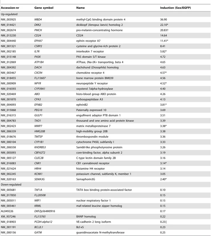
Table 1.Top SOX18 regulated genes.
Accession-nr Gene symbol Name Induction (Sox/EGFP)
Up-regulated
NM_003925 MBD4 methyl-CpG binding domain protein 4 36.90
NM_014421 DKK2 dickkopf (Xenopus laevis) homolog 2 22.10*
NM_002674 PMCH pro-melanin-concentrating hormone 20.83{
NM_013230 CD24 CD24 14.64
NM_004440 EPHA7 ephrin receptor A7 11.41*
NM_001321 CSRP2 cysteine and glycine-rich protein 2 8.41
NM_002185 IL7R interleukin 7 receptor 5.82*
NM_015148 PASK PAS domain S/T kinase 4.72
NM_012069 ATP1B4 ATPase, (Na+)K+transporting, beta 4 4.65
NM_004392 DACH dachshund (Drosophila) homolog 4.63
NM_003467 CXCR4 chemokine receptor 4 4.57*
NM_018455 FLJ13607 bone marrow protein BM039 4.56
NM_000909 NPYR neuropeptide Y receptor 4.52*
NM_016593 CYP39A1 oxysterol 7alpha-hydroxylase 4.40
NM_020469 ABO histo-blood group ABO protein 4.26
NM_001870 CPA3 carboxypeptidase A3 4.13
NM_004093 EFNB2 ephrinB2 3.81*
NM_015068 PEG10 Paternally expressed 10 3.69
NM_016315 GULP1 engulfment adaptor PTB domain 1 3.51
NM_004783 TAO1 thousand and one amino acid protein kinase 3.39
NM_002423 MMP7 matrix metalloproteinase 7 3.38*
NM_006339 HMG20B high-mobility group 20B 3.38
NM_018676 TMTSP thrombospondin module 3.36
NM_000104 CYP1B1 cytochrome P450, subfamily I 3.33
NM_006558 KHDRBS3 Sam68-like phosphotyrosine protein 3.26
NM_005093 CBFA2T2 core-binding factor, alpha subunit 2 3.19
NM_005127 CLEC2B C-type lectin domain family 2B 3.16
NM_016083 CNR1 CB1 cannabinoid receptor 3.14*
NM_021624 HRH4 histamine H4 receptor 3.14
NM_002245 KCNK1 potassium channel, subfamily K, member 1 3.05
NM_020163 SEMA3G Semaphorin3G 2.40*
Down-regulated
NM_005681 TAF1A TATA box binding protein-associated factor 0.10
NM_017850 FLJ20508 0.15
NM_005011 NRF1 nuclear respiratory factor 1 0.15
NM_005461 KRML maf-related leucine zipper homolog 0.15
AL049226 DKFZp564M0916 0.17
XM_937246 FLJ13765 BANP homolog 0.22
NM_018903 PCDH-alpha12 VE-cadherin 2 long isoform 0.23{
NM_001191 BCL2L1 Bcl-xS 0.23
NM_000156 GATM guanidinoacetate N-methyltransferase 0.25
*verified by real-time PCR; {
induction by real-time PCR was 1.5-fold;
{
PA1-24474, and Chemicon AB-3207) or rabbit IgG over-night, precipitated with blocked protein A sepharose, and washed with buffer containing increasing salt concentrations. The antibody was eluted from the DNA-protein complex in 1% SDS/0.1 M sodium bicarbonate, the agarose removed by centrifugation, and cross-linking reversed by addition of NaCl to a final conc. of 0.2 M and incubation at 65uC for 4 hours. DNA was purified over Qiagen columns, and analyzed by PCR using the primers given in Table S1. The anti-Sox18 antibody recognized Sox17 10–20 times less efficiently, no detectable cross-reactivity with Sox7 was observed (Figure S2).
Immunohistochemistry
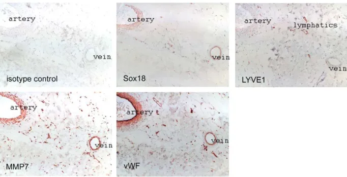
Serial sections from paraffin-embedded normal human trunk skin were stained with rabbit antibodies against SOX18 (sc-20100; 1:2500, Santa Cruz), MMP7 (ab4044, Abcam, 1:500), LYVE-1 (102PA50, Research Diagnostics, Inc., 1:100), or von Willebrand factor (A0082, Dako, 1:5000), followed by incubation with biotinylated secondary antibody (BA-1000, anti rabbit IgG, Vector Laboratories, 1:200). Bound antibodies were visualized by incubation with a streptavidin-HRP conjugate (Dako) and 3-amino-9-ethylcarbazole resulting in a red reaction product. Sections were counterstained with haematoxylin.
Results
Identification of SOX18 target genes
Our strategy to identify SOX18 target genes was to ectopically express the transcription factor in primary endothelial cells followed by microarray analysis of the resulting gene repertoire. In order to achieve high transfection efficiency we generated a SOX18 adenoviral vector containing an IRES-EGFP cassette. The control virus contained the IRES-EGFP cassette alone. As judged by EGFP expression, transduction efficiency was close to 100%. As assayed by real-time PCR, expression of Sox18 was approx. 70-fold higher as compared to controls (Figure S1), and did not alter Sox7 and -17 levels. Expression of selected candidate genes was confirmed by real-time PCR. Knock-down ofSOX18by RNA interference was then used as a complementary approach to verify the dependence of selected genes on the transcription factor. In order to obtain primary target genes and avoid the isolation of genes that are up-regulated secondary to the induction of others, we harvested the SOX18 expressing HUVEC at an early time point after transduction. SOX18 protein could be detected as early as 16 hours (not shown). RNA from SOX18-, EGFP-, and non-transduced HUVEC was subjected to microarray analysis. Using a threshold of 3-fold changes, 30 genes were found to be
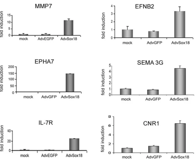
Figure 1. Real-time PCR analysis of SOX18 target genes.RNA was isolated from non-transduced (mock), control virus (AdvEGFP) and SOX18 adenovirus (AdvSOX18) transduced HUVEC and analyzed for the expression of matrix metalloproteinase 7 (MMP7), ephrinB2 (EFNB2), EPHA7, semaphorin 3G(SEMA3G), interleukin 7 receptor (IL-7R), and cannabinoid receptor 1 (CNR1). Values were normalized for ß2 microglobulin expression, and are expressed as fold induction compared to control cells.
upregulated, and 13 down-regulated (0.23% of the analyzed transcripts, Table 1). Selected genes were further analyzed using real-time PCR, and induction was confirmed forMMP7,
ephrin-B2 (EFNB2), EPHA7, semaphorin 3G (SEMA3G), interleukin 7 receptor(IL-7R), cannabinoid receptor 1(CNR1) (Figure 1), and others (Table 1). Whereas the published expression of VCAM-1
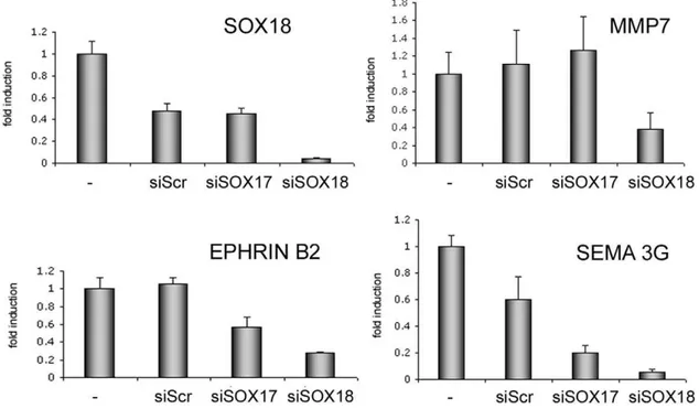
Figure 2. Knock-down of SOX18 diminishes the expression of target genes.HUVEC were transfected with siRNAs directed againstSOX17or
SOX18, or with a scrambled control (Scr), and expression of target genes analyzed by real-time PCR. Values were normalized for ß2 microglobulin levels, and are expressed as fold induction compared to control cells.
doi:10.1371/journal.pone.0030982.g002
Figure 3. Promoter analysis ofMMP7.A, Schematic representation of the twoMMP7promoter-luciferase reporter constructs containing either only the proximal or both potential SOX18 binding sites (black diamonds). B, the wild type (wt) constructs were cotransfected together with increasing amounts of SOX18, SOX7 or SOX17 expression vectors into HEK293 cells as indicated. Luciferase values were normalized for ß-Gal expression. C, in the shorter construct, the single potential SOX18 binding site was mutated. D, The shortMMP7promoter construct containing the mutated SOX18 binding site was analyzed as in (B).
could not be confirmed, both them-opioid receptor and claudin-5
were found to be 2-fold increased in the array (not shown). The entire data set has been deposited in the GEO data base (NCBI) under the accession nr. GSE8952.
In a complementary approach we then knocked down SOX18 in HUVEC (and also SOX17 which has been reported to have partially redundant function) using siRNA, and analyzed a subset of genes by real-time PCR. Of these transcripts,MMP7,EFNB2, andSEMA3Gwere reduced by SOX18 knock-down (Figure 2), the last two also responded to SOX17 knock-down. Two genes (CNR1
and EPHA7) responded weakly, whereas IL-7R expression was enhanced (data not shown).
MMP7is a direct target gene of SOX18
Based on its multiple functions in angiogenesis [33–37] and clear response to SOX18 in the overexpression and knock-down experiments, we selected MMP7 for further analysis. A set of experiments was performed to investigate whether it is a primary target or expressed indirectly, i.e., secondary to other regulators that may have been induced by SOX18. A direct target gene is supposed to contain one or more functional SOX18 binding site(s) in the promoter. We therefore analyzed theMMP7promoter by reporter gene analysis including mutation of the potential SOX18 binding site, binding of SOX18 in vitro using electrophoretic mobility shift assay (EMSA), and in vivo using chromatin immunoprecipitation (ChIP).
First, bioinformatic analysis using the program TOUCAN [38] revealed the presence of two potential SOX18 binding sites. We isolated two promoter fragments that contained either both or only the proximal site (Figure 3A), and cloned them into the luciferase reporter vector UBT-Luc [28]. Cotransfection of these constructs together with SOX18 resulted in a strong dose-dependent induction of both MMP7 promoter constructs. In contrast, cotransfected SOX7 or SOX17, the other members of the subgroup F, had only negligible effects (Figure 3B). Since cotransfection experiments showed that the proximal SOX18 binding site is sufficient to induce a strong response only this site was chosen for mutation experiments. Its mutation (Figure 3C) resulted in strongly diminishedMMP7 induction, demonstrating that this site is functional (Figure 3D). Some activity was observed with high amounts of co-transfected Sox18, possibly reflecting non-physiological effects. Furthermore, the shortMMP7promoter construct did not respond to cotransfected SOX7 or SOX17, the latter being in line with the lack of response to knock-down of SOX17 (Figure 3D).
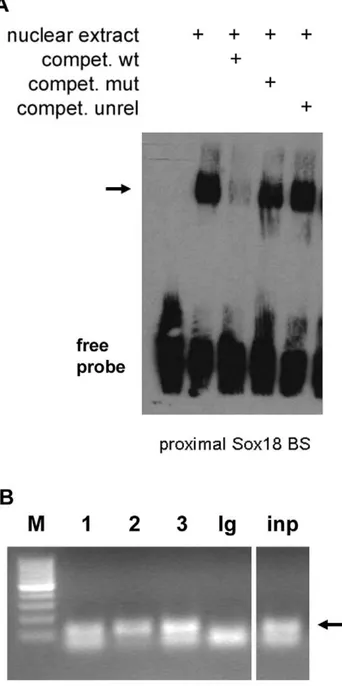
Next, we determined whether SOX18 binds to the proximal site
in vitro and in vivo. EMSA revealed that nuclear extracts from HUVEC contained a protein binding to the SOX18 binding site, and competition experiments using unlabelled wild type (wt) and mutated oligonucleotides demonstrated the specificity of binding (Figure 4A). However, probably due to technical reasons we were not able to perform a conclusive supershift to directly demonstrate the presence of Sox18 in the complex. HUVEC also express Sox-7 and -17, so in order to assess whether this site was indeed occupied by SOX18in vivo, we performed ChIP. Using primers flanking the proximal SOX18 binding site in theMMP7promoter, a specific band could be detected by PCR after precipitation with three different SOX18 antibodies, but not with a control antibody (Figure 4B). Since the Sox18 antibody from SantaCruz recognizes a region with some homology to the other SoxF group members we tested its specificity and found a weak cross-reactivity (1/10–1/ 20) with Sox17, and none with Sox7 (Figure S2). The Sox18 antibody from Chemicon recognizes an N-terminal 17 aa peptide sequence specific for Sox18. The correct identity of the amplified
bands was confirmed by sequencing. Therefore, and in accor-dance with the reporter and EMSA data, binding of SOX18 to the proximalMMP7SOX18 binding site could be demonstrated. We conclude that, using the criteria ofin vitroandin vivobinding and of reporter gene analysis, MMP7 contains at least one functional SOX18 binding site and represents a direct SOX18 target gene.
Figure 4. SOX18 binds to the proximal site in the MMP7
promoterin vitroandin vivo.A, Electrophoretic mobility shift assay (EMSA). Binding of SOX18 to its proximal site in theMMP7promoter was analyzed using nuclear extracts from HUVEC. Specificity of binding was demonstrated by competition experiments using the same (wt), a mutated (mut) or an unrelated (unrel) oligonucleotide as indicated, the specific complex is indicated by an arrow. B, The same site was analyzed by chromatin immunoprecipitation in HUVEC. Lane 1–3: differenta
-Sox18 antibodies (SantaCruz, Thermo, Chemicon, respectively) as described in Materials and Methods, Ig: IgG control antibody; inp: input, M: 100 bp marker. The 120 bp PCR fragments obtained after amplification ofDNA precipitated by the anti-SOX18 antibodies and input DNA are marked by an arrow.
Co-expression ofSOX18 andMMP7in skin vessels
SOX18has previously been found to be expressed in endothelial and smooth muscle cells [19]. To correlateSOX18expression with its target gene we performed immunohistochemical analysis of serial sections of normal adult human skin samples (Figure 5). We found SOX18 expressed in arteries, veins and some small capillaries, the latter all being LYVE1 negative and thus belonging to the blood vessel tree. SOX18 positive vessels co-expressed
MMP7. Moreover, SOX18 stained positive in the muscularis of arteries and veins, and these also expressedMMP7. The lack of a complete correlation of SOX18 and MMP7 expression might be due to the fact that MMP7 is also expressed in other cells and in response to stimuli that do not utilize SOX18 (e.g., FGF regulates MMP7 via AP1) [39], or due to diffusion of MMP7 as a secreted protein. Sections were stained with von Willebrand factor to identify all vessels, and LYVE1 for lymphatics. Of note, all lymphatics were negative for SOX18. Furthermore, we have analyzed tissue arrays containing 300 skin samples from basal cell carcinomas of a total of 100 different patients. Similar to the situation in normal skin, all LYVE1 positive lymphatics were negative for SOX18 (data not shown). Although these findings are in contrast with the notion that mutations in SOX18 affect the lymphatic vasculature [2], it may well be that these alterations occur during development and persist during adulthood without the necessity for continued expression ofSOX18.
Discussion
The notion that SOX18 plays an important role in angiogenesis is well supported by the occurrence of mutations in humans and mice that result in vascular (including lymphatic) defects; however, mechanistic insight how this transcription factor exerts its function is still very limited. As a first step to answer this question we have identified SOX18 target genes; one of them,MMP7, was analyzed in more detail, and we could show that it represents a direct SOX18 target gene. A recent study described lower levels of
Mmp7 and Il-7r inraggedmice [40]; while the former is supported by our findings, our data were controversial about the latter: Although expression of IL-7R was upregulated upon SOX18 overexpression, it was not reduced, for reasons unknown, upon knock-down in our experimental system.
In vascular cells, multiple functions have been described for MMP7. In general, MMPs degrade a broad variety of proteins, including extracellular matrix proteins. MMP7 substrates include proteoglycans, fibronectin, elastin and casein. SOX18 dependent expression ofMMP7during angiogenesis could therefore function to promote the sprouting process. However, MMP7 appears to have more functions than just clearing barriers: It has been implicated in ectodomain shedding of several cell surface molecules, including heparin-binding epidermal growth factor precursor, membrane-bound Fas ligand, E-cadherin, and TNFa
precursor, all of which are cleaved to release the respective soluble forms [35]. Moreover, VEGF-165, which is bound by connective tissue growth factor, can be released from this inactive complex by MMP7 [34]. Similarly, KC, a member of the CXC family of small chemokines can be liberated from its complex with syndecan-1, thereby generating a KC gradient capable of directing neutrophil migration [33]. These biological activities would suggest a pro-angiogenic function.
However, also anti-angiogenic properties have been attributed to MMP7. First, it can cleave plasminogen and collagen XVIII to generate angiostatin, as well as a 28 kD endostatin-spanning fragment, respectively [41]. Moreover, it cleaves decorin, a proteoglycan that can serve as a reservoir for TGF-ß, a potent anti-angiogenic factor in the extracellular matrix [36]. In a mouse model of corneal wound healing, MMP7 served as an inhibitor of corneal avascularity [37]. Therefore, the net outcome of SOX18 dependent MMP7 expression cannot be predicted, and may depend on the specific site and context of the angiogenic process. BesideMMP7, we have identified several other potential target genes of SOX18. Combining SOX18 overexpression with the more stringent criterion of SOX18 knock-down, EFNB2 and
Figure 5. SOX18 and MMP7 are co-expressed in arteries and veins, but not in lymphatic vessels.Serial sections from human skin were analyzed by immunohistochemical staining with the indicated antibodies. Endothelial cells were revealed by von Willebrand factor (vWF) staining, lymphatic vessels by LYVE-1.
SEMA3Gturned out to be reliable candidates. It should be noted thatMMP7responded only to SOX18 knock-down, whereas the other two were dependent (however, to a lesser degree) on SOX17 levels. Both EFNB2 and SEMA3G serve as guidance molecules in endothelial cells. EFNB2 and its counter-receptor EPHB4 are expressed on developing arteries and veins respectively, and repulsive signaling between the two molecules has been suggested to maintain the boundaries between the two types of vessels during network formation [42]. The identification ofEFNB2as a SOX18 target fits well to the recent observations that SOX18 knock-down in the zebrafish results in impaired arteriovenous specification [15,16]. Less information exists for SEMA3G, a member of larger family of phylogenetically conserved secreted and membrane-bound proteins. SEMA3G is a secreted molecule that binds to Neuropilin-2, but not Neuropilin-1, receptors that are also bound by certain VEGF forms. In neuronal development, SEMA3G induced the repulsion of sympathetic, but not dorsal root ganglion axons, indicating that it can selectivity repel specific types of axons [43]. Semaphorin class 3 family members have been suggested as modulators of angiogenesis [44]. Together, and also considering the additional upregulation of EPHA7 (which, however, did not clearly respond to SOX18 knock-down), suggests that one function of SOX18 may be the control of directed vessel formation. For reasons unknown, we did not identify VCAM-1, a published Sox18 target gene; one explanation could be that our experimental setup of Sox18 overexpression as a primary screen would allow solely the identification of genes for which Sox18 is alone sufficient, but not those where it is necessary (in combination with other factors). We also did not find Prox1 in HUVEC, however Sox18 could induce its expression in endothelial progenitor cells (not shown). This is consistent with the finding that Coup-TFII has been identified as a cofactor for Prox1 initial expression and maintainance [22].
Since a single transcription factor usually participates in the regulation of several genes, mutations that impair its function can be expected to affect a broad spectrum of genes and result in a complex phenotype. DiminishedMMP7expression as a result of a mutated SOX18 may already affect several aspects of angiogen-esis, both in the developing as well as the adult organism. In addition, the identification of EFNB2 and SEMA3G suggests a role for SOX18 in vessel guidance.
Taken together, the present work represents a functional way to identify potential SOX18 target genes. With the identification of
MMP7and performed functional analysis, the direct regulatory relationship between SOX18 and MMP7 was demonstrated. Future studies will focus on other potential target genes identified by this approach, to further investigate the role of SOX18 and its target genes in vessel formation. With the more detailed characterization of additional direct Sox18 target genes a better understanding of the molecular mechanisms of SOX18 in the
raggedphenotype in mice and the HLT syndrome in humans will be gained.
Supporting Information
Figure S1 Overexpression of Sox18 by a recombinant adenovirus. HUVEC were either mock transfected or trans-fected with a recombinant adenovirus for expression of GFP or of Sox18 as indicated, and mRNA isolated 16 hours later and analyzed by real-time PCR for expression of Sox7, -17 and -18. Values were normalized to ß2-microglobulin.
(TIF)
Figure S2 Crossreactivity of Sox18 antibody.A, HEK293 cells were transfected with myc-tagged expression vectors for Sox7, -17, and -18, or empty vector (EV), and protein extracts analyzed by Western blotting using anti-myc and anti-Sox18 (SantaCruz). B, repetition of the experiment using Sox17 and -18 and different times of exposure.
(TIF)
Table S1 Primers used for constructs, real-time PCR, EMSA, ChIP, and knock-down experiments.
(DOC)
Acknowledgments
The authors wish to thank the members of the Austrian Angiogenesis Network for continuous support and discussion, and Y. Schichl for proof-reading of the manuscript.
Author Contributions
Conceived and designed the experiments: OW PP RdM EH. Performed the experiments: MH HN RHW MB UR CL. Analyzed the data: HM RdM. Wrote the paper: RdM.
References
1. Irrthum A, Devriendt K, Chitayat D, Matthijs G, Glade C, et al. (2003) Mutations in the transcription factor gene SOX18 underlie recessive and dominant forms of hypotrichosis-lymphedema-telangiectasia. Am J Hum Genet 72(6): 1470–1478.
2. Francois M, Caprini A, Hosking B, Orsenigo F, Wilhelm D, et al. (2008) Sox18 induces development of the lymphatic vasculature in mice. Nature 456(7222): 643–647.
3. Hosking BM, Wang SC, Chen SL, Penning S, Koopman P, et al. (2001) SOX18 directly interacts with MEF2C in endothelial cells. Biochem Biophys Res Commun 287(2): 493–500.
4. James K, Hosking B, Gardner J, Muscat GE, Koopman P (2003) Sox18 mutations in the ragged mouse alleles ragged-like and opossum. Genesis 36(1): 1–6.
5. Pennisi D, Gardner J, Chambers D, Hosking B, Peters J, et al. (2000) Mutations in Sox18 underlie cardiovascular and hair follicle defects in ragged mice. Nat Genet 24(4): 434–437.
6. Sandholzer J, Hoeth M, Piskacek M, Mayer H, de Martin R (2007) A novel 9-amino-acid transactivation domain in the C-terminal part of Sox18. Biochem Biophys Res Commun 360(2): 370–374.
7. Pennisi D, Bowles J, Nagy A, Muscat G, Koopman P (2000) Mice null for sox18 are viable and display a mild coat defect. Mol Cell Biol 20(24): 9331–9336. 8. Hosking B, Francois M, Wilhelm D, Orsenigo F, Caprini A, et al. (2009) Sox7
and Sox17 are strain-specific modifiers of the lymphangiogenic defects caused by Sox18 dysfunction in mice. Development 136(14): 2385–2391.
9. Matsui T, Kanai-Azuma M, Hara K, Matoba S, Hiramatsu R, et al. (2006) Redundant roles of Sox17 and Sox18 in postnatal angiogenesis in mice. J Cell Sci 119(Pt 17): 3513–3526.
10. Sakamoto Y, Hara K, Kanai-Azuma M, Matsui T, Miura Y, et al. (2007) Redundant roles of Sox17 and Sox18 in early cardiovascular development of mouse embryos. Biochem Biophys Res Commun 360(3): 539–544.
11. Lioubinski O, Muller M, Wegner M, Sander M (2003) Expression of Sox transcription factors in the developing mouse pancreas. Dev Dyn 227(3): 402–408.
12. Olsson JE, Kamachi Y, Penning S, Muscat GE, Kondoh H, et al. (2001) Sox18 expression in blood vessels and feather buds during chicken embryogenesis. Gene 271(2): 151–158.
13. Stanojcic S, Stevanovic M (2000) The human SOX18 gene: cDNA cloning and high resolution mapping. Biochim Biophys Acta 1492(1): 237–241.
14. Zhang C, Basta T, Klymkowsky MW (2005) SOX7 and SOX18 are essential for cardiogenesis in Xenopus. Dev Dyn 234(4): 878–891.
15. Cermenati S, Moleri S, Cimbro S, Corti P, Del Giacco L, et al. (2008) Sox18 and Sox7 play redundant roles in vascular development. Blood 111(5): 2657–2666.
16. Herpers R, van de Kamp E, Duckers HJ, Schulte-Merker S (2008) Redundant Roles for Sox7 and Sox18 in Arteriovenous Specification in Zebrafish. Circ Res 102(1): 12–15.
18. Darby IA, Bisucci T, Raghoenath S, Olsson J, Muscat GE, et al. (2001) Sox18 is transiently expressed during angiogenesis in granulation tissue of skin wounds with an identical expression pattern to Flk-1 mRNA. Lab Invest 81(7): 937–943. 19. Garcia-Ramirez M, Martinez-Gonzalez J, Juan-Babot JO, Rodriguez C, Badimon L (2005) Transcription factor SOX18 is expressed in human coronary atherosclerotic lesions and regulates DNA synthesis and vascular cell growth. Arterioscler Thromb Vasc Biol 25(11): 2398–2403.
20. Young N, Hahn CN, Poh A, Dong C, Wilhelm D, et al. (2006) Effect of disrupted SOX18 transcription factor function on tumor growth, vasculariza-tion, and endothelial development. J Natl Cancer Inst 98(15): 1060–1067. 21. Luo M, Guo XT, Yang W, Liu LQ, Li LW, et al. (2008) Inhibition of tumor
angiogenesis by cell-permeable dominant negative SOX18 mutants. Med Hypotheses 70(4): 880–882.
22. Srinivasan RS, Geng X, Yang Y, Wang Y, Mukatira S, et al. (2010) The nuclear hormone receptor Coup-TFII is required for the initiation and early maintenance of Prox1 expression in lymphatic endothelial cells. Genes Dev 24(7): 696–707.
23. Hosking BM, Wang SC, Downes M, Koopman P, Muscat GE (2004) The VCAM-1 gene that encodes the vascular cell adhesion molecule is a target of the Sry-related high mobility group box gene, Sox18. J Biol Chem 279(7): 5314–5322.
24. Im HJ, Smirnov D, Yuhi T, Raghavan S, Olsson JE, et al. (2001) Transcriptional modulation of mouse mu-opioid receptor distal promoter activity by Sox18. Mol Pharmacol 59(6): 1486–1496.
25. Fontijn RD, Volger OL, Fledderus JO, Reijerkerk A, de Vries HE, et al. (2008) SOX-18 controls endothelial-specific Claudin-5 gene expression and barrier function. Am J Physiol Heart Circ Physiol 294(2): H891–900.
26. Nitta T, Hata M, Gotoh S, Seo Y, Sasaki H, et al. (2003) Size-selective loosening of the blood-brain barrier in claudin-5-deficient mice. J Cell Biol 161(3): 653–660.
27. Samant GV, Schupp MO, Francois M, Moleri S, Kothinti RK, et al. (2011) Sox factors transcriptionally regulate ROBO4 gene expression in developing vasculature in zebrafish. J Biol Chem 286(35): 30740–30747.
28. de Martin R, Strasswimmer J, Philipson L (1993) A new luciferase promoter insertion vector for the analysis of weak transcriptional activities. Gene 124(1): 137–138.
29. Wrighton CJ, Hofer-Warbinek R, Moll T, Eytner R, Bach FH, et al. (1996) Inhibition of endothelial cell activation by adenovirus-mediated expression of I kappa B alpha, an inhibitor of the transcription factor NF-kappa B. J Exp Med 183(3): 1013–1022.
30. McGrory WJ, Bautista DS, Graham FL (1988) A simple technique for the rescue of early region I mutations into infectious human adenovirus type 5. Virology 163(2): 614–617.
31. Mayer H, Bilban M, Kurtev V, Gruber F, Wagner O, et al. (2004) Deciphering regulatory patterns of inflammatory gene expression from
interleukin-1-stimulated human endothelial cells. Arterioscler Thromb Vasc Biol 24(7): 1192–1198.
32. Nowak DE, Tian B, Brasier AR (2005) Two-step cross-linking method for identification of NF-kappaB gene network by chromatin immunoprecipitation. Biotechniques 39(5): 715–725.
33. Ding K, Lopez-Burks M, Sanchez-Duran JA, Korc M, Lander AD (2005) Growth factor-induced shedding of syndecan-1 confers glypican-1 dependence on mitogenic responses of cancer cells. J Cell Biol 171(4): 729–738.
34. Hashimoto G, Inoki I, Fujii Y, Aoki T, Ikeda E, et al. (2002) Matrix metalloproteinases cleave connective tissue growth factor and reactivate angiogenic activity of vascular endothelial growth factor 165. J Biol Chem 277(39): 36288–36295.
35. Ii M, Yamamoto H, Adachi Y, Maruyama Y, Shinomura Y (2006) Role of matrix metalloproteinase-7 (matrilysin) in human cancer invasion, apoptosis, growth, and angiogenesis. Exp Biol Med (Maywood) 231(1): 20–27. 36. Imai K, Hiramatsu A, Fukushima D, Pierschbacher MD, Okada Y (1997)
Degradation of decorin by matrix metalloproteinases: identification of the cleavage sites, kinetic analyses and transforming growth factor-beta1 release. Biochem J 322(Pt 3): 809–814.
37. Kure T, Chang JH, Kato T, Hernandez-Quintela E, Ye H, et al. (2003) Corneal neovascularization after excimer keratectomy wounds in matrilysin-deficient mice. Invest Ophthalmol Vis Sci 44(1): 137–144.
38. Aerts S, Van Loo P, Thijs G, Mayer H, de Martin R, et al. (2005) TOUCAN 2: the all-inclusive open source workbench for regulatory sequence analysis. Nucleic Acids Res 33: W393–396.
39. Holnthoner W, Kerenyi M, Groger M, Kratochvill F, Petzelbauer P (2006) Regulation of matrilysin expression in endothelium by fibroblast growth factor-2. Biochem Biophys Res Commun 342(3): 725–733.
40. Downes M, Francois M, Ferguson C, Parton RG, Koopman P (2009) Vascular defects in a mouse model of hypotrichosis-lymphedema-telangiectasia syndrome indicate a role for SOX18 in blood vessel maturation. Hum Mol Genet 18(15): 2839–2850.
41. Patterson BC, Sang QA (1997) Angiostatin-converting enzyme activities of human matrilysin (MMP-7) and gelatinase B/type IV collagenase (MMP-9). J Biol Chem 272(46): 28823–28825.
42. Fuller T, Korff T, Kilian A, Dandekar G, Augustin HG (2003) Forward EphB4 signaling in endothelial cells controls cellular repulsion and segregation from ephrinB2 positive cells. J Cell Sci 116(Pt 12): 2461–2470.
43. Taniguchi M, Masuda T, Fukaya M, Kataoka H, Mishina M, et al. (2005) Identification and characterization of a novel member of murine semaphorin family. Genes Cells 10(8): 785–792.