J of Evolution of Med and Dent Sci/ eISSN- 2278-4802, pISSN- 2278-4748/ Vol. 3/ Issue 58/Nov 03, 2014 Page 13182
LEFT ATRIAL APPENDAGE ANEURYSM: A CASE REPORT
Ashish Ashtekar1, Abhijit Patil2, Sahil Garg3
HOW TO CITE THIS ARTICLE:
Ashish Ashtekar, Abhijit Patil, Sahil Garg. Left Atrial Appendage Aneurysm: A Case Report . Journal of Evolution of Medical and Dental Sciences 2014; Vol. 3, Issue 58, November 03; Page: 13182-13187, DOI:10.14260/jemds/2014/3752
ABSTRACT: Aneurysm of left atrium may involve the left atrial wall or left atrial appendage. Aneurysm of left atrial appendage is usually acquired in etiology secondary to mitral valve disease, whereas its congenital variant is a rare developmental anomaly. Congenital left atrial appendage aneurysm is very rarely reported entity. They lead to complications like systemic emboli, arrhythmias and worst as death. They are usually misdiagnosed most of the times as a mediastinal mass or cardiac tumour on Chest X-ray. USG provides an easily accessible modality in providing a provisional diagnosis, in confirming the location and its communication with the mediastinal vascular structures. Cardiac and coronary CT and MR help in providing a conclusive diagnosis and earlier surgical intervention thereby preventing further mortality or morbidity. We report a case of 20 year old female incidentally diagnosed with left atrial appendage aneurysm.
KEYWORDS: Left Atrial appendage aneurysm (LAAA), Sinus of valsalva aneurysm, Conventional Radiography. USG, Cardiac CT, Cardiac MRI.
INTRODUCTION: Aneurysm refers to a bulge or ‘pocketing of the wall or lining of a vessel. When it concerns the heart muscle it is referred to as cardiac aneurysm. They include following entities 1.Ventricular aneurysm (Most common) 2.Atrial septal aneurysm 3.Atrial appendage aneurysm (very rare).Left atrial appendage is derived from the left wall of the primary atrium (formed during the fourth week of embryonic development). It acts as a decompression chamber during ventricular systole or when the atrial pressure is high.
Owing to stasis due to trabeculations and its shape it is more prone to aneurysm and thromboembolism.1 Aneurysm of left atrial appendage occurs mostly due to increase in left atrial pressure secondary to mitral valve disease. Its congenital variant is a rare condition with only 75 cases reported as far.2 Congenital dysplasia of musculi pectinati has been attributed to congenital variant.8Untreated cases may develop fatal complications of systemic emboli, fatal arrhythmias, congestive heart failure, thrombus formation and eventually stroke. Due to this sequelae surgical management is indicated even in asymptomatic cases.2,5
CASE REPORT: A 20 year old female presented with fever, palpitations, on and off breathlessness with chest pain since 5 years. There was associated history of headache and weakness with pain in the lower limbs.
J of Evolution of Med and Dent Sci/ eISSN- 2278-4802, pISSN- 2278-4748/ Vol. 3/ Issue 58/Nov 03, 2014 Page 13183 Routine radiograph was performed. USG was performed using curvilinear probe. Cardiac MRI was performed using 1.5 T Siemens Avanto scanner.
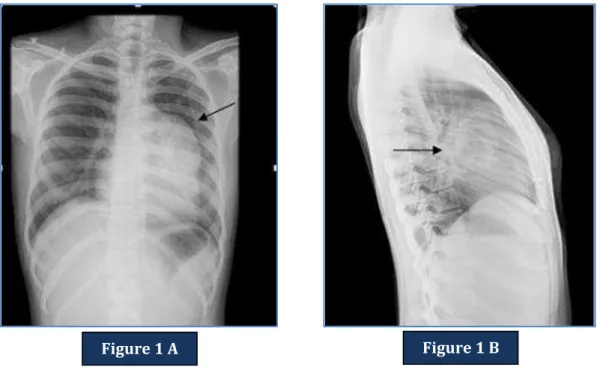
X ray chest PA and left lateral view revealed convex opacity between inferior borders of the aortic knob up to upper half of left cardiac border with peripheral curvilinear calcification. The lateral
projection revealed the hilar location of the lesion below the left main stem bronchus [Figure. 1A & 1B ].
Figure 2A &2B: Ultrasonography which revealed partially thrombosed vascular structure communicating with the left side of the heart. The residual lumen showed pulsatile flow.
Figure 1 A Figure 1 B
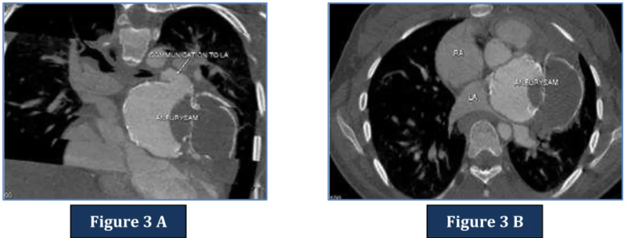
J of Evolution of Med and Dent Sci/ eISSN- 2278-4802, pISSN- 2278-4748/ Vol. 3/ Issue 58/Nov 03, 2014 Page 13184 Figure 3 (A and B): CardiacCTrevealed a partially thrombosed lobulated aneurysm of the left atrial appendage with marginal calcification.
Figure 4 (A, B, C and D)- Cardiac MRI Axial and sagittal images revealed a partially thrombosed bilobed aneurysm of the left atrial appendage.
Figure 3 A Figure 3 B
Figure 4 A Figure 4 B
J of Evolution of Med and Dent Sci/ eISSN- 2278-4802, pISSN- 2278-4748/ Vol. 3/ Issue 58/Nov 03, 2014 Page 13185
DISCUSSION: Left atrial appendage is a small pouch located high in the left atrium and within the pericardium. It has a wall adjacent to the left ventricle and acts a decompression chamber during left ventricular systole and during other periods when left atrial pressure is high.
Cardiac aneurysms (left ventricular aneurysm, atrial septal aneurysm and atrial appendage aneurysm) can be congenital or acquired. Congenital atrial septal aneurysm and atrial appendage aneurysm are rare. Acquired causes include post myocardial infarction, different interatrial pressure gradients and secondary to mitral valve disease.
In 1938, Semans and Tausig reported a saccular dilatation of atrium in 5 year old girl without appendage involvement.2,3,9 Parmley in 1962, reported dilatation of left atrial appendage in 2 children.4Williams in 1963 reported a case of 27 year old with thin walled gross dilatation of Left atrial appendage.5Palacaio and associates reported a case of 32 year old woman with cerebral embolism and atrial arrhythmia with excision of left atrial appendage.6 In 1999 Zhao and others reported case of 27 year old man with left atrial appendage aneurysm which was excised in 1996 without cardiopulmonary bypass.3,9
Pathophysiology behind acquired causes is known to be weakening of muscle in patients of myocardial infarction leading to ventricular aneurysm, compensatory atrial enlargement secondary to mitral valve disease leading to atrial appendage aneurysm and different interatrial pressure gradients in atrial septal aneurysm.6
Congenital aneurysm of atrial appendage is attributed to congenital dysplasia of musculipectinati in the left atrial appendage and from the bands of atrial muscle from which they arise.8 They may also involve the left atrial wall but are usually confined to atrial appendage only.7 They are of 2 types: Intrapericardial and extrapericardial. Definitive criteria for intrapericardial type are intact pericardium, normal mitral valve and left atrium, blood flow between the aneurysm and atrium and histological diagnosis of atrial appendage.12,13
Congenital left atrial appendage aneurysm, although known as a congenital entity usually presents in the second decade of life whereas the acquired cases presents later in life usually at the age of 40-50 years in cases of mitral valve disease and in elderly in cases of myocardial infarction. The clinical presentations includes intracardiac thrombus, supraventricular arrhythmias(mostly atrial fibrillation), heart failure due to compression of pulmonary venous drainage, chest pain secondary to compression of coronary arteries and systemic embolism.3Supraventricular thromboembolism and systemic embolism are the most common presentation of congenital left atrial appendage aneurysm.
Diagnostic modalities like conventional radiography, echocardiography, cardiac CT, cardiac MRI and conventional angiography are routinely used for its early detection, assess its complications and prevent further morbidity and mortality caused by it.
On conventional radiography features of mediastinal mass with obtuse angles and absence of lung markings are noted. Sharply defined soft tissue density lesion is noted adjacent to left heart border and on the lateral projection appears adjacent to the hilum. Other conditions that show same features on radiography include a mediastinal mass, a pericardial cyst, a bronchogenic cyst, an epicardial lipoma, a cardiac or paracardiac tumor, a pericardial defect, or valvular heart disease. Further investigations with CT and MRI are required for its diagnosis.14
J of Evolution of Med and Dent Sci/ eISSN- 2278-4802, pISSN- 2278-4748/ Vol. 3/ Issue 58/Nov 03, 2014 Page 13186 Cardiac/Coronary CT is valuable in evaluating the anatomy of coronary artery and its compression caused by the aneurysm and differentiates from sinus of valsalva aneurysm, being its close differential.14
MRI has been known to be the superior most diagnostic modality. MRI evaluates surrounding structures, cardiac anatomy and systolic and diastolic dysfunctions with adequate detection of associated lesions and their complications. It can be effectively and judiciously used in young adults and children due to absence of ionizing radiation. Intracardial and extracardial aneurysms can be differentiated and smaller size aneurysm can be easily picked up on MRI.
Different signal characteristics help in coming to a conclusive diagnosis. Central bronchogenic cyst shows high signal T2 without any contact with pericardium, pericardial cyst shows high signal T2 when it is not in contact with pericardium, Cardiac tumours typically shows signal flow void and epicardial lipomas shows typically high T1 and T2 signal intensity. Atrial appendage aneurysm can be diagnosed as communicating the left atrial appendage. It may show flow void if thrombus is present within it.14
Left atrial appendage aneurysm are usually silent in origin and may produce life threatening complications of angina pectoris, thrombus formation, stroke due to embolism and eventually death. There are increased chances of complications on increasing size of aneurysm. Hence its early detection and treatment may prevent the significant mortality caused by it4,8,10
There are extremely rare associations of LAAA with other congenital disease such as septal defect, persistent left superior vena cava, anomalies of renal arteries or anomalous pulmonary venous return.11
Treatment of choice is surgical aneuresctomy particularly in children, generally with extracorporeal circulation after thoracotomy and left atrial reconstruction. According to the literature arrhythmias usually disappear immediately in the post-operative period and heart failure is controlled and patient shows marked improvement without any recurrence. Endovascular aneurysm repair (EVAR) has recently been under investigation for treatment of atrial appendage aneurysm.
The aim of this study was to delineate the radiological features of left atrial appendage aneurysm and prevent serious sequelae including various systemic emboli and arrhythmias and eventually death. Earlier diagnosis and surgical intervention may prevent this mortality and morbidity and is presently indicated even in asymptomatic cases.
REFERENCES:
1. Saddy NM, Obel OA, Camm AJ: Left atrial appendage: structure, function, and role in thromboembolism, Am Heart J. 1999 Nov; 82 (5): 547-54.
2. Chowdhury UK, Seth S, Govindappa R, Jagia P et al: Congenital left atrial appendage aneurysm: A case report and review of literature: Heart Lung Circ 2009:18: 412-16.
3. Keith A. The evolution and action of certain muscular structures of the heart. Lancet 1904; 1 (12): 703-7.
4. Gold JP, Afifi HY, Ko W: Congenital giant aneurysm of left atrial appendage: Diagnosis and management. J Card Surg 1996; 11:147-150.
J of Evolution of Med and Dent Sci/ eISSN- 2278-4802, pISSN- 2278-4748/ Vol. 3/ Issue 58/Nov 03, 2014 Page 13187 6. Mügge A, Werner G. Daniel, Christiane Angermann, ChristophSpes, Bijoy K. Khandheria: Atrial
septal aneurtysm in adult patients1995; 91: 2785-2792.
7. Bilge M, Yasar AS, Bozkurt M, Karakas F, Bilen E, Yuksel IO. Left atrial appendage aneurysm secondary to eccentric severe ischemic mitral regurgitation. Echocardiography.2009; 26 (10): 1225–7.
8. Dean A Bramlet, Jesse E Edwards: Congenital aneurysm of left atrial appendage: Br Heart J 1981; 45: 97-100.
9. Victor S and Nayak V: Aneurysm of the Left Atrial Appendage: Tex Heart Inst J.2001: 28 (2): 111-118.
10.Park JS, Lee DH, Han SS et al. incidentally found growing congenital aneurysm of the left atrium: J Korean Med Sci 2003; 18 (2): 262-266.
11.Wagshal AB, Applebaum A, Crystal P et al: Atrial tachycardia as the presenting sign of left atrial appendage aneurysm: Pacing Clinelectrophysiol 2000:23:283-285.
12.Wilson D, Kalra N, Brody EA, Van Dyk H, Sorrell VL. Left atrial appendage aneurysm A rare anomaly with an atypical presentation. Congenit Heart Dis 2009; 4: 489-93.
13.Ulucum M, Muderrisoglu H, SezginA: Giant left atrial appendage aneurysm: the Third ventricle. Int J Cardiovasc Imaging 2005; 25:225-30.
14.Hoffmann U., Hamed N., Herold C., Globits S.: Radiological signs of a left atrial aneurysm. EurRadiol 2000; 10: 1332-1334.
AUTHORS:
1. Ashish Ashtekar 2. Abhijit Patil 3. Sahil Garg
PARTICULARS OF CONTRIBUTORS:
1. Assistant Professor, Department of Radiology, Dr. D. Y. Patil Medical College and Hospital, Pimpri.
2. Associate Professor, Department of Radiology, Dr. D. Y. Patil Medical College and Hospital, Pimpri.
3. Senior Resident, Department of Radiology, Dr. D. Y. Patil Medical College and Hospital, Pimpri.
NAME ADDRESS EMAIL ID OF THE CORRESPONDING AUTHOR:
Dr. Ashish Ashtekar, A 203, Windsor 1, Mahindra Royale, Pimpri, Pune-411018.
Email: [email protected]