RESEARCH ARTICLE
Differential Translocation of Host Cellular
Materials into the
Chlamydia trachomatis
Inclusion Lumen during Chemical Fixation
Marcela Kokes, Raphael H. Valdivia*
Department of Molecular Genetics and Microbiology and Center for the Genomics of Microbial Systems, Duke University Medical Center, Durham, North Carolina, United States of America
Abstract
Chlamydia trachomatismanipulates host cellular pathways to ensure its proliferation and survival. Translocation of host materials into the pathogenic vacuole (termed‘inclusion’) may facilitate nutrient acquisition and various organelles have been observed within the inclusion, including lipid droplets, peroxisomes, multivesicular body components, and mem-branes of the endoplasmic reticulum (ER). However, few of these processes have been documented in living cells. Here, we survey the localization of a broad panel of subcellular elements and find ER, mitochondria, and inclusion membranes within the inclusion lumen of fixed cells. However, we see little evidence of intraluminal localization of these organelles in live inclusions. Using time-lapse video microscopy we document ER marker translocation into the inclusion lumen during chemical fixation. These intra-inclusion ER elements resist a variety of post-fixation manipulations and are detectable via immunofluorescence micros-copy. We speculate that the localization of a subset of organelles may be exaggerated dur-ing fixation. Finally, we find similar structures within the pathogenic vacuole ofCoxiella burnettiinfected cells, suggesting that fixation-induced translocation of cellular materials may occur into the vacuole of a range of intracellular pathogens.
Introduction
Chlamydia trachomatisis a human pathogen of global significance–it is the most common
sex-ually-transmitted bacterial pathogen and the leading cause of preventable blindness worldwide [1]. During infection of mammalian cells, bacteria remain sequestered within a membrane-bound compartment termed an inclusion. The inclusion is fundamental to the intracellular lifestyle ofC.trachomatisas it represents a first line of defense against immune surveillance and anti-microbial effectors [2]. In addition, as an obligate intracellular pathogen,C. trachoma-tisrequires vital nutrients obtained from its infected host cell (reviewed in [3]) and interactions between the inclusion and host cellular components play an important role in this process (reviewed in [4]).
Many subcellular organelles reside in close proximity to the inclusion, some of which have been reported to interact intimately with inclusions (reviewed in [4]). In a well-characterized example, endoplasmic reticulum (ER) tubules closely appose toC.trachomatisinclusion
OPEN ACCESS
Citation:Kokes M, Valdivia RH (2015) Differential Translocation of Host Cellular Materials into the Chlamydia trachomatisInclusion Lumen during Chemical Fixation. PLoS ONE 10(10): e0139153. doi:10.1371/journal.pone.0139153
Editor:Deborah Dean, University of California, San Francisco, University of California, Berkeley, and the Children's Hospital Oakland Research Institute, UNITED STATES
Received:January 16, 2015
Accepted:September 8, 2015
Published:October 1, 2015
Copyright:© 2015 Kokes, Valdivia. This is an open access article distributed under the terms of the
Creative Commons Attribution License, which permits unrestricted use, distribution, and reproduction in any medium, provided the original author and source are credited.
Data Availability Statement:All relevant data are within the paper and its Supporting Information files.
membranes [5,6] to form membrane contact sites that facilitate the transfer of lipids directly between the ER and the pathogenic vacuole [7,8]. The Golgi apparatus fragments into mini-stacks [9] in an actin and bacterial effector-dependent manner [10] which closely surround the inclusion [11,12]. Lipid droplets are recruited to the inclusion periphery [13] and can translo-cate into the inclusion lumen [14] while lysosomes closely surround the inclusion to presum-ably enhance the ability of bacteria to acquire amino acids from degraded proteins [15]. Multivesicular bodies (MVBs) are enriched at the inclusion periphery and inhibition of MVB biogenesis can disruptC.trachomatislipid uptake [16,17]. Recycling endosomes closely associ-ate with the inclusion [18–20] but are thought to interact with the inclusion in a
fusion-inhib-ited state [19,21]. Mitochondria localize around the inclusion of someChlamydiaspecies [6,22], and mitochondrial protein import is important for replication ofC.caviae[23].
In several instances, interactions with the inclusion extend beyond recruitment to the periphery and entire organelles translocate into the inclusion lumen. Briefly, protein markers of sphingolipids [11], cholesterol [24], lipid droplets[14], and the ER [25] have been reported within the inclusion of living cells. Components of the ER [25,26], MVBs [16], and peroxi-somes [27], have been observed within the inclusion of fixed cells, as well as Rab14 [28], apoA-1 and phosphatidylcholine [29] and proteins that interact with acyl-CoA or each other includ-ing ASCL3, ACBD6, and ZNF23 [30]. Additionally, some subcellular components and markers such as MVBs [31] or phosphatidylcholine [12] have been only observed in inclusions within only fixed cells.
In this study, we sought to characterize organelle interactions with the inclusion by system-atically surveying organelle markers for localization within infected cells. We found a subset of the many inclusion-proximal subcellular elements within inclusions, including components of the ER, mitochondria, and markers of the inclusion membrane. We document the striking translocation of ER markers into the inclusion lumen during the process of chemical fixation. These internalized structures persisted through the process of sample processing for indirect immunofluorescence, indicating that the localization of a subset of organelles to the inclusion may be exaggerated by the fixation process rather than accurately reflecting organelle localiza-tion prior to manipulalocaliza-tion. Finally, we observed similar structures within the pathogenic vacu-ole ofCoxiella burnettii-infected cells, suggesting that fixation-induced translocation of subcellular elements may constitute a more widespread phenomenon in the study of intracellu-lar pathogens residing within intracellu-large vacuoles.
Results
A Subset of Inclusion-Proximal Subcellular Organelles Localize to the
Lumen of Inclusions
We surveyed interactions between host organelles and mid-to-late cycle inclusions by express-ing a panel of fluorescent protein-tagged markers of various subcellular organelles in cells infected for 30 hr (Fig 1A). We fixed cells and quantified the frequency of cells with fluorescent material in the inclusion lumen (Fig 1B). To enhance our ability to accurately define the inclu-sion edge in three dimeninclu-sions without interference from light above and below each plane of focus, we used confocal rather than widefield microscopy. Furthermore, since most markers had much lower intensity within the inclusion compared to cellular structures, we used spin-ning disk rather than laser scanspin-ning confocal microscopy to reduce photobleaching while imaging cells in three dimensions.
Of all intracellular compartments tested, we found markers of the ER lumen and membrane, mitochondrial matrix and outer membrane [32], and proteins which, in addition to differen-tially localizing to a range of subcellular compartments, also associate with the inclusion
(FAPP1-PH-GFP is a marker of phosphatidylinositol 4-phosphates [33] which are enriched on the inclusion [34]) [35,36] within a significant proportion (over 50%) of inclusions (Fig 1B) appearing as bleb-like structures (Fig 1A, arrowheads). Notably, we found markers of the cyto-sol and other inclusion-proximal subcellular organelles including recycling endosomes, MVBs, the Golgi apparatus, and lysosomes [9,12,16,18,21,31] also as bleb-like structures within only approximately one-quarter of inclusions, but did not observe a marker of the more distal plasma membrane [37] within inclusions. These findings reveal a selectivity to which inclu-sion-proximal subcellular elements are found within inclusions in fixed cells.
ER Components Appear as Large Structures inside the Inclusion of
Fixed but Not Living Cells
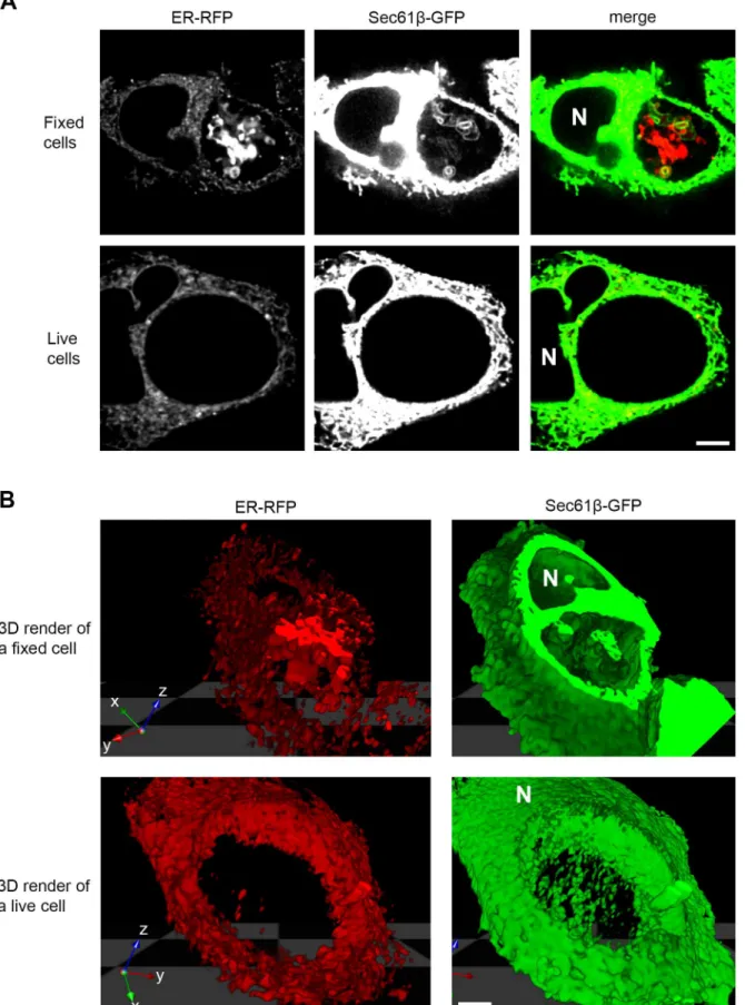
The protein markers that were most notably visible within the lumen of inclusions, such as ER-RFP and Sec61β-GFP, typically formed an expansive network of large blebs and tubules within much of the three-dimensional space of the inclusion lumen in fixed cells (Fig 2A and 2B, top panel,S1,S3, andS4Movies). However, within the limits of our experimental design and number of cells analyzed, we were unable to detect these structures within living infected cells (Fig 2A and 2B, bottom panel,S2,S5andS6Movies).
Chemical Fixation Induces ER Internalization into the Inclusion Lumen
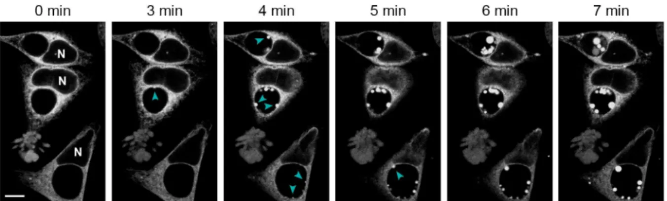
Because of the differences in ER localization patterns between living and fixed cells, we consid-ered the possibility that the process of fixation exaggerates the degree of translocation of this organelle into the inclusion lumen. To test this, we used laser scanning confocal microscopy on living infected cells to monitor changes to ER-RFP localization during paraformaldehyde fixa-tion (Fig 3andS7 Movie). Within minutes of the addition of fixative, we observed the forma-tion of blebs of ER-RFP material expanding into the inclusion lumen. These blebs appeared at random sites along the inclusion periphery and enlarged over time as new sites of inward bleb-bing (Fig 3, arrowheads) emerged. Many blebs remained attached to the inclusion edge but some appeared to detach into the center of the inclusion. By ten minutes, most new ER bleb-bing and expansion had stopped and existing structures varied in fluorescence intensity. We also noted the formation of a few smaller and less distinct bleb-like aggregates of ER-RFP in other areas of the cell, mostly at cell edges. These findings demonstrate that chemical fixation can induce a dramatic translocation of ER material into the inclusion lumen.Chemical Fixation Influences the Degree to Which Organelles Are
Observed within the Inclusion
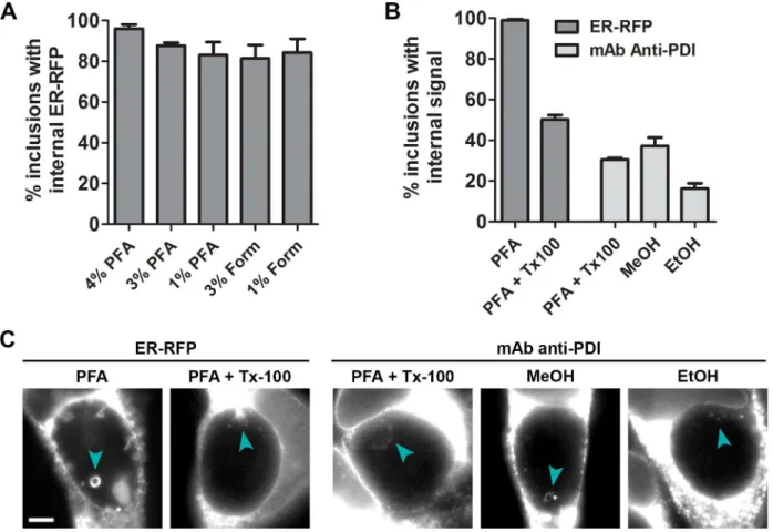
To assess whether the organelle internalization process can occur during various fixation tech-niques used previously in the study of host cellular material withinChlamydiainclusions [12,14,16,28,29], we quantified the frequency of ER-RFP structures in inclusions after fixation of infected cells (Fig 4A). Fixation with different concentrations of formaldehyde prepared
Fig 1. Mitochondria, ER, and inclusion membranes are found within the lumen ofC.trachomatisinclusions.HeLa cells were infected withC. trachomatisLGV L2, transfected with the indicated plasmids, and fixed at 30 hpi for serial spinning disk laser confocal analysis. Note the presence of markers of the ER, mitochondrial matrix and outer membranes, and inclusion membranes within the inclusion lumen (A, cyan arrowheads). Images portray a single z-section from the center of an inclusion, and inclusions are visually identified as large black centered ovals or outlined with a dashed line. Cellular-localized markers appear saturated because material within the inclusion was often significantly dimmer. (B) The frequency of internalized structures within the entire 3D space of each inclusion was assessed. Plasmids are categorized as markers of the ER, mitochondria, inclusion, cytosol, recycling endosomes, or other as indicated. Within the other category, GFP-GalT localizes to the Golgi, CD63-GFP to MVBs, LAMP1-GFP to lysosomes, and KRphi-mRFP to the plasma membrane. A dashed line at 50% distinguishes between high and low frequencies of intraluminal structures within inclusions. 12–20 inclusions were assessed in each experiment, and the mean±SEM for three independent experiments is shown. Scale bar represents 5μm.
Fig 2. ER markers reveal expansive structures within the inclusion lumen of fixed but not living cells.HeLa cells were infected withC.trachomatis LGV L2, co-transfected with ER-RFP (red) and Sec61β-GFP (green) and at 30 hpi were either fixed or imaged in three dimensions while living. Images were acquired with a laser scanning confocal microscope. (A) A single xy micrograph towards the center of a cell. SeeS1andS2Movies for a progression of xy
from either paraformaldehyde or liquid formalin (which contains small amounts of methanol) resulted in similarly high frequencies of inclusions with internal ER-RFP structures, indicating that formaldehyde concentrations and trace levels of methanol in formalin do not affect the frequency of internalization. Since gluteraldehyde fixation as used for electron microscopy causes high levels of autofluorescence across the visible light spectrum [38], we could not reli-ably distinguish any fluorescent marker or dye tested from background fluorescence under these conditions (data not shown). Furthermore, we could not assess the frequency of ER-RFP structures within inclusions after methanol fixation since mRFP fluorescence was quenched by this treatment.
To compare the effect of denaturing fixatives such as alcohols to cross-linking fixatives such as aldehydes, we monitored ER internalization into the inclusion via immunofluorescence microscopy under different conditions. Because alcohol fixation simultaneously fixes and per-meabilizes membranes, we first assessed how well ER-RFP intra-inclusion structures resist detergent permeabilization. Using a range of conditions previously employed in the study of host cellular material within inclusions [12,14,16], we found that ER-RFP structures persisted at a reduced number per inclusion (Fig 4C) and typically in only half of all inclusions (Fig 4B
andS1 Fig). We next performed immunofluorescence on paraformaldehyde-fixed and deter-gent-permeabilized cells and compared with methanol or ethanol fixed/permeabilized cells using an antibody to the ER-resident protein PDI (Fig 4B and 4C). Notably, we observed PDI-positive structures under all conditions within one-quarter to one-half of inclusions. These findings suggest that both formaldehyde and alcohol-based chemical fixation result in the translocation of endogenous ER materials into the inclusion lumen.
ER Material Is Internalized into the Vacuole of Another Intracellular
Pathogen
TheChlamydiainclusion is an unusually spacious organelle and the luminal space is largely devoid of electron-dense material compared to the host cytoplasm as assessed by transmission
micrographs along the z-axis. (B) Images were used to render volumes in 3D. 3D render of a fixed cell is limited in the z-axis to allow viewing within the inclusion. SeeS3–S6Movies for QuickTime Virtual Reality files to rotate and view these 3D volumes. Note the presence of an expansive network of material within the inclusion lumen of fixed cells. N marks the nucleus. Scale bars represent 5μm.
doi:10.1371/journal.pone.0139153.g002
Fig 3. ER-RFP translocates into the inclusion lumen during chemical fixation.HeLa cells were infected withC.trachomatisLGV L2 and transfected with ER-RFP. At 30 hpi, living cells were placed in 4% paraformaldehyde and imaged over time with a laser scanning confocal microscope. Note the genesis of ER-RFP blebs into the inclusions lumen (cyan arrowheads) during fixation. SeeS7 Moviefor a time-lapse video including more cells in a larger field of view. N marks the nuclei. Scale bar represents 10μm.
electron microscopy [39]. We speculated that this spacious nature may be conducive to fixa-tion-induced translocation of materials that might otherwise not occur elsewhere in the cell. To assess this, we asked whether another similarly spacious pathogenic vacuole occupied by
Coxiella burnettii(reviewed in [40]) would display similar internalized structures. When we transfectedCoxiella burnettii-infected cells with ER-RFP and fixed after 52 hr of infection, we found ER-RFP structures within the lumen of the pathogenic vacuole with similar appearance and frequency (90%) as those found withinChlamydiainclusions (Fig 5).
Discussion
In this study, we report that fluorescently tagged markers of the ER, mitochondria, and inclu-sion membranes are readily observed within the lumen of fixedChlamydia trachomatis inclu-sions. However this may not be an accurate reflection ofin vivoprocesses as we determined that chemical fixation can induce the translocation of ER components into the inclusion. These internalized structures resist many common immunostaining procedures and were detected by immunofluorescence microscopy, indicating that select cellular materials detected within the inclusions of fixed cells may overestimate the true localization in living cells.
Fig 4. ER structures are detected via immunofluorescence microscopy within inclusions after both formaldehyde and alcohol-based chemical fixation.HeLa cells were infected withC.trachomatisLGV L2. (A) Infected cells were transfected with ER-RFP, fixed at 30 hpi with different concentrations of either paraformaldehyde (PFA) or formaldehyde (Form), and assessed for the frequency of ER-RFP within inclusions. (B and C) Infected cells were transfected with ER-RFP as indicated. At 30 hpi, cells were fixed with PFA, methanol, or ethanol, and subsequently permeabilized with Triton X-100 (Tx100) as indicated. Treated cells were processed for immunofluorescence with an antibody to the ER protein PDI and assessed for the frequency of PDI-positive structures within inclusions. Scale bar represents 5μm.
doi:10.1371/journal.pone.0139153.g004
Our findings indicate that fixation-induced internalization into the inclusion lumen is selec-tive amongst subcellular components that reside in close proximity to the inclusion. We rou-tinely found markers of the inclusion membrane, ER lumen, ER membrane, mitochondrial matrix and mitochondrial outer membrane within the inclusion lumen of fixed cells–organelles
which all closely associate with the inclusion. However, markers of other inclusion-proximal subcellular elements, including the Golgi, recycling endosomes, lysosomes, and the cytosol, did not. Similarly, we did not observe a marker of the more distal plasma membrane within inclu-sions. As lipids are directly transferred between the ER and inclusion at membrane-contact sites between the two [7,8], we speculate that fixation-induced internalization may occur spe-cifically at inclusion-organelle interaction areas of direct material transfer. Indeed, we infre-quently observed what appeared to be the translocation of ER markers across the plasma membrane outward from cells during fixation, and membrane-contact sites between the ER and the plasma membrane have been reported (reviewed in [41]). Some organelles have been suggested to directly transfer into the inclusion, including lipid droplets [14] and peroxisomes [27], with the translocation of lipid droplets (14) and ER (25) confirmed in living cells. In gen-eral, it remains unclear how fixation-induced translocations reflectbona fideinteractions with the inclusion, although it is possible that fixation enhances the frequency ofbona fide translo-cation or interaction events.
Methanol and ethanol disrupt hydrophobic and hydrogen bonding which denatures pro-teins, and are sometimes used to fix samples for immunofluorescence microscopy due to ease of use, but can shrink and distort tissues and cells [42]. Formaldehyde reacts with and cross-links various reactive groups of biological molecules, including proteins, DNA, and sugars. It is routinely used to preserve cellular architecture and the spatial relationships of proteins in cell and tissues (reviewed in [42,43]). However, there are instances where chemical or aldehyde
Fig 5. ER-RFP within the pathogenic vacuole ofCoxiella burnettii.HeLa cells were infected withCoxiella burnettii, transfected with ER-RFP, and fixed at 52 hpi. Note the presence of ER-RFP structures with in the lumen of the pathogenic vacuole (cyan arrowheads) similar to those withinChlamydia trachomatisinclusions. N indicates the nucleus and a dashed line outlines a region of higher magnification. Scale bar represents 5μm.
fixation alters rather than preserves cellular structures. A prominent example is the mesosome, which was thought to be a separate intracellular organelle in bacteria until it was shown to be an artifact of chemical fixation [44] of exaggerated invaginations from the cell membrane [45]. There is also precedence for fixation-induced membrane blebbing in mammalian cells. Expos-ing cell monolayers to low concentrations of paraformaldehyde causes the release of vesicles from the plasma membrane [46]. These vesicles range in size from 0.5μm to 15μm and appear
at cell edges similarly to the infrequent blebbing of ER-RFP that we observed during fixation. The kinetics of large membrane blebs forming outwardly from cells fixed under standard para-formaldehyde concentrations [42] are similar to that of structures we observed forming within inclusion lumens and may share the same mechanism of genesis.
By transmission electron microscopy, the inclusion lumen appears spacious with large dis-tances between bacteria particularly in the center, and except for glycogen, is largely devoid of electron-dense material [22,39], particularly in comparison to the host cell cytosol. This rela-tively empty lumenal space could be particularly conducive to the formation of formaldehyde-induced blebs, just as is the extracellular milieu. Consistent with this model, we also observed ER-RFP structures within fixed pathogenic vacuoles ofCoxiella burnettiiat 52 hpi–a time
when this vacuole appears similarly spacious by electron microscopy (reviewed in [40]). Our findings indicate that the process of chemical fixation can lead to the accumulation of subcellular components in the lumen of the pathogenic vacuole from two intracellular patho-gens–Chlamydia trachomatisandCoxiella burnettii. The discrepancy between the relative effi-ciency with which ER membranes are found in the lumen of inclusions between fixed and live cells suggest that caution should be exercised when interpreting events observed in fixed cells. As imaging technologies surpass the diffraction-limit of light and capture images at super-reso-lution, sub-micron level differences in protein localization and aggregation are becoming apparent between chemically fixed and living cells [47]. Alternative fixation techniques that better preserve structures, such cryofixation should also reduce subcellular distortions [48] as has been used to confirm the presence of ER fragments within inclusions [25]. Fixation is unlikely to perfectly preserve the internal architecture of cells and thus observing subcellular components in living, intact cells should remain the gold standard when assessing the signifi-cance of any observed interactions. Ideally, by assessing the interaction between the inclusion and host organelles by a combination of multiple techniques, a more accurate picture of what occurs in unperturbedC.trachomatis-infected cells will emerge.
Materials and Methods
Cell Culture,
Chlamydia
Infections, Transfection, Antibodies and
Plasmids
HeLa cells (ATCC CCL-2) were grown in high glucose DMEM supplemented with L-gluta-mine, sodium pyruvate (Gibco, Life Technologies) and 10% FBS (Mediatech, CellGro), at 37°C in a 5% CO2humidified incubator.C.trachomatisLGV biovar L2 434/Bu [13] was propagated
in Vero cells (ATCC CCL-81) and purified as previously described [49]. EB titers were deter-mined by infecting Vero cell monolayers seeded in a 96 well plate. At 24 hpi cells were fixed and stained with anti-MOMP antibodies. Inclusion forming units (IFUs) were counted using a Cellomics ArrayScan automated fluorescence imaging system (Thermo Scientific). Cells were infected at an MOI of 1, synchronized by centrifugation (2,500 x g for 30 min at 10°C) onto HeLa cell monolayers, and incubated for 30 hr. As indicated, cells were transfected at the time of infection with jetPRIME (Polyplus transfection) according to manufacturer directions with a fresh media exchange after 4 hr. Antibody and plasmid sources: rabbit anti-Chlamydia
MOMP (Kenneth Fields, University of Kentucky), mouse anti-PDI (Abcam ab2792) ER-RFP
and DsRed-Mito (Richard Youle, NIH), Sec61β-GFP (Addgene 15108, Tom Rapoport, Har-vard Medical School), GFP-GalT (Addgene 11929), Matrix-YFP, YFP-Mitocb5TM and YFP--Prohibitin (Jennifer Lippincott-Schwartz, Eunice Kennedy Shriver NICHD), GFP-Rab1 (Craig Roy, Yale), GFP-RhoA, CD63-GFP, and LAMP1-GFP (Soman Abraham, Duke University), Chy-Arf1(Q71L) and mRFP (Micheal Ehlers, Pfizer Neuroscience, formerly Duke University), FAPP1-PH-GFP (Tamas Balla, Eunice Kennedy Shriver NICHD), tdTomato (Marc Caron, Duke University), KRphi-mRFP (Addgene 17276, Sergio Grinstein, University of Toronto), GFP (pcDNA3.1-CT, Invitrogen, Life Technologies), Syntaxin13-GFP (William Trimble, Uni-versity of Toronto).
Imaging of Subcellular Organelles and Quantitation of Intraluminal
Structures in Fixed Inclusions
HeLa cells grown on glass coverslips to 50% confluence were infected withC.trachomatisLGV L2 and transfected with the indicated (Fig 1) plasmids. At 30 hpi, cells were fixed with 4% para-formaldehyde (PFA) in PBS at pH 7.4 for 20 minutes at RT, incubated with 1μg/mL Hoescht
33258 (Life Technologies) in PBS for 20 minutes at RT, mounted to slides in 5μl SlowFade
Gold (Life Technologies), and sealed with nail polish. Images were acquired using a Marianas system (Intelligent Imaging Innovations) equipped with an inverted microscope (Zeiss, Axio-Observer using a 100x 1.4 NA oil objective) and a Yokogawa spinning disk confocal unit (model CSU-22). All the hardware was controlled by SlideBook version 4.2 (Intelligent Imag-ing Innovations). Z-sections were acquired from above to below each cell, with optimal spacImag-ing between z-sections to meet the Nyquist resolution criterion. To assess the frequency of fluores-cent blebs within inclusions, both the DNA staining signal (to define the inclusion and nucleus) and fluorescent protein signal in each individual z-section (approximately 30 per cell) from a z-stack of images of each inclusion were viewed with Slidebook version 4.2 or 5.5 (Intelligent Imaging Innovations). At least 12 inclusions for each marker in each independent experiment were assessed. Values from three independent experiments were averaged and standard errors were calculated. Calculations and graphs were prepared with Prism (GraphPad Software) and images were processed for display with Photoshop CS6 (Adobe).
Comparison of Intraluminal Structures in Living and Fixed Cells
For fixed cells, HeLa cells were prepared as above. For living cells, HeLa cells grown on 35 mm #1.5 glass-bottom dishes (MatTek) to 50% confluence were infected withC.trachomatisLGV L2 and cotransfected with ER-RFP and Sec61β-GFP. At 30 hpi, cells were imaged in a phenol red-free DMEM HG (Gibco, Life Technologies) media supplemented with 10% FBS and 10μM
HEPES (Gibco, Life Technologies) in a humidified chamber maintaining 37°C and 5% CO2.
Images were captured on a Leica SP5 laser scanning confocal inverted microscope equipped with a 100x 1.4 NA oil objective. Z-sections were acquired at optimal spacing to meet the Nyquist resolution criterion. Images were deconvolved using Huygens Essential (SVI) and pro-cessed with ImageJ (NIH) to create video.AVI files and Photoshop CS6 (Adobe) for presenta-tion. To create 3D-rendered volumes, deconvolved images were further processed with Volocity (PerkinElmer), with the z-axis expanded three-fold to reduce flatness. 3D volumes of each channel acquired of living and fixed cells were exported into the QuickTime (Apple Inc.) Virtual Reality format for viewing (S3–S6Movies).
hpi, cells were bathed in a phenol red-free DMEM HG (Gibco, Life Technologies) media sup-plemented with 10% FBS and 10μM HEPES in a humidified chamber maintaining 37°C and
5% CO2, and 8% paraformaldehyde in PBS was added to a final concentration of 4%. Images
were captured on a Leica SP5 laser scanning confocal inverted microscope equipped with a 63x 1.2 NA water objective. Images were acquired every 3.4 sec and the z-position was adjusted manually as needed to offset focal drift due to thermal changes. Images were compiled for dis-play with ImageJ (NIH) and Photoshop CS6 (Adobe).
Quantitation of ER-RFP Intraluminal Structures in Inclusions after
Various Fixation and Permeabilization Conditions
HeLa cells grown on glass coverslips to 50% confluence were infected withC.trachomatisLGV L2 and transfected with ER-RFP as indicated. At 30 hpi, cells were processed in one of three major ways.
Fixatives. Cells were fixed with either 4%, 3%, 1% PFA, 3%, or 1% formaldehyde in PBS pH 7.4 (the latter prepared from a formalin stock containing trace methanol) for 20 min at RT, or pre-chilled 100% methanol or ethanol for 20 min, mounted to slides in 5μl SlowFade Gold
(Life Technologies), and sealed with nail polish.
Permeabilization methods. As indicated, cells were first fixed with 4% PFA for 20 min at RT, then incubated with just PBS (untreated) or pre-chilled 0.2% (Fig 4B and 4C), 0.1% Tx-100, 0.2% Saponin on ice for 10 min, ice-cold 1:1 mix of methanol and ethanol for 5 min on ice, or 2 mg/mL, 1 mg/mL, or 0.5 mg/mL of Zwittergent 3–12 (all detergents in PBS) for 1 min on ice,
mounted to slides in 5μl SlowFade Gold (Life Technologies), and sealed with nail polish.
Samples were double-blinded and viewed on an Axioskop 2 (Zeiss) inverted widefield fluores-cence microscope or an Axio Observer Z1 (Zeiss) (Fig 4B and 4C) with a 63X 1.4 NA oil objective (Zeiss) and the frequency of inclusions containing ER-RFP intraluminal structures was quanti-fied in 50–100 cells for each experiment. Values from three independent experiments were
aver-aged and standard errors were calculated. Statistically significant differences were assessed by a one-way ANOVA followed by Dunnett's Multiple Comparisonpost hocanalysis comparing each condition to untreated with a p-value<0.05 considered significant. Statistics and graphs were
prepared with Prism (GraphPad Software) and Photoshop CS6 (Adobe).
Imaging and Quantitation of ER-RFP Structures within Fixed
Coxiella
burnettii
Pathogenic Vacuoles
HeLa cells grown on glass coverslips to 50% confluence were infected withCoxiella burnetii
Nine Mile RSA439 (phase II, clone 4) and synchronized by centrifugation (3,000 rpm for 30 min at 10°C). At 24 hpi, cells were transfected with ER-RFP. At 52 hpi, cells were fixed with 4% paraformaldehyde (PFA) in PBS for 20 minutes at RT, incubated with 1μg/mL Hoescht 33258
(Life Technologies) in PBS for 20 minutes at RT, mounted to slides in 5μl SlowFade Gold (Life
Technologies), and sealed with nail polish. Images were captured on a Leica SP5 laser scanning confocal inverted microscope equipped with a 100x 1.4 NA oil objective and over 50 infected cells were assessed for the frequency of ER-RFP structures within the pathogenic vacuole. Images were minimally processed with Photoshop CS6 (Adobe) for presentation.
Supporting Information
S1 Fig. Fixation-induced intralumenal ER-RFP structures within inclusions persist through many post-fixation manipulations.HeLa cells were infected withC.trachomatis
LGV L2 and transfected with ER-RFP for 30 hr, fixed with 4% paraformaldehyde, treated with
the indicated permeabilization solutions, and assessed for the frequency of ER-RFP within inclusions. Treatments included the nonionic detergent Triton X-100 (Tx-100), Saponin, an amphipathic glucoside, a 1:1 mix of methanol and ethanol, or Zwittergent 3–12, a dipolar ionic
detergent for various times. 50–100 inclusions were enumerated in each experiment, and the
mean ± SEM for three independent experiments is shown.indicates P<0.05 by one-way
ANOVA and Dunnett's Multiple Comparisonpost hocanalysis comparing each condition (gray bars) to the control (black bars).
(TIF)
S1 Movie. Z-sections through a fixedC.trachomatisinfected cell expressing ER-RFP and
Sec61β-GFP at 30 hpi.
(AVI)
S2 Movie. Z-sections through a livingC.trachomatisinfected cell expressing ER-RFP and
Sec61β-GFP at 30 hpi.
(AVI)
S3 Movie. QuickTime Virtual Reality file of 3D volume of ER-RFP within a fixed cell infected withC.trachomatisfor 30 hr.
(MOV)
S4 Movie. QuickTime Virtual Reality file of 3D volume of Sec61β-GFP within a fixed cell infected withC.trachomatisor 30 hr.
(MOV)
S5 Movie. QuickTime Virtual Reality file of 3D volume of ER-RFP within a living cell infected withC.trachomatisfor 30 hr.
(MOV)
S6 Movie. QuickTime Virtual Reality file of 3D volume of Sec61β-GFP within a living cell infected withC.trachomatisfor 30 hr.
(MOV)
S7 Movie. Time-lapse video microscopy of livingC.trachomatisinfected cells expressing
ER-RFP undergoing chemical fixation.White arrowheads in first frame indicateC. tracho-matisinclusions. Time is displayed in each frame as min:sec.
(AVI)
Acknowledgments
We thank the Duke Light Microscopy Core Facility and the Vann Bennett and Robert Lefko-witz labs for access to their microscope. We thank members of the Valdivia lab for valuable discussions.
Author Contributions
Conceived and designed the experiments: MK RHV. Performed the experiments: MK. Ana-lyzed the data: MK RHV. Contributed reagents/materials/analysis tools: MK RHV. Wrote the paper: MK RHV.
References
2. Kumar Y, Valdivia RH. Leading a sheltered life: intracellular pathogens and maintenance of vacuolar compartments. Cell Host Microbe. 2009; 5: 593–601. doi:10.1016/j.chom.2009.05.014PMID: 19527886
3. Saka HA, Valdivia RH. Acquisition of nutrients byChlamydiae: unique challenges of living in an intracel-lular compartment. Curr Opin Microbiol. 2010; 13: 4–10. doi:10.1016/j.mib.2009.11.002PMID: 20006538
4. Kokes M, Valdivia RH. Cell Biology of the Chlamydial Inclusion. In: Tan M, Bavoil PM, editors. Intracel-lular Pathogens I:Chlamydiales. Washington, DC: ASM Press; 2012. pp. 170–191.
5. Giles DK, Wyrick PB. Trafficking of chlamydial antigens to the endoplasmic reticulum of infected epithe-lial cells. Microbes Infect Inst Pasteur. 2008; 10: 1494–1503. doi:10.1016/j.micinf.2008.09.001
6. Peterson EM, de la Maza LM.Chlamydiaparasitism: ultrastructural characterization of the interaction between the chlamydial cell envelope and the host cell. J Bacteriol. 1988; 170: 1389–1392. PMID: 3343223
7. Derré I, Swiss R, Agaisse H. The Lipid Transfer Protein CERT Interacts with theChlamydiaInclusion Protein IncD and Participates to ER-ChlamydiaInclusion Membrane Contact Sites. PLoS Pathog. 2011; 7: e1002092. doi:10.1371/journal.ppat.1002092PMID:21731489
8. Elwell CA, Jiang S, Kim JH, Lee A, Wittmann T, Hanada K, et al.Chlamydia trachomatisco-opts GBF1 and CERT to acquire host sphingomyelin for distinct roles during intracellular development. PLoS Pathog. 2011; 7: e1002198. doi:10.1371/journal.ppat.1002198PMID:21909260
9. Heuer D, Rejman Lipinski A, Machuy N, Karlas A, Wehrens A, Siedler F, et al.Chlamydiacauses frag-mentation of the Golgi compartment to ensure reproduction. Nature. 2009; 457: 731–735. doi:10.1038/ nature07578PMID:19060882
10. Kokes M, Dunn JD, Granek JA, Nguyen BD, Barker JR, Valdivia RH, et al. Integrating Chemical Muta-genesis and Whole-Genome Sequencing as a Platform for Forward and Reverse Genetic Analysis of Chlamydia. Cell Host Microbe. 2015; 17: 716–725. doi:10.1016/j.chom.2015.03.014PMID:25920978
11. Hackstadt T, Scidmore MA, Rockey DD. Lipid metabolism inChlamydia trachomatis-infected cells: directed trafficking of Golgi-derived sphingolipids to the chlamydial inclusion. Proc Natl Acad Sci U S A. 1995; 92: 4877–4881. PMID:7761416
12. Hackstadt T, Rockey DD, Heinzen RA, Scidmore MA.Chlamydia trachomatisinterrupts an exocytic pathway to acquire endogenously synthesized sphingomyelin in transit from the Golgi apparatus to the plasma membrane. EMBO J. 1996; 15: 964–977. PMID:8605892
13. Kumar Y, Cocchiaro J, Valdivia RH. The obligate intracellular pathogenChlamydia trachomatistargets host lipid droplets. Curr Biol CB. 2006; 16: 1646–1651. doi:10.1016/j.cub.2006.06.060PMID: 16920627
14. Cocchiaro JL, Kumar Y, Fischer ER, Hackstadt T, Valdivia RH. Cytoplasmic lipid droplets are translo-cated into the lumen of theChlamydia trachomatisparasitophorous vacuole. Proc Natl Acad Sci U S A. 2008; 105: 9379–9384. doi:10.1073/pnas.0712241105PMID:18591669
15. Ouellette SP, Dorsey FC, Moshiach S, Cleveland JL, Carabeo RA.Chlamydiaspecies-dependent dif-ferences in the growth requirement for lysosomes. PloS One. 2011; 6: e16783. doi:10.1371/journal. pone.0016783PMID:21408144
16. Beatty WL. Trafficking from CD63-positive late endocytic multivesicular bodies is essential for intracel-lular development ofChlamydia trachomatis. J Cell Sci. 2006; 119: 350–359. doi:10.1242/jcs.02733 PMID:16410552
17. Beatty WL. Late endocytic multivesicular bodies intersect the chlamydial inclusion in the absence of CD63. Infect Immun. 2008; 76: 2872–2881. doi:10.1128/IAI.00129-08PMID:18426873
18. Van Ooij C, Apodaca G, Engel J. Characterization of theChlamydia trachomatisvacuole and its interac-tion with the host endocytic pathway in HeLa cells. Infect Immun. 1997; 65: 758–766. PMID:9009339
19. Scidmore MA, Fischer ER, Hackstadt T. Sphingolipids and glycoproteins are differentially trafficked to theChlamydia trachomatisinclusion. J Cell Biol. 1996; 134: 363–374. PMID:8707822
20. Taraska T, Ward DM, Ajioka RS, Wyrick PB, Davis-Kaplan SR, Davis CH, et al. The late chlamydial inclusion membrane is not derived from the endocytic pathway and is relatively deficient in host pro-teins. Infect Immun. 1996; 64: 3713–3727. PMID:8751921
21. Scidmore MA, Fischer ER, Hackstadt T. Restricted Fusion ofChlamydia trachomatisVesicles with Endocytic Compartments during the Initial Stages of Infection. Infect Immun. 2003; 71: 973–984. doi: 10.1128/IAI.71.2.973–984.2003PMID:12540580
22. Matsumoto A, Bessho H, Uehira K, Suda T. Morphological studies of the association of mitochondria with chlamydial inclusions and the fusion of chlamydial inclusions. J Electron Microsc (Tokyo). 1991; 40: 356–363.
23. Derré I, Pypaert M, Dautry-Varsat A, Agaisse H. RNAi Screen inDrosophilaCells Reveals the Involve-ment of the Tom Complex inChlamydiaInfection. PLoS Pathog. 2007; 3: e155. doi:10.1371% 2Fjournal.ppat.0030155
24. Carabeo RA, Mead DJ, Hackstadt T. Golgi-dependent transport of cholesterol to theChlamydia tracho-matisinclusion. Proc Natl Acad Sci U S A. 2003; 100: 6771–6776. doi:10.1073/pnas.1131289100 PMID:12743366
25. Dumoux M, Clare DK, Saibil HR, Hayward RD.Chlamydiaeassemble a pathogen synapse to hijack the host endoplasmic reticulum. Traffic Cph Den. 2012; doi:10.1111/tra.12002
26. Majeed M, Krause KH, Clark RA, Kihlström E, Stendahl O. Localization of intracellular Ca2+ stores in HeLa cells during infection withChlamydia trachomatis. J Cell Sci. 1999; 112 (Pt 1): 35–44.
27. Boncompain G, Müller C, Meas-Yedid V, Schmitt-Kopplin P, Lazarow PB, Subtil A. The intracellular bacteriaChlamydiahijack peroxisomes and utilize their enzymatic capacity to produce bacteria-specific phospholipids. PloS One. 2014; 9: e86196. doi:10.1371/journal.pone.0086196PMID:24465954
28. Capmany A, Damiani MT.Chlamydia trachomatisintercepts Golgi-derived sphingolipids through a Rab14-mediated transport required for bacterial development and replication. PloS One. 2010; 5: e14084. doi:10.1371/journal.pone.0014084PMID:21124879
29. Cox JV, Naher N, Abdelrahman YM, Belland RJ. Host HDL biogenesis machinery is recruited to the inclusion ofChlamydia trachomatis-infected cells and regulates chlamydial growth. Cell Microbiol. 2012; 14: 1497–1512. doi:10.1111/j.1462-5822.2012.01823.xPMID:22672264
30. Soupene E, Rothschild J, Kuypers FA, Dean D. Eukaryotic protein recruitment into theChlamydia inclu-sion: implications for survival and growth. PloS One. 2012; 7: e36843. doi:10.1371/journal.pone. 0036843PMID:22590624
31. Ouellette S, Carabeo RA. A Functional Slow Recycling Pathway of Transferrin is Required for Growth ofChlamydia. Front Cell Infect Microbiol. 2010; 1. Available:http://www.frontiersin.org/cellular_and_ infection_microbiology/10.3389/fmicb.2010.00112/full.
32. Hailey DW, Rambold AS, Satpute-Krishnan P, Mitra K, Sougrat R, Kim PK, et al. Mitochondria Supply Membranes for Autophagosome Biogenesis during Starvation. Cell. 2010; 141: 656–667. doi:10.1016/ j.cell.2010.04.009PMID:20478256
33. Balla A, Tuymetova G, Tsiomenko A, Varnai P, Balla T. A Plasma Membrane Pool of Phosphatidylinosi-tol 4-Phosphate Is Generated by PhosphatidylinosiPhosphatidylinosi-tol 4-Kinase Type-III Alpha: Studies with the PH Domains of the Oxysterol Binding Protein and FAPP1. Mol Biol Cell. 2005; 16: 1282–1295. doi:10. 1091/mbc.E04-07-0578PMID:15635101
34. Moorhead AM, Jung J-Y, Smirnov A, Kaufer S, Scidmore MA. Multiple Host Proteins That Function in Phosphatidylinositol-4-Phosphate Metabolism Are Recruited to the Chlamydial Inclusion. Infect Immun. 2010; 78: 1990–2007. doi:10.1128/IAI.01340-09PMID:20231409
35. Kumar Y, Valdivia RH. Actin and Intermediate Filaments Stabilize theChlamydia trachomatisVacuole by Forming Dynamic Structural Scaffolds. Cell Host Microbe. 2008; 4: 159–169. doi:10.1016/j.chom. 2008.05.018PMID:18692775
36. Rzomp KA, Scholtes LD, Briggs BJ, Whittaker GR, Scidmore MA. Rab GTPases are recruited to chla-mydial inclusions in both a species-dependent and species-independent manner. Infect Immun. 2003; 71: 5855–5870. PMID:14500507
37. Yeung T, Terebiznik M, Yu L, Silvius J, Abidi WM, Philips M, et al. Receptor Activation Alters Inner Sur-face Potential During Phagocytosis. Science. 2006; 313: 347–351. doi:10.1126/science.1129551 PMID:16857939
38. Lee K, Choi S, Yang C, Wu H-C, Yu J. Autofluorescence generation and elimination: a lesson from glu-taraldehyde. Chem Commun Camb Engl. 2013; 49: 3028–3030. doi:10.1039/c3cc40799c
39. Stokes GV.Chlamydia psittaci: inclusion vacuole ultrastructure. Can J Microbiol. 1980; 26: 396–401. PMID:6250694
40. Voth DE, Heinzen RA. Lounging in a lysosome: the intracellular lifestyle ofCoxiella burnetii. Cell Micro-biol. 2007; 9: 829–840. doi:10.1111/j.1462-5822.2007.00901.xPMID:17381428
41. Rowland AA, Voeltz GK. Endoplasmic reticulum-mitochondria contacts: function of the junction. Nat Rev Mol Cell Biol. 2012; 13: 607–625. doi:10.1038/nrm3440PMID:22992592
42. Fox CH, Johnson FB, Whiting J, Roller PP. Formaldehyde fixation. J Histochem Cytochem Off J Histo-chem Soc. 1985; 33: 845–853.
43. Thavarajah R, Mudimbaimannar VK, Elizabeth J, Rao UK, Ranganathan K. Chemical and physical basics of routine formaldehyde fixation. J Oral Maxillofac Pathol JOMFP. 2012; 16: 400–405. doi:10. 4103/0973-029X.102496PMID:23248474
45. Higgins ML, Tsien HC, Daneo-Moore L. Organization of mesosomes in fixed and unfixed cells. J Bac-teriol. 1976; 127: 1519–1523. PMID:821934
46. Scott RE. Plasma membrane vesiculation: a new technique for isolation of plasma membranes. Sci-ence. 1976; 194: 743–745. PMID:982044
47. Ji N, Shroff H, Zhong H, Betzig E. Advances in the speed and resolution of light microscopy. Curr Opin Neurobiol. 2008; 18: 605–616. doi:10.1016/j.conb.2009.03.009PMID:19375302
48. Pearse AGE. Histochemistry: Theoretical and Applied. Churchill Livingstone; 1980.
49. Caldwell HD, Kromhout J, Schachter J. Purification and partial characterization of the major outer mem-brane protein ofChlamydia trachomatis. Infect Immun. 1981; 31: 1161–1176. PMID:7228399