Fabrication of Multiple Layered Scafolds with Controlled Porous Micro-Architecture
Chunrong Yanga,b*, Huazhong Wuc, Jianhua Wangd
Received: September 21, 2016; Revised: November 22, 2016; Accepted: December 19, 2016
A multi-layer scafold that incorporates bioglass, phosphatidylserine and steroidal saponins loaded collagen microparticles was prepared in this study. Combinatorial processing techniques involving porogen leaching protocol and freeze-drying were used. Drug distribution and microstructure could be controlled mainly by the concentrations of drug-loaded collagen microparticles and porogen as well as freezing temperature for diferent layers of the scafolds. Morphological observation, measurement of swelling properties, examination of drug release kinetics, and analysis of cell bioactivity showed that the resultant scafolds had highly interconnected pores, gradient drug distribution and graded porous micro-architecture, similar swelling ratio for diferent layers, better drug release kinetics in view of lower initial release and slower average release rate compared to the scafolds with homogeneous structure, as well as approving support of cell ingrowth. This study suggests that the optimized scafolds have promising potential for applications in bone tissue engineering.
Keywords: multi-layer scafold, microstructure, drug distribution
* e-mail: [email protected]
1. Introduction
To date, most approaches related to tissue engineering involve delivery of drugs via porous scafolds for regeneration of tissues, and thus, the architecture of scafolds has a decisive efect on the load and release of drugs. Complex and controlled architectures are often a necessary alternative to loading drugs in a porous scafold, as control over structure is critical for gaining adequate release function. Recent novel techniques have been reported for achieving these complex architectures, including electrospinning, phase separation, and precipitation as well as gas foaming/particulate leaching1-4. Besides that, several groups have also investigated the layering of 2-D sheets to build up a complex 3-D architectures with porous features5-8. Most of these techniques result in scafold with order structure.
However, it has been increasingly recognized that these delivery systems should deliver drugs in a spatiotemporal way, including that (1) burst release should be lessened and (2) stable release should sustain a relatively long time to obtain the desired efects. It is therefore critical to design the scafold to provide inhomogeneous structure for safe and stable drug release.
Based on the compositions of nature bone, naturally derived collagen (COL) has the potential advantages
of specific cell interactions and hydrophilicity. Phosphatidylserine (PS) has been implicated in variety of cellular processes such as cell migration, cell adhesion, tumor metastasis, cell morphogenesis and differentiation, and wound healing. Selected compositions of silicate and phosphate glasses have been proved to react with physiological fluid and form tenacious bonds to hard and soft tissues through cellular activity. Therefore, bioglass (BG), COL and PS, when processed into biomaterial, will result a bone scaffold with excellent mechanical properties, appropriate bio-degradation, and well biocompatibility and bioactivity, which has been demonstrated in our previous work9.
In this report, we applied combinatorial processing techniques involving porogen leaching protocol and freeze-drying to develop a novel, multi-layered scaffold using BG, PS and steroidal saponins (SS)-loaded collagen microparticles. As the major pharmacological constituents of ophiopogon japonicus, a traditional Chinese medical herb, SS are osteoinductive and can be used to treat bone and joint diseases10. The resulting scaffolds were designed to have gradient pore structure and drug distribution. Additionally, the construct was evaluated with respect to microstructure, swelling parameters, drug release kinetics, and cell bioactivity to demonstrate the potential utility for bone tissue engineering.
a School of Materials Science and Engineering, Fujian University of Technology, Fuzhou, Fujian
Province, China
b Fujian Provincial Key Laboratory of Advanced Materials Processing and Application, Fuzhou, Fujian
Province, China
c Department of Chemistry and Chemical Engineering, University of Minjiang, Fuzhou, Fujian
Province, China
d Institute of Biomedical and Pharmaceutical Technology, Fuzhou University, Fuzhou, Fujian Province,
2 Materials and methods
2.1. Materials
58s-BG were synthesized through sol-gel method and consisted of 60 wt% SiO2, 35 wt% CaO and 5 wt% P2O5. The steroidal saponins-loaded collagen microspheres were prepared using a water-in-oilin-water emulsion/solvent evaporation technique11. PS and NuSieve GTG agarose gel were supplied by Boao Medical Ltd. (Shanghai, China) and Liyuan Medical Ltd. (Xiamen, China), respectively. Lyophilized steroidal saponins were obtained from the College of Pharmacology, Shandong University of Traditional Chinese Medicine (Jinan, China). Dulbecco’s minimum essential medium (DMEM) and fetal bovine serum (FBS) were purchased from Sigma (St. Louis, MO, USA). 1-ethyl-3-(3-dimethyl aminopropyl) carbodiimide (EDC), N-hydroxysuccinimide (NHS), morpholine ethanesulfonic acid (MES) and other reagents were of analytical grade.
2.2 Construction of multiple layered BG-COL-PS
scafolds
BG-COL-PS scafolds were prepared as follows. In brief, parain spheres of 180-250 μm diameter and steroidal saponins-loaded collagen microspheres of around 100 μm diameter were illed into cylindrical syringe prior to heating the spheres at 37°C. After annealing to room temperature, nano-bioglass powder and phosphatidylserine solution were respectively added to the syringe and carefully aspirated to ill the interspace between the spheres. The mixture were then shock-frozen as a thin layer (around 2 mm in thickness). After the layer was nearly solidiied, laminated assembly was repeated and a new layer containing a same amount of BG nanoparticles, lower weight ratio of PS, higher weight ratio of drug-loaded collagen and parain microspheres were spread onto the top of the solidiied layer. Then, the third layer was prepared with same composition ratio to the irst layer. The triple layered scafolds were attained via such additional layer-supraposition. Then the scafolds were freeze dried at -30°C for 45h.
After being lyophilized, the scafolds were treated with 90% (v/v) methanol solution for 45 min and then treated with hexane for 20h to extract the parain spheres. Hexane was replaced after 24 h and the resulting scafolds were dried under vacuum for 10 h in order to completely dry the scafolds and minimize residual hexane. Then, cross-linking was performed in MES bufer (pH 5.5) with EDC. NHS was added in an EDC:NHS ratio of 4:1. After crosslinking with a reaction time of 4 h the samples were washed twice with 0.1 M Na2HPO4 for 1 h and 4 times with deionized water for 30 min. Finally, the scafolds were dried in vacuum.
For the purpose of comparison, another type of scafolds (Type 2) with homogeneous microstructure and durg distribution
were also prepared following the same procedure. The total amounts of compositions and parain microspheres were same to the inhomogeneous scafolds (Type 1), which consisted of 65% parts of inorganic component BG by weight and 35% part of organic components, including 80 wt.% drug-loaded collagen microspheres, and 20 wt.% PS. The weight ratio of parain microspheres to scafolds was 1:3.
2.3 Characterization
2.3.1 Morphology observationTo observe the morphology of each layer of the scafolds, disk samples (10 mm in diameter) were cut into approximately isopachous sheets with 2 mm of thickness. Scanning electron microscopy (SEM) (S-3400N, Hitachi Co., Tokyo, Japan) was used and the sheets were sputter-coated with gold for observation at 10 kV and varying levels of magniication.
2.3.2 Porosity testing
To determine porosity of each layer, disk sheets with 10 mm in diameter and 2 mm in thickness were tested using amercury intrusion porosimeter (AutoPore IV).
2.3.3 Swelling kinetics measurements
Swelling kinetics measurements were based on the determination of the volume of retained low-molecular penetrating agent (0.5 M solution of acetic acid) dependent on the time of swelling at a temperature 25°C. Disk samples were used, and each weighed 1.0 g.
2.3.4 Examination of drug release kinetics
The drug release experiment of the two types of scafolds was performed in a continuous low system which was made in our own laboratory. Phosphate bufered saline (PBS) was employed for the release medium.
After speciic time periods, the specimens were removed from the PBS luids and the supernatant was collected. The drug content of supernatant for each interval was determined by using the ultra performance liquid chromatography (UPLC, Waters Co., Milford, USA).
2.3.5 Analysis of cell bioactivity
Prior to cell culture assays, all samples were soaked in PBS overnight under UV to wet and sterilize them followed by a 20 min soak in media to allow for the deposition of serum proteins.
The proliferation kinetics of MC3T3-E1 cells on diferent layers of the scafolds was assessed using a 3-(4,5-dimethyl-2thiazolyl)-2,5-diphenyl-2H tetrazolium bromide (MTT) (Sigma Co., Saint Louis, USA) assay[9]. Briely, the cells and samples were remain undisturbed in a humidiied incubator (37 0C and 5% CO2) for 1, 3 ,7, 14 and 21 days. After incubation, the sheets were rinsed twice with PBS and 200μL MTT, 1.8 ml medium was added to each sheet for incubation at 37 0C for 4 h. Then the MTT solution was removed and insoluble blue formazan crystal was dissolved in dimethyl sulfoxide (DMSO). One hundred microlitres of solution from each sheet was aspirated and poured in a 96-well plate for absorbance measurement using a microplate spectrophotometer at a wavelength of 540 nm. Non-seeded sheets were applied as negative control. The MTT absorbance data were converted into cell numbers based on the standard curve obtained at the same time.
The behaviors of cell attachment and growth were observed using a phase-contrast microscope (Olympus IX 71, Tokyo, Japan).
2.3.6 Statistical analysis
The data were reported as the mean ± SD. Two-factor analyses of variance (ANOVAs) with Bonferroni–Dunn and Fisher’s post hoc tests for multiple comparisons as well as Student’s t test were performed using Sigma StatTM Software for Windows Version 2.03, SPSS Inc. Signiicance was accepted at a level of P<0.05.
3. Result and discussion
3.1 Fabrication of multi-layer scafolds
To achieve multiple layered scafolds with controlled porous micro-architecture while lessening possibly burst release of drugs, combinatorial processing techniques involving freeze-drying and adjustable gradients of parain spheres and compositions were developed. In the process of fabrication, pores inside scafolds were usually generated by cryogenically removing ice crystallite formed at a ixed freezing temperature, and porosity and average pore size were mainly controlled by concentration of parain spheres, freezing temperature and freezing rate.
By changing concentration of parain spheres, vertical pore gradients from the top layer to the bottom layer inside scafolds were achieved, and this would potentially create multi-scale freezing cubicles for multiple layers. Therefore, combining variable concentrations of parain spheres with freeze-processing method, micro-architecture of each layer would be efectively limited and controlled.
It is known that bone implantation should can resist against enzymatic degradation with enough strength during bone regeneration. In terms of this scafolds, the disadvantages of using high content of collagen are low biomechanical stifness and rapid biodegradation. The high enzymatic turnover rate of natural collagen in vivo makes stabilization of collagen-based biomaterials necessary. This could be achieved by chemical crosslinking methods, which could provide this scafolds with desired mechanical properties for implantation and defect repair. In this study, the scafolds have been crosslinked with EDC/NHS. Use of EDC and NHS could yield this scafolds with good biocompatibility, higher cellular diferentiation potential and with increased resistance against enzymatic degradation.
3.2 Layered scafolds with gradient pore
structures
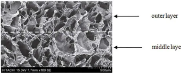
In this study, a type of triple layered and drug-loaded BG-COL-PS scafolds was constructed. Scanning electron microscopy observation of the triple layers of network composite scafolds showed a continuous structure of irregular interconnected pores (Figure 1). The pore sizes ranged from several microns up to about 400 μm. In addition, there were no any obvious clefts were observed in the interface zone between adjacent layers.
Figure 1: SEM image for interface inside the porous multi-layer scafolds. Dotted white line indicates interface zone.
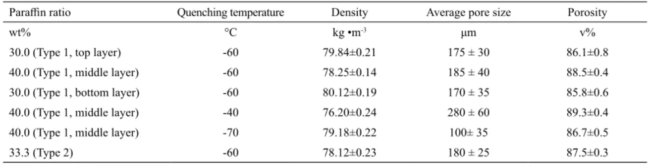
The top layer and bottom layer were similar in compositions and pore structure, but the middle layer was endowed with greater average pore size, higher porosity and drug content. The inner porous structure of the scafolds can be controlled by varying the solid content of the parain spheres and freeze-drying conditions (Table 1). As the ratio of parain spheres and the freezing temperature lowered, the density of freeze-dried scafolds increased, and the porosity and average pore size lowered.
Table 1: Characteristic of composite scafolds
Parain ratio Quenching temperature Density Average pore size Porosity
wt% °C kg •m-3 μm v%
30.0 (Type 1, top layer) -60 79.84±0.21 175 ± 30 86.1±0.8
40.0 (Type 1, middle layer) -60 78.25±0.14 185 ± 40 88.5±0.4
30.0 (Type 1, bottom layer) -60 80.12±0.19 170 ± 35 85.8±0.6
40.0 (Type 1, middle layer) -40 76.20±0.24 280 ± 60 89.3±0.4
40.0 (Type 1, middle layer) -70 79.18±0.22 100± 35 86.7±0.5
33.3 (Type 2) -60 78.12±0.23 180 ± 25 87.5±0.3
crystals push to expand biopolymer chains to a greater extent, the pore size of scafolds will be increased. On the contrary, it is possible that a rapid cooling causes the formation of many ice crystals nuclei, resulting in the formation of smaller-sized pores.
3.3. Swelling ratio
The homogeneous scafolds and diferent layers of inhomogeneous scafolds were compared morphologically with respect to their formal stability using swelling ratios and their volume changes after being soaked with 0.5 M solution of acetic acid. Swelling is the process in which collagen and phosphatidylserine receives liquid and simultaneously enlarges its volume. Swelling of the composite scafolds was generally observed in two steps. Acetic acid attacked irst of all (it was saturation of the structure), and the second step can be explained by the structure opening after break of physical bonds, particularly in a free collagen structure, but not in conjugate between collagen and phosphatidylserine due to crosslinking. We can state that the conjugate remains stable in the acidic environment, which was especially aggressive for collagen. The conjugate provided anchoring of biogalss particles in the structure of the composite, and cohesion of the material.
The behavior of scafolds during swelling can be the result of simultaneous inluenced by at least two antagonistic factors: the polarity of the composite material and the formation of hydrogen bonds between the macromolecular components. The polarity of the bioglass material inluences swelling positively. In the other case, hydrogen bonds inluence the swelling negatively, which is the result of the creation of ion complexes between the carboxylate groups of phosphatidylserine and amino groups of collagen.
It can be seen from Figure 2 that there were not signiicant diferences in swelling ratio for all sheets, which may be ascribed to two contradictory factors: on the one hand, higher porosity and greater pore size shoud mean more liquid for the matching layer, which leaded to greater swelling; on the other hand, both collagen and phosphatidylserine are hydrophilic biomolecules because of their polar groups. Therefore, even though a certain amount of crosslinker was
Figure 2: Swelling ratio of two types of scafolds with diferent microstructure and drug distribution
used, the two types scafolds still swelled to some extent. However, the contents of organic components, including collagen and phosphatidylserine were set to decrease with the increase of parain spheres which caused higher porosity and greater pore size inside the scafolds. These results indicate that presently developed inhomogeneous or homogeneous scafolds have similar swelling characteristics.
3.4 Drug release from the multiple layered
scafolds
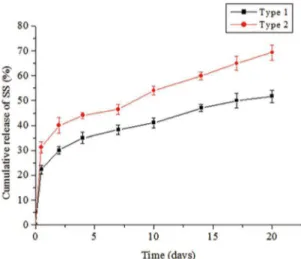
The efect of the structure of the scafolds on the release kinetics of drugs were evaluated as shown in Figure 3. The release proile in the homogeneous scalods was triphasic consisting of (1) a typical burst release within the irst 12 h, followed by (2) a period of low release rates until day 7, and inally (3) a marked increase in release from these days on.
3.5. Cell growth and proliferation inside scafolds
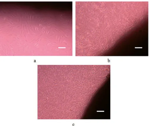
The inhomogeneous scafolds were used to co-culture with MC3T3-E1 cells to assess if the scafolds support the growth of cells. The cell attachment, spreading, and growth are important criteria to judge the biocompatibility of a biomaterial. This process is inluenced by many properties of biomaterials, including composition, the surface chemical states, and morphology.
It can be observed that cell size was around 5~30 μm and cell number notably increased after extended culture (Figure 4), which conirms that the scafolds could well support the cell growth. ECM adhesive molecules are concerned with the speciic attachment and proliferation of cells on material surface. Collagen and PS are main components of ECM. The interaction of the polar carboxyl, hydroxyl, and amino groups of surface hydrophilic and porous scafolds with the cells is one possible mechanism to explain the MC3T3-E1 cell ainity. In addition, porosity and pore size in each layer of the scafolds should be also suited for the cell attachment and migration.
Besides that, it can also be observed from Figure 5 that there were signiicant diferences in the number of proliferated cells between the outer layer and middle layer of scafolds. As indicated previously, drug-loaded collagen content changed in a increase trend from the outer layer to the middle layer of the scafolds, and on the other hand, scafolds had looser pore structures in the middle layer. More cells would grow inwards the layer with both higher porosity and suitable pore size. Therefore, the synergetic efects arisen from composition, structure and certain properties of scafolds result in signiicant diferences in the number of proliferated cells among layers of scafolds.
4. Conclusions
We developed the inhomogeneous and gradient BG-COL-PS scafolds embedding the SS using porogen leaching protocol and freeze drying technique. Drug distribution and microstructure could be controlled mainly by regulating the concentrations of drug-loaded collagen microparticles and parain spheres as well as freezing temperature for diferent layers of the scafolds. It was found that the scafolds have graded average pore size and porosity, gradient drug distribution, and similar swelling ratio for diferent layers. In comparision to the scafolds with homogeneous structure, burst release of drug from inhomogeneous scafolds was lower and the average release rate was slower after burst release because of gradient drug distribution and graded porous micro-architecture. As well as the achieved scafolds could well support the cell growth.
Figure 3: Cumulative release of SS from two types of scafolds with difrent microstructure and drug distribution
from the homogeneous scafolds was faster signiicantly than that of the inhomogeneous scafolds. The cumulative release of drug from the inhomogeneous scafolds was 51.75±0.41%% in 20 days, while drug released from the homogeneous scafolds reached 69.34±0.25%%.
The diferent release proiles of drugs from the two types of scafolds have been largely afected by the structure and drug distribution. This outcome was explained to result from the difusional barrier through the scafold matrix. In the case of the scafolds with gradient structure, when PBS luid lowed from the top layer to bottom layer, drug difusion was driven from low to high resistance, which induced slow release. In contrast, difusion from the homogeneous structure was faster. Additionally, the encapsulated drugs were absorbed on the middle layer much more than the outer two layers. The form would enhance the therapy eiciency and vary release proile in comparison with traditional incorporation methods.
Such diferences in release kinetics may have distinct efects on bone regeneration. A key goal of sustained delivery techniques is achieving therapeutic levels of released factors over biologically relevant time scales. Longer-lasting and lower release amount within the safe range could avoid potential toxicity and reduce undesirable side-efects, such as metaplasia, hyperplasia, hypertrophy, unwanted vessel growth, or ectopic bone formation. This leads to the conclusion that the constructed inhomogeneous scafolds incorporating gradient drug-loaded microparticles could allow spatially efective release of drug, a prerequisite for the regeneration of bone defects.
Figure 4: Optical micrographs of cells after 1 d (a), 7 d (b) and 14 d (c) of culturing with the inhomogeneous scafolds (scale bar 20 μm). Shadow indicates materials.
Figure 5: Cell proliferation in diferent layers of the inhomogeneous scafolds
5. Acknowledgments
This work was inancially supported by natural science foundation project of Fujian (Grant no. 2014J05054), scientiic research development fundation project of Fujian university of technology (Grant no. GY-Z15093) and scientiic research starting foundation project of Fujian university of technology (Grant no. GY-Z0854).
6. References
1. Zhu Y, Wan Y, Zhang J, Yin D, Cheng W. Manufacture of layered collagen/chitosan-polycaprolactone scafolds with biomimetic microarchitecture. Colloids and Surfaces B: Biointerfaces. 2014;113:352-360.
2. Koens MJ, Faraj KA, Wismans RG, van der Vliet JA, Krasznai AG, Cuijpers VM, et al. Controlled fabrication of triple layered and molecularly deined collagen/elastin vascular grafts resembling the native blood vessel. Acta Biomaterialia. 2010;6(12):4666-4674.
3. Knight T, Basu J, Rivera EA, Spencer T, Jain D, Payne R. Fabrication of a multi-layer three-dimensional scafold with controlled porous micro-architecture for application in small intestine tissue engineering. Cell Adhesion & Migration. 2013;7(3):267-274.
4. Shalumon KT, Chennazhi KP, Nair SV, Jayakumar R. High thick layer-by-layer 3D multiscale ibrous scafolds for enhanced cell iniltration and it’s potential in tissue engineering. Journal of
Biomedical Nanotechnology. 2013;9(12):2117-2122.
5. Sarkar S, Isenberg BC, Hodis E, Leach JB, Desai TA, Wong JY. Fabrication of a layered microstructured polycaprolactone construct for 3-D tissue engineering. Journalof Biomaterials
Science. Polymer Edition. 2008;19(10):1347-1362.
6. Lee W, Lee V, Polio S, Keegan P, Lee JH, Fischer K, et al. On-demand three-dimensional freeform fabrication of multi-layered hydrogel scafold with luidic channels. Biotechnology and
7. Liang C, Li H, Li C, Yang Z, Zhou X, Tao Y, et al. Fabrication of a layered microstructured polymeric microspheres as a cell carrier for nucleus pulposus regeneration. Journal of Biomaterials
Science. Polymer Edition. 2012;23(18):2287-2302.
8. Neal RA, Jean A, Park H, Wu PB, Hsiao J, Engelmayr GC Jr, et al. Three-dimensional elastomeric scafolds designed with cardiac-mimetic structural and mechanical features. Tissue
Engineering. Part A. 2013;19(5-6):793-807.
9. Yang CR, Wang YJ, Chen XF. Mineralization regulation and biological inluence of bioactive glass-collagen-phosphatidylserine composite scafolds. Science China. Life Sciences. 2012;55(3):236-240. 10. Wang YZ, Wang JJ, Liang JC, Chen Y. Efects of diosgenin on
cell proliferation, diferentiation and OPG/RANKL mRNA expression of rat osteoblasts cultured in vitro. China Journal of
Traditional Chinese Medicine and Pharmacy. 2010;25(1):134-136.
11. Yang C, Wang J. Preparation and characterization of collagen microspheres for sustained release of steroidal saponins.