Recebido para publicação: Janeiro de 2009 • Aceite para publicação: Novembro de 2009
1145
predição de eventos arrítmicos
em adultos com tetralogia
de Fallot corrigida
[77]
RUBENRAMOS, LUÍSABRANCO, ANAAGAPITO, JOSÉALBERTOOLIVEIRA, LÍDIASOUSA, ANAGALRINHO,
ANTÓNIOFIARRESGA, ALEXANDRATOSTE, ANALOUSINHA, MÁRIOOLIVEIRA, J. NOGUEIRA DASILVA, RUICRUZFERREIRA
Serviço de Cardiologia, Hospital de Santa Marta, Lisboa, Portugal
Rev Port Cardiol 2010; 29 (07-08): 1145-1161
RESUMO
Introdução: Os adultos com Tetralogia de
Fallot corrigida (TFC) podem estar em risco de dilatação, disfunção progressiva do ventrículo direito e eventos arrítmicos ventriculares e supraventriculares. Em doentes com cardiopatia congénita a ecocardiografia com Doppler tecidular (TDI) demonstrou ser especialmente útil na avaliação da função ventricular direita. No entanto, é desconhecido se esses parâmetros derivados do TDI têm valor na predicção de eventos clínicos nesta população.
Objectivo: Estudar se a função ventricular
direita avaliada por Doppler tecidular está associada a eventos arrítmicos supraventri-culares em doentes com TFC.
Materiais e métodos: Entre 2007 e 2008
efectuaram-se exames ecocardiográficos exaustivos a 40 D consecutivos com TFC enviados para avaliação de rotina (idade 35 ± 11 anos, sexo masculino 62%). Foram considerados os seguintes parâmetros ecocardiográficos 1) Clássicos: fracção de ejecção, volumes telesistólico e telediastólico ventriculares esquerdos, área fraccional ventricular direita (VD), volumes auriculares, velocidades ventriculares diastólicas E e A, índice Tei do VD, TAPSE
Usefulness of tissue Doppler imaging to predict arrhythmic events in adults with repaired tetralogy of Fallot
ABSTRACT
Introduction: Adults with repaired tetralogy
of Fallot (TOF) may be at risk for progressive right ventricular (RV) dilatation and dysfunc-tion, which is commonly associated with arrhythmic events. In frequently volume-overloaded patients with congenital heart disease, tissue Doppler imaging (TDI) is par-ticularly useful for assessing RV function. However, it is not known whether RV TDI can predict outcome in this population.
Objective: To evaluate whether RV TDI
parameters are associated with supraventric-ular arrhythmic events in adults with repaired TOF.
Methods: We studied 40 consecutive patients
with repaired TOF (mean age 35±11 years, 62% male) referred for routine echocardio-graphic exam between 2007 and 2008. The following echocardiographic measurements were obtained: left ventricular (LV) ejection fraction, LV systolic volume, LV end-diastolic volume, RV fractional area change, RV end-systolic area, RV end-diastolic area, left and right atrial volumes, mitral E and A
1146
Recebido para publicação: ????????????? • Aceite para publicação: ?????????
VD e área de regurgitação pulmonar, 2) TDI: E’, A´e S’ ao nível dos segmentos basais do septo e paredes laterais de ambos os ventrículos, strain, strain rate e tissue
tracking dos mesmos segmentos, velocidade
máxima de contracção isovolumétrica (CI), duração da CI e índice de aceleração miocárdica durante a CI da parede livre do VD e 3) Electrocardiográficos: duração QRS em repouso, frequência cardíaca máxima e arritmia durante prova ergométrica. Os doentes foram posteriormente divididos em dois subgrupos: Grupo 1 (n = 12) – doentes com eventos arrítmicos prévios: taquicardia auricular e fibrilhação/flutter auricular; Grupo 2 (n = 28) doentes sem eventos prévios. Todos se encontravam em classe funcional NYHA 1 ou 2. Foi estudada a associação estatística entre os parâmetros avaliados e a ocorrência de arritmias por análise uni e multivariada.
Resultados: Comparativamente ao grupo
controlo, os doentes com história de arritmias supraventriculares eram mais velhos (41± 14 versus 31± 6 anos, p = 0.005)), tinham maior duração do QRS em repouso (173 ± 20 versus 140 ± 32 mseg., p = 0.01) e menor frequência cardíaca máxima na prova de esforço (69± 35 versus 92 ± 9 %, p = 0.03). As características clínicas como a idade à data da cirurgia correctiva, presença de cirurgia paliativa prévia ou defeitos residuais não diferiram. Da mesma forma, as dimensões das cavidades cardíacas, a função ventricular esquerda e direita avaliada pelos parâmetros ecocardiográficos convencionais ou o grau de obstrução e regurgitação valvular foram semelhantes. Os parâmetros de deformação miocárdica (strain e strain rate) também não variaram de forma significativa entre os dois grupos. No entanto, os índices e velocidades miocárdicas ventriculares direitas sistólicos (Sa: 5.4 ± 2 versus 8.5 ± 3, p = 0.004) e diastólicos (Ea, Aa, E/Ea septal,
tissue tracking tricúspide) avaliados por TDI
encontravam-se significativamente reduzidos nos doentes com história de arritmias. No modelo de regressão linear multivariado, apenas a velocidade diastólica inicial da
velocities, RV myocardial performance index (Tei index), tricuspid annular plane systolic excursion (TAPSE), myocardial iso-volumic acceleration (IVA), pulmonary regurgitation color flow area, TDI basal lat-eral, septal and RV lateral peak diastolic and systolic annular velocities (E’ l, A’ l, S’ l, E’ s, A’ s, S’ s, E’ rv, A’ rv, S’ rv), strain, strain rate and tissue tracking of the same segments. QRS duration on resting ECG, total duration of Bruce treadmill exercise stress test and presence of exercise-induced arrhythmias were also analyzed. The patients were subsequently divided into two groups: Group 1 – 12 patients with previous documented supraventricular arrhythmias (atrial tachycardia, fibrillation or flutter) and Group 2 (control group) – 28 patients with no previous arrhythmic events. Univariate and multivariate analysis was used to assess the statistical association between the studied parameters and arrhythmic events.
Results: Patients with previous events were
older (41±14 vs. 31±6 years, p=0.005), had wider QRS (173±20 vs. 140±32 ms, p=0.01) and lower maximum heart rate on treadmill stress testing (69±35 vs. 92±9%, p=0.03). All patients were in NYHA class I or II. Clinical characteristics including age at cor-rective surgery, previous palliative surgery and residual defects did not differ signifi-cantly between the two groups. Left and right cardiac chamber dimensions and ventricular and valvular function as evaluated by con-ventional Doppler parameters were also not significantly different. Right ventricular strain and strain rate were similar between the groups. However, right ventricular myocardial TDI systolic (Sa: 5.4±2 vs. 8.5±3, p=0.004) and diastolic indices and velocities (Ea, Aa, septal E/Ea, and RV free wall tissue tracking) were significantly reduced in patients with arrhythmias com-pared to the control group. Multivariate lin-ear regression analysis identified RV lin-early diastolic velocity as the sole variable inde-pendently associated with arrhythmic histo-ry (RV Ea: 4.5±1 vs. 6.7±2 cm/s, p=0.01). A cut-off for RV Ea of <6.1 cm/s identified
INTRODUÇÃO
N
os doentes com tetralogia de Fallot a cirurgia correctiva é actualmente efectua-da de forma precoce e com bons resultados. No entanto, a sobrevida desses doentes a longo prazo é menor do que a da população geral(1). Os defeitos residuais (obstáculopul-monar, regurgitação pulmonar e tricúspide) não são raros, podendo condicionar sobrecar-ga e consequente dilatação e disfunção das cavidades direitas. As arritmias auriculares, ventriculares, insuficiência cardíaca direita e morte súbita são mais frequentes nos doentes em que se documentam regurgitação valvular significativa e compromisso da função ven-tricular direita(2). Assim, a avaliação objectiva
da função ventricular direita é primordial no seguimento clínico dos doentes com tetralogia de Fallot corrigida (TFC). No entanto a com-plexa geometria ventricular direita, sobretudo quando co-existe regurgitação tricúspide e/ou
INTRODUCTION
S
urgical repair of tetralogy of Fallot (TOF) is now performed early and with good results. However, long-term survival of these patients is worse than for the general population(1).Residual defects (pulmonary obstruction and pulmonary and/or tricuspid regurgitation) are not uncommon, leading to overload and hence dilatation and dysfunction of the right cham-bers. Atrial and ventricular arrhythmias, right heart failure and sudden death are more com-mon in patients with significant valvular regur-gitation and right ventricular (RV) dysfunction
(2). Objective assessment of RV function is
therefore essential in the clinical follow-up of patients with repaired TOF. However, the com-plex geometry of the right ventricle, particular-ly in the presence of significant tricuspid and/or pulmonary regurgitation, limits the use-fulness of conventional echocardiography. Magnetic resonance imaging (MRI) and 1147
Recebido para publicação: ????????????? • Aceite para publicação: ?????????
parede da parede livre do VD (Ea tricúspide: 4.5 ± 1 versus 6.7 ± 2 cm/s, p = 0.01]) se associou de forma independente à ocorrência de eventos arrítmicos. Um valor Ea tricúspide <6.1 cm/s identificou com uma sensibilidade de 86 % e especificidade 59 % os doentes com arritmias prévias (AUC = 0.8).
Conclusão: Os resultados sugerem que a
ecocardiografia com Doppler tecidular poderá detectar disfunção ventricular direita em doentes com função aparentemente conservada por ecoDoppler convencional. A redução da velocidade miocárdica diastólica (E’) inicial a nível do anel lateral da válvula tricúspide parece ser uma alteração precoce e está associada à ocorrência de eventos arrítmicos supraventriculares. Os índices TDI poderão ser um método útil na estratificação de risco de doentes com tetralogia de Fallot corrigida.
Palavras-chave:
Tetralogia de Fallot; Doppler tecidular; Arritmias supraventriculares
patients in the arrhythmic group with 86% sensitivity and 59% specificity (AUC=0.8).
Conclusions: Our results suggest that TDI
may detect RV dysfunction in patients with apparently normal function as assessed by conventional echocardiographic parameters. Reduction in RV early diastolic velocity appears to be an early abnormality and is associated with occurrence of arrhythmic events. TDI may be useful in risk stratifica-tion of patients with repaired tetralogy of Fallot.
Key words
Repaired tetralogy of Fallot; Tissue Doppler; Supraventricular arrhythmias
1148
Recebido para publicação: ????????????? • Aceite para publicação: ?????????
pulmonar significativa, limita o papel da eco-cardiografia convencional. A ressonância magnética ou a ventriculografia radionuclear são métodos de referência mas são onerosos,
time-consuming e requerem logística
especi-alizada(3, 4). A medição das velocidades
miocárdicas por Doppler tecidular (TDI) demonstrou ser um instrumento poderoso na avaliação da função sistólica e diastólica ven-tricular esquerda(5) e ultimamente tem sido
usada também no estudo da função ventricular direita(6). A determinação das velocidades
miocárdicas do anel tricúspide por TDI é um método relativamente volume independente, que permite estimar tanto a função sistólica como a diastólica e tem sido recentemente apontado como um marcador prognóstico nos doentes com e sem cardiopatia congénita(6, 7).
Atendendo ao exposto, colocámos a hipó-tese de a função ventricular direita avaliada por TDI ser um marcador prognóstico inde-pendente mesmo quando considerados os parâmetros convencionais de avaliação não invasiva como a duração do QRS, a capaci-dade de esforço ou as dimensões e contrac-tilidade ventricular direita estimada por ecocardiografia convencional. Testámos essa hipótese em doentes TFC com e sem eventos arrítmicos documentados.
MÉTODOS
População
Examinámos prospectivamente 40 doentes consecutivos com TFC que haviam sido enviados para avaliação ecocardiográfica de rotina entre 2007 e 2008 no laboratório de ecocardiografia do Serviço de Cardiologia do Hospital de Santa Marta. Todos os doentes foram submetidos a cirurgia correctiva (encer-ramento da comunicação interventricular e remoção do obstáculo pulmonar) sob circu-lação extra corporal e hipotermia profunda. Quinze doentes (37%) haviam sido submeti-dos previamente a pelo menos uma cirurgia paliativa (criação de um shunt sistémico-pul-monar tipo Blalock- Taussig). À data da inclu-são no estudo (em média 23 + 11 anos após a
radionuclide ventriculography are the refer-ence methods but they are costly, time-con-suming and require specialized facilities(3, 4).
Measurement of myocardial velocities by tis-sue Doppler imaging (TDI) is a powerful tool for assessing left ventricular (LV) systolic and diastolic function(5) and TDI has also been
used to study RV function(6). Tricuspid annular
velocities assessed by TDI are relatively vol-ume-independent, enabling estimation of both systolic and diastolic function, and have recently been put forward as prognostic mark-ers in patients with and without congenital heart disease(6, 7).
Against this background, we set out to determine whether RV function assessed by TDI is an independent prognostic marker com-pared to the standard parameters of noninva-sive assessment such as QRS duration, exer-cise capacity and RV dimensions and contrac-tility as estimated by conventional echocardio-graphy. We tested this hypothesis in patients with repaired TOF, with and without docu-mented arrhythmic events.
METHODS
Population
We prospectively studied 40 consecutive patients with repaired TOF referred for routine assessment between 2007 and 2008 at the echocardiographic laboratory of the cardiology department of Hospital de Santa Marta. All patients had undergone corrective surgery (ventricular septal defect closure and removal of pulmonary obstruction) under cardiopul-monary bypass and deep hypothermia. Fifteen patients (37%) had previously undergone at least one palliative surgery (creation of a Blalock-Taussig systemic-pulmonary shunt). At the time of inclusion in the study (on aver-age 23±11 years after corrective surgery), all patients were in NYHA functional class I or II and in sinus rhythm, with right bundle branch block. Twelve patients (Group 1) had had pre-vious arrhythmic events: atrial tachycardia in three, atrial flutter in five, atrial fibrillation in three, and one had had atrial flutter and at
1149
Recebido para publicação: ????????????? • Aceite para publicação: ?????????
cirurgia correctiva), todos os doentes se encontravam em classe funcional NYHA I ou II e em ritmo sinusal com padrão de bloqueio de ramo direito. Doze doentes (Grupo 1) tinham tido eventos arrítmicos prévios: taquicardia auricular - 3, flutter auricular - 5, fibrilhação auricular - 3 e um doente tinha tido flutter auricular e pelo menos um episódio de taquicardia ventricular mantida, sendo desde então portador de um cardiodes-fibrilhador implantável. Dez doentes deste grupo (83%) encontravam-se sob medicação com amiodarona e/ou betabloqueantes na altura do exame ecocardiográfico. Os restan-tes 28 doenrestan-tes sem quaisquer registos de ar-ritmias serviram de grupo controlo (Grupo 2) (Tabela 1).
least one episode of sustained ventricular tachycardia, following which an implantable cardioverter defibrillator was implanted. Ten patients in this group (83%) were under med-ication with amiodarone and/or beta-blockers at the time of the echocardiographic exam. The other 28 patients, without arrhythmic events, made up the control group (Group 2) (Table I).
Echocardiography
Patients underwent exhaustive echocardio-graphic study by an experienced operator who was unaware of their status in terms of previous arrhythmic events. All exams were performed using a Vivid 7 scanner (GE, USA) equipped with a 3.5-5 MHz transducer and recorded for subsequent analysis. Measurements were made
Total Com Arritmias Sem Arritmias p
(n=40) (n=12) (n=28)
Idade (anos) 35±11 41±14 31±6 0.005
Sexo masculino (%) 25 (62) 7 (64) 18 (58) 0.7
Cirurgia paliativa (CP) 15 (37%) 5 (41) 10 (35) 0.7
Idade na CP (anos) 1.1±0.3 1.4±2.4 0.8±1.5 0.2
Idade à data da Cirurgia
correctiva (anos) 13±12 15±13 10.3±9 0.3
Follow-up médio (anos) 23±11 26±13 21±7 0.03
Tabela 1: Características basais da população
Total Arrhythmia No arrhythmia p
(n=40) (n=12) (n=28)
Age, years 35±11 41±14 31±6 0.005
Male (%) 25 (62) 7 (64) 18 (58) 0.7
Palliative surgery (PS) (%) 15 (37%) 5 (41) 10 (35) 0.7
Age at PF, years 1.1±0.3 1.4±2.4 0.8±1.5 0.2
Age at corrective surgery, years 13±12 15±13 10.3±9 0.3
Mean follow-up, years 23±11 26±13 21±7 0.03
Table I: Baseline characteristics of the population
Ecocardiografia
Os doentes foram submetidos a um exame ecocardiográfico exaustivo por um operador experiente que desconhecia o status dos doentes quanto à existência de eventos arrítmicos prévios. Todos os exames foram efectuados num ecógrafo VIVID 7 (GE, EUA) equipado com um transdutor de 3,5 – 5 MHz e gravados para análise subsequente. As
off-line by a second operator who was also unaware of the patients’ status. Left ventricular end-systolic and end-diastolic volumes, LV ejection fraction (EF), and maximum left and right atrial volumes were calculated using Simpson’s method of disks(8, 9). The right
ven-tricular endocardial border was traced manual-ly and RV fractional area change was calculat-ed using the formula: (diastolic area - end-systolic area) / end-diastolic area(10). All
two-medições foram efectuadas em off-line por um segundo operador também ignorando o status dos doentes. Os volumes telesistólico e telediastólico do ventrículo esquerdo, a frac-ção de ejecfrac-ção do VE, o volume máximo da aurícula esquerda e direita foram obtidos pelos métodos dos discos de Simpson(8,9) . O
bordo endocárdico do ventrículo direito (VD) também foi traçado manualmente e a área fraccional do VD foi calculada usando a fórmula (área telediastólica – área telesis-tólica) / área telediastólica(10). Todas as
medições em 2D foram indexadas à área de superfície corporal. A velocidade de enchi-mento diastólico inicial e tardio do ventrículo esquerdo (E e A), foi obtida por Doppler pulsado na incidência de 4 câmaras com o cursor localizado a nível do topo dos folhetos valvulares mitrais. O índice de performance miocárdico do ventrículo direito (índice Tei) foi obtido pela fórmula (duração de regurgitação tricúspide – duração de ejecção pulmonar) / duração da ejecção pulmonar(11).
A excursão longitudinal sistólica do anel tricúspide (TAPSE) foi calculada em modo M orientado por imagem 2D(12). A regurgitação
pulmonar foi estimada semi-quantitativa-mente segundo a determinação da maior área-cor na câmara de saída do ventrículo direito durante a diástole ventricular(13). As medições
TDI foram obtidas por Doppler codificado a cor e obtidas na incidência apical de 4 câ-maras com a amostra do Doppler pulsado de 2 mm colocado 1 cm apical ao ângulo lateral do anel tricúspide, anel mitral e septo inter-ventricular no local de inserção das válvulas aurículo-ventriculares (Figura 1b). O índice E/Ea foi estimado pela divisão da velocidade de enchimento inicial pela velocidade tecidular mitral (Ea l) ou septal (Ea s)(14,15) O
índice de aceleração miocárdica durante a contracção isovolumétrica (IVA) foi calculado dividindo a velocidade máxima durante a contracção isovolumétrica (IVV) pelo inter-valo de tempo decorrido do início da contrac-ção isovolumétrica até ao pico da mesma onda (ICT) (Figura 1a)(16). O strain (ou deformação),
strain rate (taxa de deformação) e tissue tra-cking (deslocamento) dos segmentos basais do
dimensional (2D) measurements were indexed to body surface area. Mitral early and late dias-tolic velocities (E and A) were obtained by pulsed Doppler in 4-chamber view with the cursor positioned at the top of the mitral valve leaflets. The RV myocardial performance index (Tei index) was calculated by the formu-la: (duration of tricuspid regurgitation - dura-tion of pulmonary ejecdura-tion) / duradura-tion of pul-monary ejection(11). Tricuspid annular plane
systolic excursion (TAPSE) was calculated in M-mode guided by 2D images(12). Pulmonary
regurgitation was estimated semi-quantitative-ly based on the largest color flow area in the RV outflow tract during ventricular diastole(13).
TDI measurements were obtained by color Doppler in apical 4-chamber view with a 2-mm pulsed Doppler sample positioned 1 cm apical to the lateral angle of the tricuspid annulus, mitral annulus and ventricular septum at the point of insertion of the atrioventricular valves (Figure 1b). The E/Ea ratio was estimated by dividing early filling velocity by mitral lateral (Ea l) or septal (Ea s) tissue velocity(14, 15).
Myocardial isovolumic acceleration (IVA) was calculated by dividing peak myocardial isovo-lumic velocity (IVV) by isovoisovo-lumic contraction time (ICT) (Figure 1a) (6). Strain, strain rate and
tissue tracking of the basal septum and RV lat-eral wall were obtained in the same way and analyzed off-line by color tissue Doppler(16).
Electrocardiographic parameters
QRS duration at rest measured in V1 on a standard 12-lead electrocardiogram, and func-tional capacity determined by maximal dura-tion of Bruce treadmill exercise testing, per-centage of maximum predicted heart rate dur-ing exercise and occurrence of exercise-induced arrhythmias have all been suggested as important prognostic markers in patients with TOF(17-19), and accordingly were also
assessed in this study. The results of exams closest in time to the echocardiographic assessment were included in the analysis.
Statistical analysis
Continuous variables were expressed as means ± standard deviation and categorical
1150
septo e parede lateral do ventrículo direito foram obtidos da mesma forma e analisados em off-line por Doppler tecidular codificado a cor(16).
Parâmetros electrocardiográficos
A duração do QRS em repouso medido em na derivação V1 em electrocardiograma clássico de 12 derivações, a capacidade funcional avaliada pela duração máxima de exercício segundo o protocolo de Bruce, a percentagem da frequência cardíaca máxima
variables as percentages. Differences between patients with and without arrhythmic events were analyzed using the Mann-Whitney test for continuous variables with a non-normal distri-bution and the Student’s t test for those with a normal distribution. Categorical variables were compared by the chi-square test or Fisher’s exact test as appropriate. The correlation between two parametric variables was calcu-lated using Pearson’s coefficient. Variables with p<0.1 on univariate analysis were includ-ed in a multivariate linear regression model to determine the variables independently
associ-1151
Recebido para publicação: ????????????? • Aceite para publicação: ?????????
Total Com Arritmias Sem Arritmias p
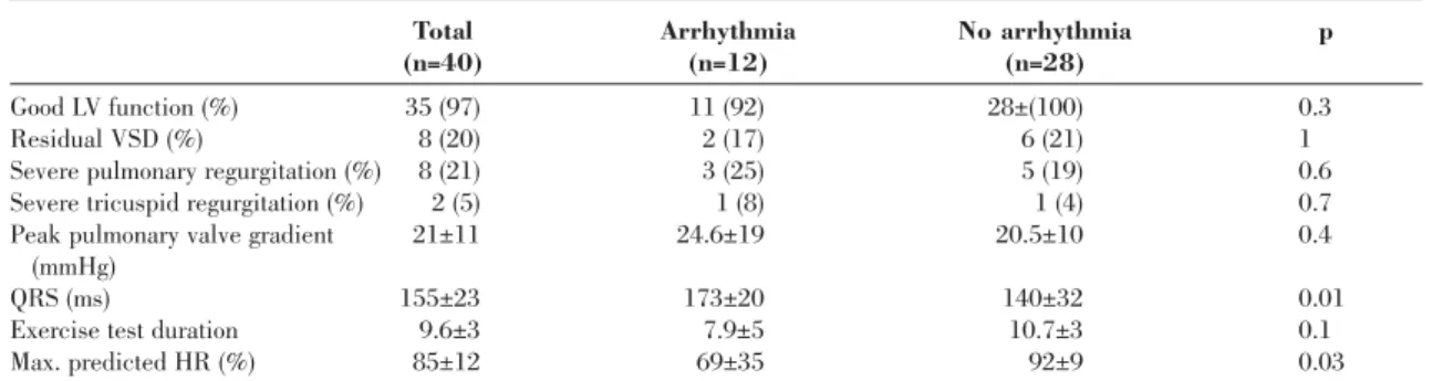
(n=40) (n=12) (n=28) Boa função VE (%) 35 (97) 11 (92) 28±(100) 0.3 CIV residual (%) 8 (20) 2 (17) 6 (21) 1 I. Pulmonar grave (%) 8 (21) 3 (25) 5 (19) 0.6 I. Tricúspide grave (%) 2 (5) 1 (8) 1 (4) 0.7 Grad. valvular 21±11 24.6±19 20.5±10 0.4 pulmonar max. (mmHg) QRS (mseg.) 155±23 173±20 140±32 0.01 Duração P. Esforço 9.6±3 7.9±5 10.7±3 0.1 FCMP (%) 85±12 69±35 92±9 0.03
Tabela II: Parâmetros ecocardiográficos e electrocardiográficos basais
VE - ventrículo esquerdo; CIV - comunicação interventricular: 1. pulmonar - insuficiência pulmonar: I Tricúspide - insuficiência tricúspide; Grad = gradiente: MAX. – Máximo: P. esforço – prova de esforço: FCMP - Frequência cardíaca máxima prevista
Total Arrhythmia No arrhythmia p
(n=40) (n=12) (n=28)
Good LV function (%) 35 (97) 11 (92) 28±(100) 0.3
Residual VSD (%) 8 (20) 2 (17) 6 (21) 1
Severe pulmonary regurgitation (%) 8 (21) 3 (25) 5 (19) 0.6
Severe tricuspid regurgitation (%) 2 (5) 1 (8) 1 (4) 0.7
Peak pulmonary valve gradient 21±11 24.6±19 20.5±10 0.4
(mmHg)
QRS (ms) 155±23 173±20 140±32 0.01
Exercise test duration 9.6±3 7.9±5 10.7±3 0.1
Max. predicted HR (%) 85±12 69±35 92±9 0.03
Table II: Baseline echocardiographic and electrocardiographic parameters
HR: heart rate; LV: left ventricular; Max.: maximum; VSD: ventricular septal defect
prevista durante o esforço e a presença de arritmia de esforço tem sido apontado como factores com importância prognóstica nos doentes com Tetralogia de Fallot(17, 19), pelo que
também foram avaliados neste estudo. Foram considerados os valores referentes aos exames temporalmente mais próximos da avaliação ecocardiográfica.
ated with arrhythmic events. ROC curves were constructed to relate the significant variables with occurrence of arrhythmias. Sensitivity and specificity were calculated according to the standard definitions. Values of p<0.05 were considered statistically significant. The statis-tical program used was SPSS version 15.0 (Chicago, IL, USA).
Análise estatística
As variáveis contínuas foram expressas como média ± desvio padrão e as variáveis categóricas como percentagem. As diferenças entre doentes com e sem eventos arrítmicos foram analisa-das segundo o teste de Mann-Whitney para as variáveis contínuas com distribuição não normal
RESULTS
Electrocardiographic, ergometric and conventional echocardiographic parameters
The clinical, echocardiographic and elec-trocardiographic characteristics of the study
1152
Recebido para publicação: ????????????? • Aceite para publicação: ?????????
Figura 1: A. Imagem ampliada das ondas de contração isovolumétrica do septo (amarelo) e parede livre do VD (verde) representadas dentro da caixa vermelha na figura 1B. Determinação da velocidade de contracção isovolumétrica (IVV), tempo de contração isovolumétrica (TCI) e índice de aceleração da contração isovolumétrica (IVA). IVA = IVV/ICT B. Determinação das velocidades miocárdicas ventriculares direitas usando o TDI. Sa = velocidade sistólica Ea = velocidade diastólica inicial, Aa = velocidade diastólica tardia
Figure 1. A: Enlarged image of isovolumic contraction waves of the septum (yellow) and RV free wall (green), shown within the red square in B, and determination of isovolumic velocity (IVV), isovolumic contraction time (ICT), and isovolumic acceleration (IVA). IVA = IVV/ICT; B: RV myocardial velocities by TDI.
Sa: systolic velocity; Ea: early diastolic velocity; Aa: late diastolic velocity
Total Arritmia Sem Arritmias p
(n=40) (n=12) (n=28) Volume AE (ml/m2) 35 (97) 11 (92) 28±(100) 0.3 Volume AD (ml/m2) 8 (20) 2 (17) 6 (21) 1 TDVD/TDVE 8 (21) 3 (25) 5 (19) 0.6 Area fraccional VD (%) 2 (5) 1 (8) 1 (4) 0.7 TAPSE VD (mm) 21±11 24.6±19 20.5±10 0.4 dP/dt VD (mmHg/s) 155±23 173±20 140±32 0.01 Índice Tei VD 9.6±3 7.9±5 10.7±3 0.1
Tabela III: Dados ecocardiográficos (2D e Modo M)
VE - Aurícula esquerda; AD - Aurícula direita; TDVD - Diâmetro telediastólico do ventrículo direito; TDVE - Diâmetro telediastólico do ventrículo esquerdo; VD - Ventrículo direito
Total Arrhythmia No arrhythmia p
(n=40) (n=12) (n=28) LA volume (ml/m2) 35 (97) 11 (92) 28 ± (100) 0.3 RA volume (ml/m2) 8 (20) 2 (17) 6 (21) 1 RVEDD (mm/m2) 8 (21) 3 (25) 5 (19) 0.6 RVEDD/ LVEDD 2 (5) 1 (8) 1 (4) 0.7 RV fractional area (%) 21±11 24.6±19 20.5±10 0.4 RV TAPSE (mm) 155±23 173±20 140±32 0.01 RV dP/dt (mmHg/s) 9.6±3 7.9±5 10.7±3 0.1 Tei index 0.23±0.1 0.22±0.2 0.24±0.1 0.7
Table III: Echocardiographic data (2D and M-mode)
LA: left atrial; RA: right atrial; LVEDD: left ventricular diastolic diameter; RV: right ventricular; RVEDD: right ventricular end-diastolic diameter; TAPSE: tricuspid annular plane systolic excursion
e segundo o teste T-Student para aquelas com distribuição normal. As variáveis categóricas foram comparadas pelo teste do χ2ou teste de
Fisher conforme apropriado. A correlação entre duas variáveis paramétricas foi estimada pelo cálculo do coeficiente de Pearson. As variáveis com p <0,1 na análise univariada foram in-cluídas num modelo de regressão linear multi-variado para determinar as varáveis indepen-dentes associadas aos eventos arrítmicos. Foram construídas curvas ROC para relacionar as variáveis significativas com a ocorrência de arritmias. A sensibilidade e especificidade foram calculadas de acordo com as definições
standard. A significância estatística foi
consi-derada para valores de p <0.05. O programa estatístico utilizado foi o SPSS versão 15.0 (Chicago, IL, EUA)
RESULTADOS
Parâmetros electrocardiográficos, ergométricos e ecocardiográficos convencionais
As características clínicas, ecocardio-gráficas e electrocardioecocardio-gráficas estão suma-rizadas nas Tabelas I e II. Os doentes com eventos arrítmicos prévios (Grupo 1) eram em média 10 anos mais velhos que aqueles sem eventos (Grupo 2) à data da realização do exame ecocardiográfico. Com excepção de um doente cuja função ventricular esquerda foi considerada como razoável (fracção de ejecção 48%), todos os outros doentes tinham boa função ventricular esquerda (fracção de ejecção> 55%). Foi detectada regurgitação pulmonar em todos os doentes, mas a sua gravidade não diferiu significativamente entre os dois grupos: regurgitação pulmonar grave em nove doentes (Grupo 1: três doentes e Grupo 2: cinco doentes) e regurgitação pulmonar ligeira ou moderada nos restantes 32 doentes (Grupo 1: dez doentes e Grupo 2: 22 doentes). Foi encontrado comunicação interventricular residual em oito doentes (20%) e apenas um doente do Grupo 1 apresentou obstrução grave na câmara de saída do ventrículo direito (o
population are summarized in Tables I and II. Patients with previous arrhythmic events (Group 1) were 10 years older on average than those without events (Group 2) at the time of the echocardiographic exam. With the excep-tion of one patient whose left ventricular func-tion was considered only reasonable (EF 48%), all patients had good LV function (EF >55%). Pulmonary regurgitation was detected in all patients but its severity did not differ signifi-cantly between the two groups: severe in 8 (3 in group 1 and 5 in Group 2) and mild or mod-erate in the other 32 (10 in Group 1 and 22 in Group 2). A residual ventricular septal defect was found in 8 patients (20%), but only one patient, from Group 1, presented severe RV outflow tract obstruction (pulmonary trans-valvular gradients ranged between 6.8 and 71 mmHg, mean 21±11 mmHg). All patients were in NYHA functional class I (n=38) or II. QRS duration in V1 was significantly longer in patients with previous arrhythmic events (173 vs. 140 ms, p=0.01). While exercise test dura-tion did not differ significantly between the two groups, patients without previous arrhythmic events attained a higher percentage of their maximum predicted heart rate (92 vs. 69%, p=0.03).
Right chamber dimensions in the study population were generally larger than those found in the general population: right atrial volume of 37 ml/m2 and tricuspid annular
diameter of 28.6 mm/m2, although no
statisti-cally significant differences were found between the two groups under analysis. Similarly, RV function as assessed by conven-tional echocardiographic parameters (RV frac-tional area change, TAPSE, peak dP/dt and myocardial performance index) did not differ between patients with and without arrhythmic events. However, mitral early diastolic velocity (E wave) was significantly greater in Group 1 than in Group 2: E=109±15 vs. 88±24 cm/s, p=0.01.
Myocardial velocities by TDI
Right ventricular strain and strain rates were similar in patients with and without arrhythmias: septal strain -17±15 vs. -19±9%, 1153
gradiente transvalvular pulmonar variou de 6.8 – 71 mmHg, média de 21 + 11 mmHg). Todos os doentes encontravam-se em classe funcional NYHA I (38 doentes) ou NYHA II. A duração do QRS em V1 foi significa-tivamente superior nos doentes com eventos arrítmicos prévios (173 versus 140 msec, p = 0,01). Embora o tempo de exercício durante a prova de esforço não tenha diferido de forma significativa entre os dois grupos, os doentes sem eventos arrítmicos prévios atingiram maior percentagem da frequência cardíaca máxima prevista (69 versus 92%, p = 0,03).
Em média as dimensões das cavidades direitas da população estudada foram su-periores às encontradas na população normal (volume da aurícula direita de 37 ml/m2 e
dilatação do anel tricúspide - 28.6 mm/m2).
No entanto, nenhuma diferença estatistica-mente significativa foi encontrada quando comparados entre si os dois grupos em estu-do. Da mesma forma, a função ventricular direita quando avaliada por parâmetros ecocardiográficos convencionais (área frac-cional do ventrículo direito, TAPSE, dP/dt máximo ou índice de performance miocár-dico), não foi diferente nos doentes com e sem eventos arrítmicos. A velocidade de enchimento inicial do ventrículo esquer-do (onda E) no entanto foi significativamente maior nos doentes do grupo arrítmico em relação aos doentes do grupo controle: E = 109 ± 15 versus 88 ± 24 cm/s, p = 0,01.
Velocidades miocárdicas por TDI
Os parâmetros de deformação miocárdica longitudinal do ventrículo direito, strain e
strain rate, foram relativamente coincidentes
nos doentes com e sem arritmias, respecti-vamente (strain septal -17 ± 15 versus -19 ± 9%, p = 0,4; strain do anel lateral da tricúspide -16 ± 12 versus -21 ± 10%, p = 0,4;
strain rate septal -2 ± 1 versus -0.9 ± 1 s-1, p
= 0,4; strain rate tricúspide lateral -3.4 ± 4
versus -2,2 ± 2 s-1, p =0,1). Contudo,
comparado com o grupo controlo, os doentes com TFC e arritmias revelaram signifi-cativamente menor velocidade miocárdica
p=0.4; lateral tricuspid strain 16±12 vs. 21±10%, p=0.4; septal strain rate 2±1 vs. -0.9±1 s-1, p=0.4; lateral tricuspid strain rate
-3.4±4 vs. -2.2±2 s-1, p=0.1, respectively.
However, compared to the control group, patients with repaired TOF and arrhythmias presented significantly lower septal and tricus-pid early diastolic myocardial velocities (E’ s: 5.7±2 vs. 7.3±2 cm/s, p=0.01; E’ t: 5.4±2 vs. 8.5±3 cm/s, p=0.004), as well as lower systolic contraction velocity as shown by the Sa wave (4.5±1 vs. 6.7±2, p=0.004) and tricuspid annular systolic motion measured by tissue tracking (7.6±4 vs. 11.2±4 mm). The septal E/Ea ratio was greater (19.5±9 vs. 12.7±4, p=0.03) in the group with arrhythmias.
Peak myocardial isovolumic velocity (IVV) was reduced in patients with arrhythmic events (1.4±0.9 vs. 2.6±1 cm/s, p=0.02) and time to peak myocardial isovolumic velocity was longer (50±10 vs. 40±10 ms, p=0.04). Myocardial isovolumic acceleration (IVA) did not differ significantly between the two groups (47±26 vs. 39±41 cm/s2, p=0.4).
On comparison of conventional parameters of RV function with those derived from TDI, only TAPSE showed a good correlation with tricuspid annular tissue tracking (r2=0.4,
p<0.0001) (Figure 3B). Peak RV myocardial systolic velocity (Sa rv) showed a significant but weak correlation with RV fractional area change (r2=0.1; p=0.04) (Figure 3A).
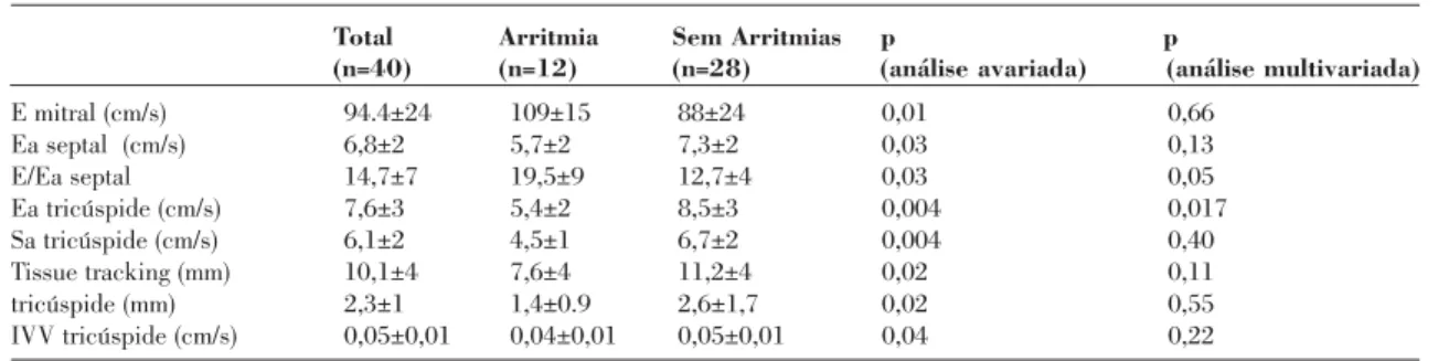
All variables with p<0.1 on univariate analysis were included in a multivariate lin-ear regression model. Only lin-early diastolic tricuspid annular velocity (p=0.01) was independently associated with occurrence of arrhythmias (Table IV). Age, QRS duration, percentage of maximum predicted heart rate during exercise testing and tricuspid annu-lar velocity were not significant on multi-variate analysis. ROC curve analysis of tri-cuspid Ea enabled a cut-off point to be determined for prediction of arrhythmic events (Figure 2). Thus, in this population, tricuspid Ea of <6.1 cm/s identified patients with arrhythmias with 86% sensitivity and 59% specificity.
1154
diastólica precoce septal- E’s (5.7 ± 2 versus 7,3 ± 2 cm/s, p = 0,01) e tricúspide - E’t (5,4 ± 2 versus 8.5 ± 3, p = 0,004) e também menor velocidade de contracção na sístole como evidenciado pela onda Sa (4,5 ± 1 versus 6,7 ± 2, p = 0,004) e pelo deslocamento longitudinal sistólico do anel tricúspide medido pelo tissue
tracking (7,6 ± 4 versus 11,2 ± 4 mm). O ratio
E/Ea septal foi superior (19,5 ± 9 versus 12,7 ± 4, p = 0,03) no grupo de doentes com ar-ritmias.
A velocidade máxima de contracção isovolumétrica (IVV) encontrava-se diminuí-da nos doentes com eventos arrítmicos (2,6 ± 1 versus 1,4 ± 0,9 cm/s, p = 0,02) e o tempo até à velocidade máxima da contracção isovolu-métrica variou de maneira inversa (40 ± 10
versus 50 ± 10 ms, p = 0,04). O índice de
aceleração miocárdica (IVA) não diferiu de forma significativa nos dois grupos (47 ± 26
versus 39 ± 41 cm/s2, p = 0,4).
Quando correlacionados os parâmetros clássicos de avaliação da função ventricular direita com os parâmetros derivados do Doppler tecidular apenas o TAPSE demons-trou correlação significativa com o tissue
tra-cking anular tricúspide (r2 = 0,4, p <0.0001).
A velocidade miocárdica máxima durante a sístole (Sa) demonstrou uma correlação significativa mas pobre com a área fraccional do ventrículo direito (r2 = 0,1; p = 0,04) (Figura 3A).
Todas as variáveis com valor de p <0,1 na análise univariada foram inseridas num mo-delo de regressão linear multivariada. Ape-nas a velocidade diastólica inicial do anel tricúspide (p = 0,01) foi associado de forma independente à ocorrência de arritmias (Tabela IV). A idade, a duração do QRS, a frequência máxima prevista durante a prova de esforço e a velocidade anular tricúspide não foram significativos na análise multiva-riada. A curva ROC da Ea tricúspide per-mitiu obter um valor cut-off para a predicção de eventos arrítmicos (Figura 2). Assim, nesta população uma Ea tricúspide inferior a 6,1 cm/s permitiu identificar com 86% de sensibilidade e 59% de especificidade os doentes com eventos arrítmicos.
DISCUSSION
In this study we assessed right ventricular function by tissue Doppler in adults with repaired tetralogy of Fallot and good function-al capacity, and established a significant corre-lation between ventricular diastolic function and previous supraventricular arrhythmic events.
The overall survival rate of patients with repaired TOF is good, with mortality of less than 6% at 25 years after corrective surgery. Nevertheless, residual defects, pressure and volume overload and right ventricular dysfunc-tion in such patients are associated with right heart failure, arrhythmias and sudden death (19).
Although it is essential to assess RV function, the geometry, and possibly the mechanism of contraction, of the right ventricle make study by 2D echocardiography difficult and likely to be inaccurate. Pulsed TDI of the tricuspid annulus is rapid, reproducible and noninva-sive and has recently been shown to be a reli-able method of assessing RV function com-pared to the gold standard of magnetic reso-nance imaging(4, 20, 21). TDI thus has potential
advantages over conventional echocardio-graphic parameters; decreased myocardial velocities may precede global dysfunction as assessed by ejection fraction or the eyeball method(22), and measurement of TDI velocities
may therefore be a more sensitive and earlier indicator of RV dysfunction(15). In our cohort,
decreased myocardial early diastolic (but not systolic) velocity of the basal lateral segment of the tricuspid annulus (Ea t) appeared to be independently associated with the occurrence of arrhythmic events. This was found even when RV dimensions and systolic function were apparently preserved, with values close to those described in the literature for a healthy population(6, 15). These findings suggest
that diastolic dysfunction may precede ventric-ular systolic dysfunction in these patients. Previous studies have demonstrated that myocardial longitudinal motion of the right ventricle during systole (Sa) and diastole (Ea and Aa) is decreased in patients with repaired TOF compared to healthy individuals (6).
1155
Meluzin et al. proposed a cut-off for Sa of <11.5 cm/s to distinguish patients with RV ejection fraction of <45%, with a sensitivity of 90% and specificity of 85% (21). Reduced
myocardial contraction velocity appears to be relatively independent of the volume overload arising from concomitant valvular regurgita-tion(23, 24). The weak correlation in our
popula-tion between RV fracpopula-tional area change calcu-lated by planimetry and systolic index
1156
Recebido para publicação: ????????????? • Aceite para publicação: ?????????
Figura 2: Curva ROC para a variável ecocardiográfica indepen-dentemente relacionada com a ocorrência de eventos arrítmicos. E a tricúspide – velocidade diastólica inicial da parede lateral do anel da tricúspide (AUC = 0.80, p = 0.003)
Figure 2. ROC curve analysis for the echocardiographic variable independently associated with occurrence of arrhythmic events. Tricuspid Ea: early diastolic velocity of the lateral wall of the tricuspid annulus (AUC=0.80, p=0.003)
DISCUSSÃO
Neste estudo avaliámos a função ventri-cular direita por Doppler tecidular em adultos com tetralogia de Fallot corrigida e boa capacidade funcional e estabelecemos uma correlação significativa entre a disfunção ven-tricular diastólica e a ocorrência de eventos arrítmicos supraventriculares prévios.
A sobrevida global dos doentes com TFC é muito boa. Têm sido descritas taxas de mortalidade inferior a 6% aos 25 anos após a cirurgia correctiva. Porém, defeitos residuais, sobrecarga de pressão/volume e disfunção ventricular direita estão associados, a insuficiência cardíaca direita, arritmias e morte súbita(19). Apesar da importância de um
estudo dirigido à função ventricular direita, a geometria e possivelmente também o me-canismo de contracção do ventrículo direito tornam a sua avaliação por ecocardiografia 2D difícil e provavelmente incorrecta. O TDI pulsado do anel tricúspide é um método rápido, reprodutível e não invasivo que tem recentemente demonstrado sua fiabilidade na avaliação da função ventricular direita usando
Figura 3 A
Figura 3 A-B: Correlação entre a área fraccional do ventrículo direito e a velocidade sistólica da parede lateral do anel tricúspide. r2= 0,1; p = 0.04 3b: Correlação entre tissue tracking
do anulo da tricúspide e o TAPSE. r2= 0.4, p < 0.0001
Figure 3A-B: Correlation between RV fractional area change and systolic velocity of the lateral wall of the tricuspid annulus, r2=0.1;
p=0.04; B: Correlation between tissue tracking of the tricuspid annulus and TAPSE, r2=0.4, p<0.0001.
TAPSE: tricuspid annular plane systolic excursion
a ressonância magnética nuclear como Gold-standard(4,20,21). Existem vantagens
poten-ciais no uso do TDI em relação aos parâmetros ecocardiográficos convencionais. A diminui-ção das velocidades miocárdicas poderá preceder a disfunção global quando avaliada pela fracção de ejecção ou Eye-ball(22). A
medição das velocidades TDI poderá assim ser um indicador mais sensível ou precoce de disfunção ventricular direita(15). No nosso
coorte, uma velocidade diastólica miocárdica precoce reduzida (mas não a velocidade sis-tólica) a nível do anel lateral basal do ventrí-culo direito (Ea t) parece estar associada de forma independente à ocorrência de eventos adversos arrítmicos. Esses resultados verifi-cam-se apesar das dimensões e função ventri-cular direita sistólica estarem aparentemente preservadas e com valores próximos aos des-critos na literatura para uma população não doente(6,15). Estes dados parecem sugerir que a
disfunção diastólica poderá preceder nestes doentes o compromisso ventricular sistólico. Estudos prévios demonstram que a velocidade
assessed by TDI (r=0.1; p=0.04) would seem to support this. The fact that myocardial systolic and early diastolic velocities were consider-ably reduced compared to a normal popula-tion, despite volume overload (severe pul-monary regurgitation in 21% of patients), sug-gests that TDI indices can unmask RV dys-function even in patients with increased pre-load.
A mitral E/septal Ea ratio of >15 has been validated as a reliable marker of left chamber pressure overload(14). In our, despite
apparent-ly greater left chamber filling pressures in the group with arrhythmias (septal E/Ea of 19.5 vs. 12.7), RV contraction (Sa of 4.5 vs. 6.7 cm/s) and relaxation (Ea of 5.4 vs. 8.5 cm/s) veloci-ties were consistently significantly lower than in those with no history of arrhythmias.
Increased afterload has also been associat-ed with decreasassociat-ed myocardial systolic and diastolic velocities. Giorgi et al. demonstrated an inverse relation between aortic transvalvu-lar gradients and mitral annutransvalvu-lar velocities in patients with aortic valve stenosis(26), but it is
1157
Total Arritmia Sem Arritmias p p
(n=40) (n=12) (n=28) (análise avariada) (análise multivariada)
E mitral (cm/s) 94.4±24 109±15 88±24 0,01 0,66 Ea septal (cm/s) 6,8±2 5,7±2 7,3±2 0,03 0,13 E/Ea septal 14,7±7 19,5±9 12,7±4 0,03 0,05 Ea tricúspide (cm/s) 7,6±3 5,4±2 8,5±3 0,004 0,017 Sa tricúspide (cm/s) 6,1±2 4,5±1 6,7±2 0,004 0,40 Tissue tracking (mm) 10,1±4 7,6±4 11,2±4 0,02 0,11 tricúspide (mm) 2,3±1 1,4±0.9 2,6±1,7 0,02 0,55 IVV tricúspide (cm/s) 0,05±0,01 0,04±0,01 0,05±0,01 0,04 0,22 Tabela IV. Resultados do Doppler tecidular
IVA: isovolumic acceleration; IVV: isovolumic velocity; ICT: isovolumic contraction time
Total Arrhythmia No arrhythmia p p
(n=40) (n=12) (n=28) (univariate analysis) (multivariate analysis)
Mitral E (cm/s) 94.4±24 109±15 88±24 0.01 0.66
Septal Ea (cm/s) 6.8±2 5.7±2 7.3±2 0.03 0.13
Septal E/Ea 14.7±7 19.5±9 12.7±4 0.03 0.05
Tricuspid Ea (cm/s) 7.6±3 5.4±2 8.5±3 0.004 0.017
Tricuspid Sa (cm/s) 6.1±2 4.5±1 6.7±2 0.004 0.40
Tricuspid tissue tracking (mm) 10.1±4 7.6±4 11.2±4 0.02 0.11
Tricuspid IVV (cm/s) 2.3±1 1.4±0.9 2.6±1.7 0.02 0.55
Tricuspid ICT (s) 0.05±0.01 0.04±0.01 0.05±0.01 0.04 0.22
Table IV. Tissue Doppler results
LA: left atrial; RA: right atrial; LVEDD: left ventricular diastolic diameter; RV: right ventricular; RVEDD: right ventricular end-diastolic diameter; TAPSE: tricuspid annular plane systolic excursion
de deslocamento longitudinal miocárdico ventricular direito na sístole (Sa) e na diástole (Ea e Aa) estão diminuídas nos doentes com TFC em relação a indivíduos saudáveis(6).
Meluzin et al. propuseram um cut-off de Sa <11,5 cm/s para descriminar doentes com fracção de ejecção ventricular direita <45% com uma sensibilidade de 90% e especifi-cidade de 85%(21). A diminuição da velocidade
de contracção miocárdica parece ser relativa-mente independente da sobrecarga de volume resultante de regurgitação valvular conco-mitante(23,24). A fraca correlação entre a área
fraccional ventricular direita calculada por planimetria e o índice sistólico avaliado por TDI nesta população (r = 0.1; p = 0.04) parece suportar essa hipótese. O facto das veloci-dades miocárdicas sistólicas e diastólicas ini-ciais estarem claramente reduzidas, em com-paração com uma população normal, apesar da existência sobrecarga de volume (regur-gitação pulmonar grave em 21% dos doentes) sugere que estes índices TDI poderão permitir desmascarar a disfunção ventricular direita mesmo nos doentes com pré-carga aumentada. A relação Emitral /Ea septal> 15 foi vali-dada como um marcador fiável de sobrecarga de pressão de enchimento nas cavidades esquerdas(14). Consistentemente, verifica-se
que no grupo de doentes com arritmias, apesar da aparente maior pressão de enchimento à esquerda (E/Ea septal 19.5 versus 12.7), as velocidades contracção (Sa 4.5 versus 6,7 cm/s) e relaxamento do ventrículo direito (Ea 5,4 versus 8.5 cm/s) são significativamente menores do que os doentes com TFC e sem história de arritmias.
Por outro lado, o aumento da pós-carga foi também relacionado com a diminuição da velocidade miocárdica sistólica e diastólica. Giorgi et al. demonstraram uma relação inversa entre o gradiente transvalvular aórtico e as velocidades miocárdicas do anel mitral em doentes com estenose valvular aórtica(25).
Não é seguro que esses resultados possam ser extrapolados para a obstrução pulmonar resi-dual nos doentes com TFC. Na nossa popu-lação, cinco doentes (12%) apresentavam gra-diente residual máximo na câmara de saída do
by no means certain that these results can be extrapolated to residual pulmonary obstruction in patients with repaired TOF. In our popula-tion, five patients (12%) presented a residual peak RV outflow tract gradient of over 40 mmHg, but even excluding these patients, the differences in TDI indices between patients with and without arrhythmias remain signifi-cant.
Tricuspid annular plane systolic excursion is one of the parameters commonly used to assess RV systolic function (12). Besides
myocardial velocities, TDI also enables quan-tification of absolute longitudinal motion from a particular point in the ventricle (tissue track-ing). In our cohort, there was a reasonably and statistically significant correlation between these two parameters (r=0.4; p<0.001). Nevertheless, Ea was the parameter that showed the strongest association with occur-rence of arrhythmic events (AUC=0.80) com-pared to tissue tracking or TAPSE (AUC=0.70 and 0.65, respectively).
Clinical implications
In patients with repaired TOF and residual defects, mainly severe pulmonary regurgita-tion, RV dysfunction is associated with increased risk for arrhythmic events and sud-den death. Pulmonary valve replacement in such cases decreases ventricular dilatation and improves function. However, if performed at a late stage, RV recovery may be compro-mised(26-28), and so in order to preserve
contrac-tile function, pulmonary valve replacement should be considered before RV function dete-riorates. Assessment of RV function by meth-ods such as TDI that are accessible, repro-ducible and noninvasive can help identify patients at increased risk, as appears to have been the case in our patients with repaired TOF and arrhythmic events.
LIMITATIONS
In our population, mean age at the time of corrective surgery was 13 years and the mean period since surgery was around 23 years; most
1158
ventrículo direito superior a 40 mmHg. Mes-mo excluindo esses doentes as diferenças nos índices TDI entre os doentes com e sem his-tória de arritmias permaneceu significativa.
O deslocamento sistólico do anel tricúspide (TAPSE) é um dos parâmetros frequentemente utilizados para avaliar a função sistólica ventricular direita(12). A análise TDI permite
também quantificar, para além das velocidades miocárdicas, o deslocamento longitudinal abso-luto de um determinado ponto do ventrículo (tissue tracking). No nosso coorte de doentes verificou-se uma correlação razoável e signifi-cativa entre os valores obtidos pelos dois parâ-metros (r = 0.4; p <0.001). No entanto, na nossa população o Ea foi o parâmetro que melhor se associou à ocorrência de eventos arrítmicos (AUC de 0,80) em comparação com o tissue
tracking ou o TAPSE (AUC de 0,70 e 0,65,
res-pectivamente)
Implicações clínicas
Em doentes com TFC e defeitos residuais, principalmente regurgitação pulmonar grave, a disfunção ventricular direita correlaciona-se com um aumento de eventos arrítmicos e morte súbita. A substituição valvular pul-monar nesses casos resulta na diminuição da dilatação ventricular e melhoria da função. Contudo, se efectuada numa fase muito tardia a recuperação ventricular direita pode ficar comprometida(26,28). Para preservar a função
contráctil, a implantação valvular pulmonar deverá ser considerada antes que a função do VD se deteriore. Assim a avaliação ventricular direita por métodos fáceis de obter, reprodutíveis e não invasivos como é o TDI poderá ajudar a identificar doentes de risco acrescido, como parece ser o caso dos doentes com TFC e eventos arrítmicos.
LIMITAÇÕES
A idade média à data da cirurgia correctiva na nossa população foi de 13 anos e o tempo médio desde a cirurgia foi de cerca de 23 anos. Actualmente a maioria dos doentes é operada em idades mais precoces. Embora o tempo
patients are now operated at younger ages. Although the time between corrective surgery and arrhythmic events did not differ between the groups (8.7 vs. 9.2 years), patients with a history of arrhythmia were significantly older at the time of the echocardiographic assessment. Although age was not an independent factor, the possibility cannot be excluded that it influ-enced the results obtained. RV function was not assessed by reference methods such as MRI or radionuclide ventriculography in our study, but TDI indices are an accepted means of assessing ventricular function. All our patients had Sa values below that established for a diag-nosis of RV systolic dysfunction (<11.5 cm/s). However, two additional factors may have con-tributed to these results. Firstly, myocardial velocities were determined by off-line analysis of color TDI, and values thus obtained are gen-erally around 20% lower than those obtained by pulsed TDI since they correspond to mean rather than peak velocities. Secondly, 83% of the patients with previous arrhythmic events were under medication with amiodarone and/or beta-blockers. It is not clear whether anti-arrhythmic drugs with negative inotropic prop-erties could have contributed to the lower myocardial velocities observed in this group.
It should also be noted that the arrhythmic events in our study population were mostly supraventricular, with only one ventricular arrhythmia. Thus, our results cannot be extrap-olated to ventricular arrhythmias, however tempting this might be.
CONCLUSIONS
Our results suggest that TDI may detect right ventricular dysfunction in patients with apparently normal function as assessed by conventional echocardiographic parameters. Reduction in RV early diastolic velocity of the lateral tricuspid annulus (Ea t) seems to be an early abnormality and is associated with occur-rence of supraventricular arrhythmic events. TDI may be useful in risk stratification of patients with repaired tetralogy of Fallot. 1159
decorrido entre a cirurgia correctiva e os eventos arrítmicos não tenha diferido entre os grupos (8.7 versus 9.2 anos), a idade dos doentes com história de arritmias era significativamente maior à data de realização do exame ecocardiográfico. Apesar de a idade não ter sido um factor independente não nos é possível excluir completamente que esse facto tenha tido influência nos resultados obtidos. No nosso estudo a função ventricular direita não foi avaliada pelos métodos de referência como a RMN ou a ventriculografia radionuclear. No entanto, os índices TDI são bem aceites e estabelecidos para a avaliação da função ventricular. Todos os nossos doentes tiveram um valor de Sa inferior ao estabelecido para o diagnóstico de disfunção sistólica ventricular direita (Sa <11.5cm/s). Contudo, dois factores adicionais podem ter contribuído para esses resultados. Primeiro, as velocidades miocár-dicas foram determinadas pela análise off-line do TDI codificado a cor. Os valores obtidos por esse método são habitualmente inferiores em cerca de 20% em relação àqueles obtidos por TDI pulsado uma vez que se trata de uma análise de velocidades médias e não de pico. Segundo, 83% dos doentes com eventos ar-rítmicos prévios encontravam-se medicados com amiodarona e/ou beta-bloqueantes. Não é claro se fármacos antiarrítmicos com proprieda-des inotrópicas negativas podem ou não ter contribuído para as menores velocidades miocárdicas verificadas nesse grupo.
De referir também que na população por nós estudada os eventos arrítmicos estudados foram supraventriculares, sendo apenas um ventricular. Assim, e embora conceptual-mente atractiva, os nossos resultados não podem ser extrapolados para eventos arrítmi-cos ventriculares.
CONCLUSÕES:
Os nossos resultados sugerem que o TDI poderá detectar disfunção ventricular direita em doentes com função aparentemente conservada por ecoDoppler convencional. A redução da velocidade miocárdica diastólica inicial a nível do anel tricúspide lateral (Ea t) parece ser uma alteração precoce e está associada à ocorrência de eventos arrítmicos supraventriculares. Os índices TDI poderão ser um método útil na estratificação de risco de doentes com tetralogia de Fallot corrigida.
Pedido de separatas para: Adress for reprints: Ruben Ramos
Serviço de Cardiologia Hospital de Santa Marta Rua de Santa Marta 1169-024 Lisboa
e-mail: [email protected]
1160
Recebido para publicação: ????????????? • Aceite para publicação: ?????????
1. Murphy JG, Gersh BJ, Mair DD, Fuster V, McGoon MD, Ilstrup DM, et al. Long-term outcome in patients undergoing surgical repair of tetralogy of Fallot. N Engl J Med 1993;329:593-9.
2. Gatzoulis MA, Balaji S, Webber SA, Siu SC, Hokanson JS, Poile C, et al. Risk factors for arrhythmia and sudden cardiac death late after repair of tetralogy of Fallot: a multicentre study. Lancet 2000;356:975-81.
3. Helbing WA, Bosh HG, Maliepaard C, Rebergen SA, van der Geest RJ, Hansen B, et al. Comparison of echocardiographic methods with magnetic resonance imaging for assessment of right ventricular function in children. Am J Cardiol 1995;76: 589-94.
BIBLIOGRAFIA / REFERENCES
4. Schwerzmann M, Samman AM, Salehian O, et al. Comparison of Echocardiographic and Cardiac Magnetic Resonance Imaging for Assessing Right Ventricular Function in Adults With Repaired Tetralogy of Fallot. Am J Cardiol 2007; 99:1593-1597.
5. Troughton RW, Prior DL, Frampton CM, Nash PJ, Pereira JJ, Martin M, Fogarty A, Morehead AJ, Starling RC, Young JB, et al. Usefulness of tissue Doppler and color M-mode indexes of left ventricular diastolic function in predicting outcomes in systolic left ventricular heart failure. Am J Cardiol 2005;96:257-262 6. Toyono M, Harada K, Tamura M, et al. Myocardial Acceleration During Isovolumic Contraction as a New Index of Right Ventricular Contractile Function and Its Relation to
1161
Recebido para publicação: ????????????? • Aceite para publicação: ?????????
Pulmonary Regurgitation in Patients After Repair of Tetralogy of Fallot. J Am Soc Echocardiogr 2004;17:332-7
7. Brili S, Alexopoulos N, Latsios G. Tissue Doppler Imaging and Brain Natriuretic Peptide Levels in Adults with Repaired Tetralogy of Fallot. J Am Soc Echocardiogr 2005;18:1149-1154 8. Quinones MA, Waggoner AD, Reduto LA, Nelson JG, Young JB, Winters WL Jr., Ribeiro LG, Miller RR. A new, simplified and accurate method for determining ejection fraction with two dimensional echocardiography. Circulation 1981;64:744-753. 9. Tsang TSM, Barnes ME, Gersh BJ, Bailey KR, Seward JB. Left atrial volume as a morphophysiologic expression of left ventricular diastolic dysfunction and relation to cardiovascular risk burden. Am J Cardiol 2002; 90:1284-1289.
10. Kaul S, Tei C, Hopkins JM, Shah PM. Assessment of right ventricular function using two-dimensional echocardiography. Am Heart J 1984;107:526-531.
11. Tei C, Ling LH, Hodge DO, Bailey KR, Oh JK, Rodeheffer RJ, Tajik AJ, Seward JB. New index of combined systolic and diastolic myocardial performance: a simple and reproducible measure of cardiac function - a study in normals and dilated cardiomyopathy. J Cardiol 1995;26:357-366.
12. Kaul S, Tei C, Hopkins JM, Shah PM. Assessment of right ventricular function using two-dimensional echocardiography. Am Heart J. 1984 Mar;107(3):526-31.
13. Zoghbi WA, Enriquez-Sarano M, Foster E, Grayburn PA, Kraft CD, Levine RA, Nihoyannopoulos P, Otto CM, Quinones MA, Rakowski H, et al. Recommendations for evaluation of the severity of native valvular regurgitation with two-dimen-sional and Doppler echocardiography. J Am Soc Echocardiogr 2003;16: 777-802.
14. Rivas-Gotz C, Manolios M, Thohan V, Nagueh SF. Impact of left ventricular ejection fraction on estimation of left ventricu-lar filling pressures using tissue Doppler and flow propagation velocity. Am J Cardiol 2002;91:780-784.
15. Dokainish H, Abbey H, Gin K, Ramanathan K, Lee PK, Jue J. Usefulness of tissue Doppler imaging in the diagnosis and prognosis of acute right ventricular infarction with inferi-or wall acute left ventricular infarction. Am J Cardiol 2005;95:1039-1042
16. Solarz DE, Witt SA, Glascock BJ, et al. Right Ventricular Strain Rate and Strain Analysis in Patients with Repaired Tetralogy of Fallot: Possible Interventricular Septal Compensation. Am Soc Echocardiogr 2004;17:338-44. 17. Balaji S, Lau YR, Case CL, Gillette PC. QRS prolongation is associated with inducible ventricular tachycardia after repair of tetralogy of Fallot. Am J Cardiol. 1997; 80(2):160-3. 18. Steeds RP, Oakley D. Predicting late sudden death from
ventricular arrhythmia in adults following surgical repair of tetralogy of Fallot. QJM. 2004 97(1):7-13
19. Luísa M Branco, M Lourdes Ferreira, Ana F Agapito, J A Oliveira, Amadeu J Pereira, Mário G Gonçalves, A Mata Antunes. O adolescente e adulto com tetralogia de Fallot oper-ada. Bol Clin HCL 1991; 48 (1-4): 107-116.
20. Oechslin EN, Harrison DA, Connelly MS, Webb GD, Siu SC. Mode of death in adults with congenital heart disease. Am J Cardiol 2000;86:1111-6.
21. Frommelt PC, Ballweg JA, Whotstone BN, Frommelt MA. Usefulness of Doppler tissue imaging analysis of tricuspid annular motion for determination of right ventricular function in normal infants and children. Am J Cardiol 2002;89:610-3. 22. Meluzin J, Spinarova L, Bakala J, Toman J, Krejci J, Hude P, et al. Pulsed Doppler tissue imaging of the velocity of tricus-pid annular systolic motion: a new, ratricus-pid, and non-invasive method of evaluating right ventricular systolic function. Eur Heart J 2001;22:340-8.
23. Yu C-M, Lin H, Yang H, Kong S-L, Zhang Q, Lee SW-L. Progression of systolic abnormalities in patients with “isolated” diastolic heart failure and diastolic dysfunction. Circulation 2002;105:1195-1201.
24. Pela G, Regolisti G, Coghi P, Cabassi A, Basile A, Cavatorta A, et al. Effects of the reduction of preload on left and right ven-tricular myocardial velocities analyzed by Doppler tissue echocardiography in healthy subjects. Eur J Echocardiogr 2004;5:262-71.
25. Ama R, Segers P, Roosens C, Claessens T, Verdonck P, Poelaert J. The effects of load on systolic mitral annular veloc-ity by tissue Doppler imaging. Anesth Analg 2004;99: 332-8. 26. Giorgi D, Di Bello V, Talini E, Palagi C, Donne MG, Nardi C, et al. Myocardial function in severe aortic stenosis before and after aortic valve replacement: a Doppler tissue imaging study. J Am Soc Echocardiogr 2005;18:8-14.
27. Warner KG, Anderson JE, Fulton DR, Payne DD, Geggel RL, Marx GR. Restoration of the pulmonary valve reduces right ventricular volume overload after previous repair of tetralogy of Fallot. Circulation 1993;88:189-97.
28. d’Udekem Y, Rubay J, Shango-Loddy P, Ovaert C, Vliers A, Caliteaux M, et al. Late homograft valve insertion after transan-nular patch repair of tetralogy of Fallot. J Heart Valve Dis 1998;7:450-4.
29. Vliegen HW, van Straten A, de Roos A, Roest AA, Schoof PH, Zwinderman AH, et al. Magnetic resonance imaging to assess the hemodynamic effects of pulmonary valve replace-ment in adults late after repair of tetralogy of Fallot. Circulation 2002; 36:1670-5.
Recebido para publicação: ????????????? • Aceite para publicação: ?????????
CICLO DE FORMAÇÃO
AVANÇADA EM CARDIOLOGIA
L
ISBOA
, C
ASA DO
C
ORAÇÃO DA
S
OCIEDADE
P
ORTUGUESA DE
C
ARDIOLOGIA
16 de Outubro de 2010
Imagem cardíaca em 2010 - Desafios e perspectivas
(Consultar pág. 1279)