Preoperative Pulmonary Valvuloplasty
Preoperative Pulmonary Valvuloplasty in
Tetralogy of Fallot with Right-To-Let Shunt
Sağdan-Sola Şantlı Fallot Tetralojisinde
Preoperatif Pulmoner Valvüloplasti
DOI: 10.4328/JCAM.1365 Received: 03.11.2012 Accepted: 20.11.2012 Printed: 01.03.2016 J Clin Anal Med 2016;7(2): 250-2
Corresponding Author: Barış Buğan, Malatya Military Hospital, Cardiology Service, Malatya, Turkey. T.: +905323476096 F.: +90 4223362043 E-Mail: bbugan@hotmail.com
Özet
Fallot tetralojisi en sık saptanan siyanotik konjenital kalp hastalığıdır ve sağ vent
-rikül çıkış yolu obstrüksiyonu, vent-riküler septal defekt, ata binen aorta ve sağ ventrikül hipertrofisi ile karakterizedir. Sağ ventrikül çıkış yolu obstrüksiyonu ve ventriküler septal defekt, sendromun majör klinik parçalarıdır. Çoğu hastaya düzel
-tici bir operasyon uygulanmasına rağmen, Fallot tetralojili hastaların önemli olan az bir kısmına preoperatif palyatif bir işlem uygulanır. Burada biz sağdan-sola şant nedeniyle, düzeltici cerrahi için ameliyat edilemez olarak düşünülen Fallot tet
-ralojili 19 yaşında bir bayan hastada, pulmoner darlığın başarılı palyatif perküta
-nöz balon valvüloplastisini sunuyoruz.
Anahtar Kelimeler
Balon Valvüloplasti; Sağ-Sol; Şant; Fallot Tetralojisi
Abstract
Tetralogy of Fallot is the most common cyanotic congenital heart disease and characterized by right ventricular outlow tract obstruction, ventricular septal defect, overriding aorta, and right ventricular hypertrophy. Right ventricular out
-low tract obstruction and ventricular septal defect are the major clinical compo
-nents of the syndrome. Although most have undergone a corrective operation, an important minority of patients with tetralogy of Fallot have had a preoperative palliative procedure. Herein we reported a succesful palliative percutaneous bal
-loon valvuloplasty of pulmonary stenosis at an 19-year-old female patient with tetralogy of Fallot who was considered as inoperable for corrective surgery due to right -to-let shunt.
Keywords
Balloon Valvuloplasty; Right-to-Let; Shunt; Tetralogy of Fallot
Barış Buğan1, Turgay Çelik2, Uygar Çağdaş Yüksel2, Serdar Fırtına3, Atila İyisoy2 1Malatya Military Hospital, Cardiology Service, Malatya, 2Gulhane Military Medical Academy, Department of Cardiology, Ankara,
3Erzincan Military Hospital, Cardiology Service, Erzincan, Türkiye
| Journal of Clinical and Analytical Medicine
Introduction
There is a wide clinical aspect of disease in tetralogy of Fallot (TOF), depending on the severity and localization of the right ventricular outlow truct obstruction (RVOTO) [1, 2]. The ven-tricular septal defect (VSD) is generally large and non-restric-tive. The direction and magnitude of low through the defect depends on the severity of the RVOTO or increased pulmonary vascular resistance causing Eisenmenger Syndrome. If obstruc-tion to the right ventricular outlow is severe, or if there is an increased pulmonary vascular resistance, a large right-to-let shunt with low pulmonary blood low and severe cyanosis are present [3].
Recently most patients could undergo complete primary repair during infancy, however balloon dilatation of RVOTO in symp-tomatic patients with diminutive pulmonary arteries has been reported as a palliative step to prevent repeated surgeries for TOF before corrective surgery [2, 4]. There are concerns regard-ing to the eicacy and safety of this technique, especially in patients with TOF and right-to-let shunt [4]. In this paper, we reported a 19-year-old female patient with TOF and right-to-let shunt who underwent balloon dilatation of pulmonary ste-nosis before complete corrective surgery.
Case Report
A 19-year-old female patient with TOF was referred to our cen-tre from Afghanistan for further evaluation. Her symptoms were fainting, dizziness, fatigue, and palpitation. On admission, she was severe cyanotic and had clubbing at her ingers. There was a loud 3/6 systolic murmur at mesocardia and pulmonary fo-cus with a splitting of second heart sound. Electrocardiogram (ECG) showed sinus tachycardia with right bundle branch block and right axis deviation. She was hypoxic at blood gases, and her oxygen (O2) saturation was almost 30 % with room air and 73 % with 5 liter O2 per minute. During mobilization, she has had fainting spells. Biochemical tests were found normal except hemoglobin (Hb) and hematocrit (Htc) values (Hb: 18.2 gr/dl, and Htc: 63.7%). Echocardiograpy revealed right ventricular hy-pertrophy, overriding aorta, VSD (Figure1) and pulmonary valve stenosis with a peak gradient of 100 mmHg. For further evalu-ation, cardiac catheterization was performed. Overriding aorta was visualized with right to let shunt ater opaque injection. Pulmonary angiography showed that major branches of pulmo-nary artery were enough developed, and distal bed was intact but thin (Figure 2). Pressure measurements were as follows; a systolic gradient of 112 mmHg at pulmonary valve (pulmonary artery; 18,12,15 mmHg; right ventricule; 130,10,50 mmHg). Pul-monary balloon valvuloplasty was performed succesfully (Tysh-sca 22mmX 4) and shunt reversed let to right (Figure 3). Her O2 saturation in blood gases raised above %90 with room air. She was referred to surgery for complete repair of TOF two weeks later.
Discussion
The natural history of TOF is variable and depends on the se-verity of RVOTO. Twenty-ive percent of infants with severe ob-struction not treated surgically die within the irst year. Let un-treated, 40% die by age 3 years, 70% by age 10 years, and 95% by age 40 years. The risk of death is greatest in the irst year
of life. Major causes of death in surgically untreated patients include hypoxic spells (62%), cerebrovascular accidents (17%), and brain abscesses (13%) [1, 3].
There are many conlicts about the optimum time and appro-priate procedure for symptomatic patients with TOF. Although complete corrective surgery should be performed immediately ater the diagnosis, the best age for repair is 3–6 months of age [3, 5]. Early age and using trans-annular patch to relieve the RVOTO are high risk predictors for postoperative mortality. Balloon dilatation is an efective and recommended palliative therapy for these patients to postpone the surgery [5]. Balloon dilatation in TOF was irst performed Lababidi in 1983 [6]. In 1991 Sreeram et al [4] reported 86% of success rate on series of patients with TOF. Balloon dilatation increases pulmonary blood low and oxygen saturation and should improve growth of pulmonary artery branches. Pulmonary arterial hypoplasia is major obstacle to perform this therapy. If the pulmonary tree
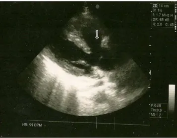
Figure 1. Echocardiogram showing ventricular septal defect and overriding aorta (arrow).
Figure 2. Right(A) and let (B) pulmonary angiography showing pulmonary arterial tree. Distal pulmonary bed is intact but thin.
Figure 3. Angiograms showing pulmonary balloon valvuloplasty. Arrows indicat
-ing the stenotic pulmonary valve (A). Ater balloon valvuloplasty stenosis was
improved (B).
Journal of Clinical and Analytical Medicine | 251
| Journal of Clinical and Analytical Medicine
does not develop enough, acute right heart failure may occur due to volume and pressure overload. So, complete assessment of the pulmonary tree and right ventricular function are very critical [2, 4]. On the other hand, detection of the shunt direc-tion is critical in especially adult patient to detect whether the patient is inoperable or not. If the right to let shunt depends on the severity of the RVOTO, it reverse with ballon dilatation therapy and complete corrective surgery may be performed successfully as our patient.
In conclusion, balloon dilatation is an efective and useful pal-liative procedure to be a bridge therapy for complete correc-tive surgery in both early age symptomatic patients and adult patients with right to let shunt, however the physicians should keep in mind that assessment of the pulmonary tree, RV func-tion and the shunt direcfunc-tion are very critical for success of the procedure.
Competing interests
The authors declare that they have no competing interests.
References
1. Starr JP. Tetralogy of fallot: yesterday and today. World J Surg 2010; 34(4):
658-68.
2. Kreutzer J, Perry SB, Jonas RA, Mayer JE, Castaneda AR, Lock JE. Tetralogy of Fallot with diminutive pulmonary arteries: preoperative pulmonary valve dilation and transcatheter rehabilitation of pulmonary arteries. J Am Coll Cardiol 1996;
27(7): 1741-7.
3. Apitz C, Webb GD, Redington AN. Tetralogy of Fallot. Lancet 2009; 374(9699):
1462-71.
4. Sreeram N, Saleem M, Jackson M, Peart I, McKay R, Arnold R, et al. Results of balloon pulmonary valvuloplasty as a palliative procedure in tetralogy of Fallot. J Am Coll Cardiol 1991; 18(1): 159-65.
5. Kirklin JW, Blackstone EH, Jonas RA, Shimazaki Y, Kirklin JK, Mayer JE, et al. Mor
-phologic and surgical determinants of outcome events ater repair of tetralogy of Fallot and pulmonary stenosis. A two-institution study. J Thorac Cardiovasc Surg 1992; 103(4): 706-23.
6. Lababidi Z, Wu JR. Percutaneous balloon pulmonary valvuloplasty. Am J Cardiol 1983; 52(5): 560-2.
How to cite this article:
Buğan B, Çelik T, Yüksel UÇ, Fırtına S, İyisoy A. Preoperative Pulmonary Valvuloplasty in Tetralogy of Fallot with Right-To-Let Shunt. J Clin Anal Med
2016;7(2): 250-2.
| Journal of Clinical and Analytical Medicine 252