Availableonlineatwww.sciencedirect.com
NeuromuscularDisorders28(2018)791–797
www.elsevier.com/locate/nmd
Case
report
Juvenile
dermatomyositis
forty
years
on:
Case
report
Inês
Rego
de
Figueiredo
a,1,∗,
Sara
Guerreiro
Castro
a,
Vera
Bernardino
a,
José Silva
Nunes
b,
Pedro
Alves
c,
Maria Francisca
Moraes-Fontes
aaUnidadedeDoençasAuto-imunes/Medicina7.2,HospitaldeCurryCabral,CentroHospitalardeLisboaCentral(CHLC),Portugal bServiçodeEndocrinologia,HospitaldeCurryCabral,CHLC,Portugal
cServiçodeRadiologia,HospitaldeDonaEstefânia,CHLC,Portugal
Received13November2017;receivedinrevisedform10April2018;accepted26June2018
Abstract
We presenta case report of a42 year old female, diagnosedat the age of 3 with JuvenileDermatomyositis. The clinicalcourse was severe and refractory to immunosuppressive therapy. Currently, she is mostly affected by severe muscle atrophy, large joint contractures, calcinosis,andalipodystrophyassociatedmetabolicsyndromewithhypertriglyceridemia,insulinresistance,hightotaltestosteroneandhepatic steatosis.ShedevelopedHodgkin´slymphomainthe courseofherdisease.Personalizedtherapeuticchoicesarediscussedasregardsjuvenile dermatomyositiscomplications.
© 2018ElsevierB.V.Allrightsreserved.
Keywords:Juveniledermatomyositis;Lipodystrophy;Calcinosis.
1. Introduction
Juvenile dermatomyositis (JDM) is an autoimmune dis-ease resulting inperivascular inflammation, perifascicular at-rophy and muscle degeneration [1,2] and represents 85% of the idiopathic inflammatory myopathies in childhood [3]. Novel autoantibodies associated to specific clinical pheno-typeshavebeendescribedinthepastdecade.Amongstthese, anti-p155/140– targetedtotranscriptionalintermediaryfactor 1gamma(TIF1-γ )andanti-p140– targetedtonuclearmatrix protein 2 (NXP2), are more likely tooccur in children with calcinosis andcutaneousulceration [4–6].
Fifty years ago, active treatment of childhood onsetJDM resulted in much improved prognosis [7]. Despite progress, standardofcarestillremainsconfinedtountargeted immuno-suppressive therapy with steroids [8]. As recently reviewed [9], the use of steroid sparing agents is recommended, usu-allywithmethotrexate,butalsoazathioprineandcyclosporine,
∗Correspondingauthor.
E-mailaddress:[email protected](I.RegodeFigueiredo).
1 Permanentaddress:UnidadedeDoençasAuto-imunes/Medicina7.2,
Hos-pitaldeCurryCabral,RuadaBeneficência,n° 8,1069-166Lisboa,Portugal.
intravenous immune globulin, tacrolimus, rituximab and cy-clophosphamideinrefractorycases.Cardiacor respiratory in-volvement lower 10–year survival rates, otherwise reported to be over 90% [10,11]. Many children therefore survive to adulthood, but there are scarce descriptions of disease activ-ity, co-morbidities and functional status after prolonged dis-ease. Aiming to contribute to disease knowledge we report an adult patient with JDM exhibiting lipodystrophy, ongo-ing calcinosis, and irreversible joint contractures, in whom a prior diagnosis of malignancy restricts therapeutic choices andwhose management remains anongoing challenge.
2. Case report
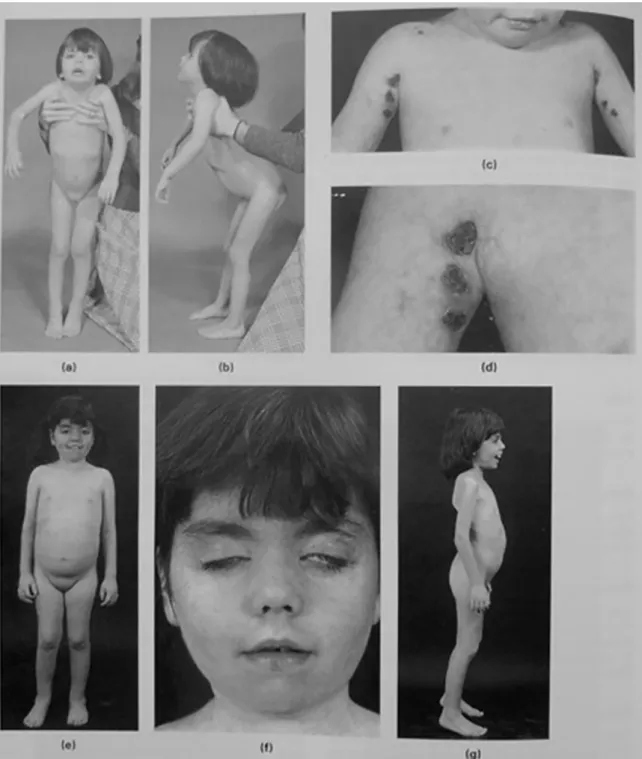
The female patient, currently 42 years-old, was admitted tothe HammersmithHospitalinLondon, atthe ageof 3. At that time, major complaintswere proximal muscle weakness with Gowers sign, unilateral facial nerve palsy and ulcerat-ing skin lesions (Fig. 1a–d). The electromyography of right deltoid, triceps and tibialis anterior muscles revealed short, small amplitude polyphasic potentials, with no spontaneous activity at rest. The diagnosis of JDM was made on the ba-sis of needle biopsy of left quadriceps displaying vacuolar
https://doi.org/10.1016/j.nmd.2018.06.011
Fig.1. Dermatomyositis;vasculiticulcers;recurrentcontractures.Thisgirlwasfirstseenat3yearsofagewithan8-monthhistoryofweakness,skinrash andmisery,withprogressiveflexioncontracturesofthehips(a)andknees(b).Sherespondedwelltosteroidsandwasrehabilitatedwithactivephysiotherapy. Shewasreadmitted fromabroad8monthslaterwith markedvasculiticulcersin theaxillae,which graduallyresolved (c)and(d).She laterhadrecurrent relapsesandremissionswithadjustmentoftherapy.At7yearsofageshestoppedwalkingafterformationofalargehaematomaintherightthigh.Shewas againrehabilitatedwithactivephysiotherapy(e).Therewasstillactiveskinrashandsomefacialweaknesswithlimitedeyeclosure(f).Shewasstillambulant 15monthslaterbuthadconsiderableweaknessandfixedequinus(g).Shehadlostweightandhadverylittlemusclebulk.TextandImagesreproducedwith permissionfromMuscleDisordersinChildhood,2ndEd.ByV.Dubowitz.549pp.,illustrated.Philadelphia:W.B.Saunders,1995.
appearance of several muscle fibers and perifascicular atro-phy(Supplementary Fig.1).
Fromthetimeofdiagnosis,treatmentconsistedofasteroid regimen, initially given alone and subsequently combined with azathioprine, followed by cyclosporine. From the on-set of the illness, periods of immobility were followed by
majorjoint contracturesandover thenext few yearsshewas regularly admitted to hospital for periods of intensive phys-iotherapy which were extremely successful (Fig. 1e–g). Of note, there was no history of consanguinity, episodic fevers, seizures, anemia, respiratory difficulties or learning disabil-ities and she successfully completed a university degree.
I.RegodeFigueiredoetal./NeuromuscularDisorders28(2018)791–797 793 Overall, response to treatment was poor and by the age of
21shewaswheelchairboundandimmunosuppressivetherapy was stopped. Even though the episodes of cutaneous ulcera-tionnolongeroccurred,severalotherfeaturesdevelopedover the next yearsnamely an erythematous pruritic and progres-sively moreindurated facial skin, generalized calcinosis and intermittentdiarrhea,thelatterattributedtointestinalbacterial overgrowth syndrome. Nodular-Sclerosis Classical Hodgkin Lymphoma (stage II, supra-diaphragmatic, category B) was diagnosed at age 31, with full remission after six cycles of chemotherapy consisting of doxorubicin, bleomycin, vin-blastin and dacarbazine. There was no serological evidence of acute Epstein-Barr virus infection (virological status in lymphoma cells was not evaluated). She did not tolerate a cyproterone/ethinylestradiol combination pill dueto dysmen-orrhoea.
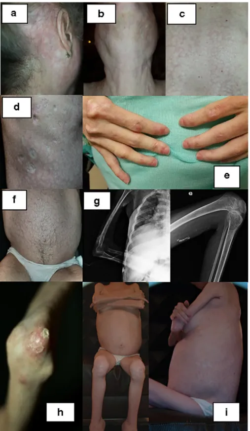
Atthe ageof 38shewasre-evaluated inourAutoimmune DiseasesUnit.Shecomplainedofmenstrualirregularities, hir-sutism and relentless calcinosis. She exhibited low stature, diffuse alopecia, poikiloderma in a photosensitive distribu-tion, symmetric parotid hypertrophy,periungueal telangiecta-sia, livedoreticularis(over thetrunkandanterior thighs)and multipleatrophicscarscorresponding toareasofprevious ul-ceration. There were fixed contractures of the major joints (shoulders, elbows and knees), lumbar scoliosis, generalized muscleatrophyandmultiplefociofcalcinosisalongthe mus-cle fascia of limbs and trunk. The abdomen was dispropor-tionately large and corresponded to a most striking lipodys-trophy with absence of fat in the face and limbs (Fig. 2). There wasno cervicalweakness, nodifficulty inspeaking or in swallowing. She was able to move her upper limbs, pick up small objects andfeedherself.
Laboratory evaluation revealednormalcreatine kinaseand aldolase,increasedtotalserumtestosterone of71ng/dL, (nor-mal values10,8–56,9ng/dL),but normallevels ofserum thy-roid stimulating hormone, prolactin, androstenedione, basal 17-hydroxy progesterone, estradiol, FSH, LH, ACTH and serumcortisol levels. Her fastingplasma glucose(69mg/dL) and Peptide C (6,5ng/mL, normal range 0,9–7.1ng/mL) were normal but insulin was increased to 65,5uUI/mL (normal range 1,9–23uUI/mL). She also had hypertriglyc-eridemia (1860mg/dL, normal range<150mg/dL) and low levels of highdensity lipoproteincholesterol(28mg/dL, nor-malrange<130mg/dL).Livertransaminaseswerenormalbut the gamma-glutamyl transferase was 3 fold elevated; ANA testingwasintermittentlypositive,withalowtitle(1/160)but nospecificpositivitywasfound;therewasnolung parenchy-mal change in the chest radiograph which displayed multi-ple calcifications in the subcutaneous tissues; the echocar-diogram was normal; no structural abnormality or pressure changesweredetectedbyDopplerechocardiographyand nail-fold video-capilaroscopy revealed a late-scleroderma pattern (Supplementary Fig. 2). Magnetic resonance imaging (MRI) showed diffuse muscle fatty atrophy (Grade III–IV) of mas-seters,shouldergirdle,deltoids,paravertebralthoracic,gluteal and thigh groups. The only relatively preserved muscles lo-calized to the neck, thighs (adductor magnus and vastus
in-termedius)andlegs(tibialisanterior andposteriorandtoe ex-tensors).There was nosign of muscle inflammation onfluid sensitivesequences (STIR andT2-weighted images). Parotid glands were heterogeneous with fatty infiltrates and diffuse calcifications.There was a marked increase in posterior cer-vical,axillary,mediastinal andintra-peritoneal fatdeposition, also asymmetrically localizedto the anterior and lateral por-tion of both thighs (Fig. 3). Small areas of subcutaneous oedema were suggestive of panniculitis in thighs and legs. Hepatomegalywasnoted withsplenomegaly,normal kidneys, uterusand ovaries. There was no lymphadenopathy.No mu-tations were found in the genes LMNA, ZMPSTE24, PTRF,
CAV-1, AGPAT2, BSCL2, PPARG, INSR, PLIN1, CIDEC,
PIK3R1, NSMCE2, POC1A, PCYT1A, POLD1 or PSMB8.
Soon after the genetic tests were performed, the patient´s serumtestedpositiveforanti-transcriptionalintermediary fac-tor gamma protein antibody (TIF1-gamma) (Euroimmun®
-Lübeck,Euroline scansoftware, patient intensity 15,control 99),supporting the original diagnosis. There was no reactiv-ityagainst the followingantigens: Mi-2(nucleosome remod-eling deacetyalse complex), MDA (melanoma-differentiation associated gene 5), SRP (54kDa, signal recognition parti-cle),NXP-2,SAE(small-ubiquitin-likemodifieractivating en-zyme), Ku, PM-Scl (75 and 100kDa), Jo-1 (histidyl-tRNA synthetase),PL-7(threonyl-tRNAsynthetase), PL-12 (alanyl-tRNA synthetase), EJ protein (glycyl-tRNA synthetase), OJ (isoleucyl-tRNAsynthetase) andRo-52KD.
Following re-assessment, weekly s.c. methotrexate (up to 20mg/week) and quarterly i.v. Pamidronate failed to have a positive impact on the rate of calcinosis deposits and range of metabolic abnormalities. The latter was associated with an episode of renal colic and both were discontin-ued after two years. Since then, painful calcium deposits have been removed surgically on three occasions. She has not tolerated metformin due to side-effects and is poorly compliant with fenofibrate. Decreased gastrointestinal ab-sorption may also contribute to a poor response to ther-apy and serum triglyceride concentrations are usually 5–6 foldelevated.Shereportsbeneficialanti-diarrhealeffectswith VSL#3®, a commercially available probiotic, taken as
re-quired. Apart from feeling that the skin around her face is tighterthere hasbeen nochange inherclinical features over thepastfouryears.Sheispartiallydependent forherhygiene needs whichrestrict her social lifebut is otherwise indepen-dent in her daily activities with an electric wheelchair and adaptedmotorizedvehicle.Sheundertakesphysiotherapy ses-sionsconsistingof passive limb mobilizationthreetimes per week.
3. Discussion
JDM is a very rare condition with an incidence of 2–4 cases per million per year, mostly affecting females. In so much as the peak incidence occurs between the ages of 5– 10,upto25%of patientsexperience diseaseonsetbeforethe age of 4 [12], similarly to our patient. The disease may re-mit,relapse or followachroniccontinuousform [13].When
Fig.2. Clinicalfeaturesofthepatientasanadult:poikilodermaandskintightness(a);parotidhypertrophy(b); livedoreticularisoverdorsum(c);atrophic scars(d); periungueal telangiectasia (e);hirsutism (f); calcinosis in fascialplanes and jointcontractures (g); cutaneouscalcinosis (h); lipodystrophy with abdominalredistributionoffatandmuscleatrophy(i).
I.RegodeFigueiredoetal./NeuromuscularDisorders28(2018)791–797 795
Fig.3. MRIT1-weightedaxialimagesfromtheabdomen(a)andthigh(b),fromahealthysubject(1)andfromthepatient(2)wherethemassiveincrease infatismainlyintraperitonealbutalsoassymmetricallydistributedthroughoutthethighcircunference.Thereismarked(GradeIII–IV)diffusethighmuscle atrophy.
compared with adult auto-antibody reactivities, anti-TIF1-γ positivechildrenaremorefrequentlyaffectedbymuscle atro-phy,contractures,calcinosisandcutaneousvasculitis[14].Of note,ourpatientreceivedappropriatetherapy,namelysteroids andcyclosporinefromdiseaseonset.Physiotherapytreatments were intense but intermittent, with prolonged periods of im-mobility. Recurrent contractures developed from early on in the course of disease.
Several complex complications developed as disease progressed. Lymphoma developed in adulthood, many years after disease onset. In a recent literature review lymphoma was very rarely diagnosed in juvenile dermato/polymyositis, occurred only after the disease was well established and all the patients suffered from the non-Hodgkin´s lymphoma type [15]. Hodgkin lymphoma (HL) has been described in non-immunosuppressed individuals but HL-like conditions have also been described in the spectrum of post-transplant lymphoproliferative disorders [16]. After a ten year-lapse, a relationship to prior immunosuppressive therapy remains an elusive possibility inourpatient.
Albeit not confirmed, the initial lack of an auto-antibody marker, the presence of marked lipodystrophy,possible pan-niculitisandlackofmuscleinflammationinthebiopsy speci-menreviewedraisedthe suspicionof analternative diagnosis butnoevidenceofanauto-inflammatorydisorderor amuscle dystrophywasfound.Whilethelipodystrophywassuggestive of Chronic Atypical Neutrophilic Dermatosis with Lipodys-trophy and Elevated temperatures (CANDLE) [17,18], lack of periodic fevers and systemic inflammation made this less
likely,whereas the presence of anti-TIF1-γ , muscle atrophy, joint contractures, calcinosis and nailfold changes clinched the diagnosis of JDM.
JDM occurs in genetically susceptible patients [19–22]. Its association with lipodystrophy (deposition of fat in ec-topiclocationssuch as insalivaryglands,the peritoneal cav-ity, liver and muscle and absence of fat deposits in other sites) [23,24] and lipodystrophy-associated metabolic abnor-malities(hypertriglyceridemia, insulinresistance,high testos-terone, hypertrichosis and probable fatty liver disease) has beendescribedwithseverecalcinosis,jointcontractures, mus-cleatrophy,chroniccontinuousillnesscourse,facialerythema andanti-TIF1-γ positivity [25,26],allof whichwere present inour patient.
Follow up of JDM patients into adulthood has demon-stratedthe overall systemic nature of the disease andits im-pactonseveralorgansonthe longterm.Even thoughstudies onaNorway cohort followedover 16,8years (2–38,1years) has showed JDM patients often maintain active disease in thelongterm,our patientmostlysuffersfromcumulative or-gandamage,whosemainpredictorisdiseaseactivity atonset [27].
Because of the loss of adipose tissue, levels of the adipocyte-secretedhormone leptinmaybe lowand leptin re-placement couldbe atherapeutic option inourpatient, in an attempttoovercomemorphologicalandmetabolic abnormali-ties.Howeverlymphomaafterleptintherapyforpatientswith acquired lipodystrophy has been described [28]. In addition, nosingle drug or associations of drugs has been effective in
thetreatmentofcalcinosisinJDM[29].Upregulationof type I interferon pathway, described as a biomarker of JDM dis-easeactivity[30]aswellasaneffectormoleculeinCANDLE [31], may bea driver of disease pathogenesis. It istempting to speculate that antagonizing type I interferon could have afavorable therapeutic impact in our patient. Overall, a his-toryof lymphoma,longstanding immune-mediateddisease, a potentiallyvulnerableimmunesystemandthedegreeof irre-versibleorgan damage lead us totake aconservative“do no harm” approach.
In conclusion we present a patient with a diagnosis of JDMattheageof 3years, withapartial responsetotherapy and several co-morbidities. Marked lipodystrophy and little inflammation in the original muscle biopsy specimen raised suspicion of an overlap with muscle dystrophy which was notconfirmed. Our key message highlightsthe extraordinary advances introduced inthe 1980´s, reducing patient mortality and side-effects of extremely high dosages of steroids. De-spite the presence of disability our patientsurvived a severe childhooddisease andleadsa meaningfullife.
Acknowledgments
Victor Dubowitzfordiagnosis andclinicalcarefrom diag-nosisuntiladulthood,clinicalinformation,imagesand discus-sions, Vanda Lúcio Bernardes for physiotherapy, Francesco Muntoni, Ana Grilo, Ana Catarina Rodrigues, Manuel Vaz Riscado,NunoRisoandAntónioPanarraforclinicalcare, An-tónio Caetano for imaging, Caroline Sewry for histopathol-ogy review, Jocelyne Demengeot, Jocelyne Magré, Corinne Vigouroux,Olivier Lascols, Pascale Richard, Annachiara De Sandre-GiovannoliandNunoCostafor genetictesting,Maria Céu Santos for auto-antibody profiling and W. B. Saunders (nowElsevier)publishers forpermissiontouseimagesofthe patientas achild [1].
Supplementarymaterials
Supplementary material associatedwiththisarticle canbe found, in the online version, at doi:10.1016/j.nmd.2018.06. 011.
References
[1] Dubowitz V. Muscle disorders in childhood.2nd ed. W.B. Saunders (nowElsevier);1995.
[2] FeldmanBM,RiderLG,ReedAM,PachmanLM.Juvenile dermato-myositisand other idiopathicinflammatory myopathies of childhood. Lancet2008;371(9631):2201–12.
[3] McCannLJ,JugginsAD,MaillardSM,WedderburnLR,DavidsonJE, Murray KJ, et al. The Juvenile Dermatomyositis National Registry andRepository(UKandIreland)–clinicalcharacteristicsofchildren re-cruitedwithinthefirst5yr.Rheumatology2006;45(10):1255–60.
[4] BetteridgeZE,GunawardenaH,McHughNJ.Novelautoantibodiesand clinicalphenotypes inadultand juvenilemyositis. ArthritisRes Ther. 2011;13(2):209.
[5] GunawardenaH,BetteridgeZE,McHughNJ.Myositis-specific autoan-tibodies:theirclinicalandpathogenicsignificanceindiseaseexpression. Rheumatology2009;48(6):607–12.
[6]Gunawardena H,WedderburnLR,ChinoyH,BetteridgeZE,NorthJ, Ollier WE, et al. Autoantibodies to a 140-kd protein in juve-nile dermatomyositis are associated with calcinosis. ArthritisRheum 2009;60(6):1807–14.
[7]MillerG,Heckmatt JZ, DubowitzV. Drugtreatmentofjuvenile der-matomyositis.ArchDisChild1983;58(6):445–50.
[8]Dubowitz V. Treatment of dermatomyositis in childhood. Arch Dis Child1976;51(7):494–500.
[9]MalikA,HayatG,KaliaJS,GuzmanMA.IdiopathicInflammatory My-opathies:ClinicalApproachandManagement.FrontNeurol.2016;7:64.
[10]DankoK,PonyiA,ConstantinT,Borgulya G,SzegediG.Long-term survival of patients with idiopathicinflammatory myopathies accord-ing to clinical features: a longitudinal study of 162 cases. Medicine 2004;83(1):35–42.
[11]PeloroTM,MillerOF,HahnTF3rd,NewmanED.Juvenile dermato-myositis: aretrospective review ofa 30-yearexperience.JAm Acad Dermatol2001;45(1):28–34.
[12]Symmons DP, Sills JA, Davis SM. The incidence of juvenile der-matomyositis: results from a nation-wide study. Br J Rheumatol 1995;34(8):732–6.
[13]Constantin T, Ponyi A, Orbán I, Molnár K, Dérfalvi B, Dicso F, et al. National registry of patients with juvenile idiopathic inflam-matory myopathies in Hungary – clinical characteristics and disease course of 44 patients with juvenile dermatomyositis. Autoimmunity 2006;39(3):223–32.
[14]EspadaG,MaldonadoCoccoJA,FertigN,OddisCV.Clinicaland sero-logiccharacterization ofan Argentinepediatricmyositiscohort: iden-tification of a novel autoantibody (anti-MJ) to a 142-kDa protein. J Rheumatol2009;36(11):2547–51.
[15]Stubgen JP. Juvenile dermatomyositis/polymyositis and lymphoma. J NeurolSci2017;377:19–24.
[16]CarboneA,SpinaM,GloghiniA,TirelliU.ClassicalHodgkin’s lym-phomaarisingindifferenthost’s conditions:pathobiologyparameters, therapeuticoptions,andoutcome.AmJHematol2011;86(2):170–9.
[17]Torrelo A, Patel S, Colmenero I, Gurbindo D, Lendinez F, Hernan-dezA,etal.Chronicatypicalneutrophilicdermatosiswithlipodystrophy andelevatedtemperature(CANDLE)syndrome.JAmAcadDermatol 2010;62(3):489–95.
[18]Liu Y, Ramot Y, Torrelo A, Paller AS, Si N, Babay S, et al. Mu-tationsin proteasomesubunitbeta type8 causechronicatypical neu-trophilicdermatosiswith lipodystrophyand elevatedtemperature with evidence of genetic and phenotypic heterogeneity. Arthritis Rheum 2012;64(3):895–907.
[19]ReedAM,PachmanL,OberC.Moleculargeneticstudiesofmajor his-tocompatibilitycomplexgenesinchildrenwithjuvenile dermatomyosi-tis:increased riskassociated withHLA-DQA1 ∗0501. HumImmunol 1991;32(4):235–40.
[20]Mamyrova G, O’Hanlon TP, Monroe JB, Carrick DM, Malley JD, AdamsS,etal.Immunogeneticriskandprotectivefactorsforjuvenile dermatomyositisinCaucasians.ArthritisRheum2006;54(12):3979–87.
[21]PachmanLM,Liotta-DavisMR,HongDK,KinsellaTR,MendezEP, Kinder JM, et al. TNFalpha-308A allele in juvenile dermatomyosi-tis:association withincreased productionoftumornecrosisfactor al-pha, disease duration, and pathologic calcifications. Arthritis Rheum 2000;43(10):2368–77.
[22]RiderLG,ArtlettCM,FosterCB,AhmedA,NeemanT,ChanockSJ, etal.Polymorphismsin theIL-1 receptorantagonist geneVNTRare possible risk factors forjuvenile idiopathic inflammatory myopathies. ClinExpImmunol2000;121(1):47–52.
[23]QuecedoE,FebrerI,SerranoG,Martinez-AparicioA,AliagaA.Partial lipodystrophy associatedwith juvenile dermatomyositis:reportoftwo cases.PediatrDermatol1996;13(6):477–82.
[24]KavanaghGM,ColacoCB,KennedyCT.Juveniledermatomyositis as-sociated with partiallipoatrophy. J AmAcad Dermatol 1993;28(2 Pt 2):348–51.
[25]MukamelM,HorevG,MimouniM.Newinsightintocalcinosisof ju-veniledermatomyositis:astudyofcompositionandtreatment.JPediatr 2001;138(5):763–6.
I.RegodeFigueiredoetal./NeuromuscularDisorders28(2018)791–797 797 [26]Bingham A, Mamyrova G, Rother KI, Oral E, Cochran E,
Premku-marA,etal.Predictorsofacquiredlipodystrophyinjuvenile-onset der-matomyositisandagradientofseverity.Medicine2008;87(2):70–86.
[27]SannerH,SjaastadI,Flatø B.Diseaseactivityandprognosticfactorsin juveniledermatomyositis:along-termfollow-upstudyapplyingthe Pae-diatricRheumatologyInternationalTrialsOrganizationcriteriafor inac-tivediseaseandthemyositisdiseaseactivityassessmenttool. Rheuma-tology2014;53(9):1578–85.
[28]Brown RJ, Chan JL,Jaffe ES,Cochran E, DePaoli AM, GautierJF, et al. Lymphoma in acquired generalized lipodystrophy. Leuk Lym-phoma2016;57(1):45–50.
[29]FrediM,BartoliF,CavazzanaI,CeribelliA,CarabelleseN,TincaniA, etal.Calcinosisin poly-dermatomyositis: clinicaland laboratory pre-dictorsandtreatmentoptions.ClinExpRheumatol2017;35(2):303–8.
[30]BaechlerEC,BilgicH,ReedAM.TypeIinterferonpathway inadult andjuveniledermatomyositis.ArthritisResTher2011;13(6):249.
[31]Brehm A, Liu Y, Sheikh A, Marrero B, Omoyinmi E, Zhou Q, etal.Additiveloss-of-functionproteasomesubunitmutationsin CAN-DLE/PRAAS patients promote type I IFN production. J Clin Invest 2015;125(11):4196–211.