www.reumatologia.com.br
REVISTA BRASILEIRA DE
REUMATOLOGIA
Original article
Different aspects of magnetic resonance imaging of muscles
between dermatomyositis and polymyositis
Soia Silveira de Castro Miranda
a, Daniel Alvarenga
b, João Carlos Rodrigues
b,
Samuel Katsuyuki Shinjo
a,c,*
a Rheumatology Service, Hospital das Clínicas, Medicine School, Universidade de São Paulo, São Paulo, SP, Brazil
b Department of Radiology, Orthopedia and Traumatology Institute, Hospital das Clínicas, Medicine School, Universidade de São Paulo, São
Paulo, SP, Brazil
c Medicine School, Universidade de São Paulo, São Paulo, SP, Brazil
a r t i c l e i n f o
Article history:
Received 28 January 2014 Accepted 7 April 2014
Keywords:
Dermatomyositis Muscle diseases Polymyositis Magnetic resonance
a b s t r a c t
Introduction: Although dermatomyositis (DM) and polymyositis (PM) share many clinical features in common, they have distinct pathophysiological and histological features. It is possible that these distinctions relect also macroscopically, for example, in muscle altera-tions seen in magnetic resonance images (MRI).
Objectives: To compare simultaneously the MRI of various muscle compartments of the thighs of adult DM and PM.
Materials: The present study is a cross-sectional that included, between 2010 and 2013, 11 newly diagnosed DM and 11 PM patients (Bohan and Peter’s criteria, 1975), with clinical and laboratory activity. They were valued at RM thighs, T1 and T2 with fat suppression, 1.5 T MRI scanner sequences.
Results: The mean age at the time of MRI, the time between onset of symptoms and the realization of the MRI distribution of sex and drug therapy were comparable between the two groups (p>0.050). Concerning the MRI, muscle edema was signiicantly found in DM, and mainly in the proximal region of the muscles. The area of fat replacement was found predominantly in PM. The partial fat replacement area occurred mainly in the medial and distal region, whereas the total fat replacement area occurred mainly in the distal muscles. There was no area of muscle ibrosis.
Conclusions: DM and PM have different characteristics on MRI muscles, alike pathophysiolo-gical and histolopathophysiolo-gical distinctions.
© 2014 Sociedade Brasileira de Reumatologia. Published by Elsevier Editora Ltda. All rights reserved.
* Corresponding author.
E-mail: [email protected] (S.K. Shinjo).
Aspectos distintos de ressonância magnética de músculos entre dermatomiosite e polimiosite
Palavras-chave:
Dermatomiosite Doença muscular Polimiosite
Ressonância magnética
r e s u m o
Introdução: Embora a dermatomiosite (DM) e a polimiosite (PM) compartilhem diversos as-pectos clínicos em comum, cada uma delas apresenta características isiopatológicas e his-tológicas próprias. É possível que estas diferenças também se relitam macroscopicamente, como, por exemplo, em imagens musculares vistas em ressonância magnética (RM).
Objetivos: Comparar simultaneamente a RM de diversos compartimentos musculares das coxas de pacientes com DM e PM adultos.
Materiais: Estudo transversal, em que foram avaliadas, entre o período de 2010 a 2013, as imagens de RM das coxas realizadas em aparelho de 1,5 Tesla (T) com sequências pon-deradas em T1 e T2 com supressão de gordura, para rastreamento, de 11 DM e 11 PM (Bohan e Peter, 1975) recém-diagnosticados, em atividade clínica e laboratorial.
Resultados: A média de idade na ocasião da RM, o tempo entre o início de sintomas e a realização das RM, a distribuição de sexos e a terapia medicamentosa foram comparáveis entre os dois grupos (p>0,050). Em termos de RM, edema muscular foi encontrado signii-cantemente em DM, e principalmente na região proximal dos músculos. A área de lipossub-stituição dos músculos foi encontrada predominantemente em PM. Essa lipossublipossub-stituição, quando de uma forma parcial, ocorreu principalmente nos terços médio e distal, enquanto que a forma total transcorreu apenas no terço distal dos músculos. Não houve nenhuma área de ibrose muscular.
Conclusões: A DM e a PM apresentam características distintas entre si em RM de músculos, a exemplo de distinções isiopatológicas e histológicas.
© 2014 Sociedade Brasileira de Reumatologia. Publicado por Elsevier Editora Ltda. Todos os direitos reservados.
Introduction
Dermatomyositis (DM) and polymyositis (PM) are part of a group of systemic autoimmune diseases characterized by symmetric and progressive proximal muscle weakness of limbs. Moreover, extramuscular manifestations (i.e. articular, heart, lung and gastrointestinal tract involvement) may oc-cur.1,2 In the case of DM, typical skin changes still occur, such
as heliotrope and/or Gottron’s papules.
Although DM and PM share many similar clinical and labo-ratory features, each of these conditions also exhibit distinct epidemiological, pathophysiological and histological charac-teristics. Thus, from the histological and physiopathological standpoint, in PM there is a focal iniltrate of CD8 (+) lympho-cytes and macrophages in the muscle ibers, which, in turn, express high levels of MHC class I antigens and release per-forin granules,3 resulting in lysis and necrosis of the muscle
ibers themselves,4 as well as fat in replacement areas and
tissue ibrosis. In the case of DM, there are several features that suggest an important role of B cells in the pathogenesis of the disease, such as the presence of autoantibodies, im-mune complex deposition in the dermo-epidermal junction in skin lesions, the presence of B cells in sore muscles,5,6 and
in perivascular areas.7,8 In addition to this, the deposition of
complement and immunoglobulin in the perifascicular endo-thelium can lead to muscle ischemia and atrophy, showing the importance of humoral immunity.9
It is plausible that these differences observed between DM and PM are also macroscopically relected, such as in muscle
images obtained from studies of magnetic resonance imaging (MRI), although to date there are no studies in the literature demonstrating these possible differences.
MRI has been used in idiopathic inlammatory myopathies as an additional instrument to assess disease activity, thera-peutic monitoring, and prognosis of disease, as well as a guide to the most likely location to ind positive areas of inlamma-tory iniltrate in a muscle biopsy.10-23 However, most of these
studies are based on juvenile DM.10,11,13,14,17,18,22 On the other
hand, there is paucity of MRI studies on adult DM patients, as well as on PM ones,12,16,23 and this served as motivation for us
to conduct this study.
Patients and methods
This was a cross-sectional study that, during the period from 2010 to 2013, assessed 11 DM patients and 11 PM patients deined by Bohan and Peter criteriaand recently diagnosed as with clinical and laboratory activity.24,25 Patients with a
di-agnosis of amyopathic DM, myopathies associated with ma-lignancy or other types of collagenosis were not included, as well as patients under the age of 18 years.
The present work is a sequel of a study previously ap-proved by the local Ethics Committee [HC 0039/10].
differential diagnoses were dismissed; and, prior to muscle biopsy for diagnostic purposes, the patients underwent a MRI of the muscles of the thighs. Searching a database of previously standardized electronic data (electronic medical record), the following data were collected: current age; eth-nicity; gender; time between onset of symptoms and MRI; limb muscle strength (grade 0: absence of muscle contrac-tion; grade I: signs of slight contractility; grade II: normal range of motion, but without overcome the action of grav-ity; grade III: normal range of motion against gravgrav-ity; grade IV: full mobility against gravity, with a certain degree of sistance; and grade V: complete mobility against strong re-sistance and against gravity);26 serum levels of muscle
en-zymes [creatine phosphokinase (normal range: 24-173 U/L) and aldolase (normal range: 1.0-7.5 U/L) determined by an automated kinetic method]; autoantibodies against cellular components, determined by indirect immunoluorescence, using Hep-2 cells as substrate; pre-MRI medicamentous his-tory (corticosteroids and/or immunosuppressants).
MRI was performed by fast spin echo technique, obtain-ing T1- and T2-weighted sequences with fat suppression, in multiplanar acquisitions, using a Philips 1.5 T unit from the Department of Radiology at our institution. Fifteen muscles were evaluated: sartorius, vastus lateralis, vastus interme-dius, vastus medialis, rectus femoris, tensor fasciae latae, adductor longus, adductor brevis, adductor magnus, pectin-eus, gracilis, gluteus maximus, semitendinosus, semimem-branosus and biceps femoris. The following parameters were evaluated for each muscle in its proximal, middle and distal thirds: presence or absence of an edema area, ibrosis, and fat replacement (partial or total). Furthermore, we evaluated the overall appearance of muscles, as being normal or hypo/ atrophic. The images were evaluated by two radiologists with proven experience in muscle MRI analysis. These profession-als worked independently and were unaware of the clinical cases.
Statistical analysis
Our data were expressed as mean ± standard deviation (SD), median (interquartils) or percentage (%), being assessed for normal distribution by Kolmogorov-Smirnov analysis. The Student t test and Mann-Whitney test were used for the anal-ysis of continuous data. The Fisher exact test was used to ana-lyze categorical data. These calculations were performed with the computer program STATA version 7.0 (STATA, College Sta-tion, TX, USA). P-values <0.050 were considered statistically signiicant.
Results
The means of age at the time of MRI, gender distribution, and ethnicity, as well as the time interval between the onset of symptoms and MRI examination of thighs, were compa-rable between DM and PM patients (p>0.050), as shown in Table 1. The intensities of muscle weakness and levels of muscle enzymes were also similar in both groups (p>0.050). In terms of drug therapy, 100.0% of DM patients and 81.8% of PM patients were already on corticosteroids and/or
immu-nosuppressants – eight patients with azathioprine (2-3 mg/ kg/day), four with methotrexate (15-25 mg/week), one with cyclosporine (3 mg/kg/day), one with lelunomide (20 mg/ day) and/or three with intravenous human immunoglobulin (2 g/kg/day), at the time of the clinical picture investigation and of MRI. Nevertheless, the cumulative dose of prednisone and the previous use of immunosuppressive drugs and of pulse therapy with methylprednisolone were similar in both groups (p>0.050).
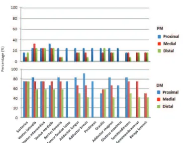
In MRI, a signiicant muscle edema was found in DM pa-tients (41.7 to 91.7% of muscle compartments) compared with PM patients (from 8.3% to 33.3% of the muscle compartments) (p<0.050). Moreover, in 50% of the examined muscles in DM patients, the edema decreases in the proximal-distal direc-tion, as shown in Fig. 1.
The area of fat replacement of muscles was predominantly observed in different muscle compartments in PM patients, when compared to DM patients. When partial, this fat re-placement affected mainly the middle and distal thirds of the muscles of PM patients (0% to 41.7% of the muscles; Fig. 2), while a total fat replacement was observed only in the distal third of muscles of PM patients (0% to 16.7% of the muscles; Fig. 3).
Table 1 – Demographic, clinical, laboratory and drug therapy characteristics of patients with dermatomyositis and polymyositis
Characteristics DM (n=11) PM (n=11) p
Age (years) 50.9±11.4 49.9±16.2 0.868 Female gender 9 (81.8) 10 (90.9) 1.000 Caucasian 9 (81.8) 8 (72.7) 1.000 Time: MRI – symptoms
(months)
16.7±22.2 29.0±20.3 0.191
Muscle strength Upper limbs
Grade V 1 (9.1) 5 (45.5) 0.155
Grade IV 5 (45.5) 4 (36.4) 1.000 Grade III 4 (36.4) 2 (18.2) 0.635
Grade II 1 (9.1) 0 1.000
Lower limbs
Grade V 0 1 (9.1) 1.000
Grade IV 5 (45.5) 7 (63.6) 0.670 Grade III 5 (45.5) 2 (18.2) 0.361
Grade II 1 (9.1) 1 (9.1) 1.000
Muscle enzymes Creatine
phosphokinase (U/L)
1340 (158-3489) 870 (207-2519) 0.768
Aldolase (U/L) 11.2 (7.4-29.0) 9.7 (6.6-20.6) 0.577 Antinuclear factor 7 (63.6) 5 (45.5) 0.670 Methylprednisolone
(pulse therapy)
3 (27.3) 2 (18.2) 1.000
Prednisone 11 (100.0) 9 (81.8) 0.476 Cumulative dosage
(mg), <1 m
1200 (180-2100) 300 (300-1500) 0.290
Cumulative dosage (g), <3 m
3.0±3.2 1.4±1.1 0.190
Immunosuppressants 3 (27.3) 6 (54.5) 0.387
Data are expressed as mean ± standard deviation, median (interquartils), or percentage (%).
DM, dermatomyositis; PM, polymyositis; MRI, magnetic resonance imaging.
In the present study, no area of muscle i brosis was identi-i ed.
Discussion
The present study showed that DM and PM, although sharing many clinical and laboratory aspects in common, have differ-ent characteristics from the point of view of muscle MRI.
Of the few studies that addressed the use of MRI in idio-pathic inl ammatory myopathies,10-23 most are restricted to
ju-venile DM.10,11,13,14,17,18,22 In the case of PM and DM in adults,12,16,23
the studies available in the literature are scarce.
Kaufman et al.23 globally analyzed i ve PM patients and
eight DM patients with a broad age range (12-77 years). The authors made no mention of the disease duration or type of
drug therapy received by their patients prior to MRI,23 and
compared the MRI i ndings according to disease activity. We chose to evaluate and compare concomitantly adult DM and PM patients in various muscle compartments of their thighs at an early stage of the disease. Furthermore, our patients had similar demographic, clinical, laboratory, and therapeutic characteristics to each other, allowing a meaningful compari-son of MRI i ndings.
Tomasová et al.16 evaluated nine PM patients and 20 DM
patients, with a mean duration of disease of 2.3 years. All pa-tients exhibited clinically and biochemically active disease, and 63.2% of these cases had never received drug therapy. These authors showed a correlation between the intensity of muscle edema observed by MRI with the degree of disease ac-tivity and the posiac-tivity for an inl ammatory ini ltrate found in muscle biopsies guided by MRI.16 However, these authors
did not make comparisons regarding possible changes on MRI in PM and DM.
Reimers et al.12 evaluated MRI studies of various muscle
compartments of lower limbs of 58 patients aged 21-83 years-old. However, in addition to the 14 DM patients and 25 PM patients, eight patients with granulomatous myositis and 11 patients with inclusion body myositis were included. More-over, Reimers et al. did not mention the type of drug treat-ment received by the participants in the study, who were arbitrarily classii ed as acute (less than one year disease), or chronic (when there is signii cant evidence of fat replacement and of muscle i brosis in muscle MRI) patients. Despite these limitations, the authors observed that the presence of areas of muscle edema and of fat replacement were respectively more and less frequent in patients with acute DM, when compared to the other diseases included in the analysis.
Our results showed that the area of muscle edema was present mainly in DM patients, when compared with PM patients. This may be a result of the previous use of cortico-steroids, which may inl uence the intensity and presence of muscle edema.16 However, we observed that both the use of
corticosteroids (cumulative dose) and immunosuppressants Fig. 3 – Distribution of total fat replacement area of different muscle compartments of the thigh by MRI in patients with dermatomyositis and polymyositis
Fig. 1 – Distribution of edema area in different muscle compartments of the thigh by MRI in patients with dermatomyositis and polymyositis
Fig. 2 – Distribution of partial fat replacement area of different muscle compartments of the thigh by MRI in patients with dermatomyositis and polymyositis
P e rc e n tag e ( % ) Sart oriu s Vast us l ater alis Vast us int
erm ediu s Vast us m edia lis Rect us f emor is Tenso r fasc
iae l atae Addu ctor long us Addu ctor bre vis Peci neus Gra cilis Addu ctor mag nus Glu teus m
axim us Sem itend inosu s Sem imem bran osu s Bice ps f emor is P e rc e n tag e ( % ) Sart oriu s Vast us l ater alis Vast us i nter med ius Vast us m edia lis Rect us f emor is Tenso r fasc
iae l atae Addu ctor long us Addu ctor bre vis Peci neus Gra cilis Addu ctor mag nus
Glute us m axim us Sem itend inosu s Sem imem bran osu s Bice ps f emor is P e rc e n tag e ( % ) Sart oriu s Vast us l ater alis Vast us int
erm ediu s Vast us m edia lis Rect us f emor is Tenso r fasc
iae l atae Addu ctor long us Addu ctor bre vis Peci neus Gra cilis Addu ctor mag nus Glu teus m
were similar in both DM and PM groups of patients. Moreover, other parameters – such as the time of drug treatment, and the time between the MRI and the onset of muscle weakness symptoms were similar in both groups, which shows that the presence of muscle edema was an inherent characteristic of patients with active and newly diagnosed DM.
In addition, the fact that the swelling is mainly located on the proximal thigh, compared to the distal region, is compat-ible with the clinical indings, in those there is objective evi-dence of increased muscle weakness in so far as we get closer to the waists.
The prevalence of presence of fat replacement areas was low in our study, probably because we evaluated only cases of newly diagnosed DM and PM. Partially impaired muscle areas (partial fat replacement) were present in most muscle com-partments, especially in PM. On the other hand, signiicantly compromised areas (total fat replacement) were only present in the muscles of PM patients, mainly in the long muscles, in the distal area, and in the posterior muscle group of thighs. These indings can be explained by the fact that these mus-cles are subjected to less traction and mechanical stimulation during the ambulation.
Previous studies have shown the beneit of physical ex-ercise in patients with idiopathic inlammatory myopathies. Resistance exercises, for example, can reduce the expression of genes involved in the inlammatory process and ibrosis in the muscle tissue.27
In the present study, no areas of muscle ibrosis were ob-served, both in DM as in PM patients. In general, the presence of areas of muscle ibrosis, as well as those of fat replacement, are commonly observed in patients with a diagnosis of chron-ic myositis unresponsive to medchron-ical therapy.28
Our study has its limitations: it has a cross-sectional design, and therefore the correlation between muscle MRI indings and clinical and laboratory manifestations of pa-tients were not evaluated. Second, ours was a small sample. Third, we evaluated only the muscle groups of the thighs and therefore did not have enough substrate to generalize this difference in muscle MRI indings to other muscle com-partments of DM and PM patients. Fourth, no correlation be-tween the muscle MRI data with possible indings of muscle biopsy was performed. And last but not least, not all patients were naïf as to drug therapy at the time of MRI, which may under or overestimate the muscle MRI indings of the pa-tients analyzed.
In summary, our results showed that DM and PM pa-tients have different characteristics from the point of view of muscle MRI, just like physiopathological and histological indings.
Conlicts of interest
The authors declare no conlicts of interest.
R E F E R E N C E S
1. Callen JP. Dermatomyositis. In: Callen JP. 2 ed. Dermatological signs of internal disease. Saunders. 1995.
2. Fathi M, Lundberg IE. Interstitial lung disease in polymyositis and dermatomyositis. Curr Opin Rheumatol. 2005;17:701-6. 3. Nyberg P, Wikman AL, Nennesmo I, Lundberg I. Increased
expression of interleukin 1 alpha and MHC Class I in muscle tissue of patients with chronic, inactive polymyositis and dermatomyositis. J Rheumatol. 2000;27:940-8.
4. Goebels N, Michaelis D, Engelhardt M, Huber S, Bender A, Pongratz D et al. Differential expression of perforin in muscle-iniltrating T cells in polymyositis and dermatomyositis. J Clin Invest. 1996;97:2905-10.
5. Emslie-Smith AM, Engel AG. Microvascular changes in early and advanced dermatomyositis: a quantitative study. Ann Neurol. 1990;27:343-56.
6. Engel AG, Arahata K. Mononuclear cells in myopathies: quantitation of functionally distinct subsets, recognition of antigen speciic cell-mediated cytotoxicity in some diseases, and implications for the pathogenesis of the different inlammatory myopathies. Hum Pathol. 1986;17:704-21. 7. Botet JC, Grau JM, Casademont J, Urbano-Marquez A, Rozman
C. Characterization of mononuclear exudates in idiopathic inlammatory myopathies. Virchows Arch Pathol Anat Histopathol. 1988;412:371-4.
8. Dalakas MC. The future prospects in the classiication, diagnosis and therapies of inlammatory myopathies: a view to the future from the “bench-to-bedside”. J Neurol. 2004;251:651-7.
9. Noss EH, Hausner-Sypek DL, Weinblatt M. Rituximab as therapy for refractory polymyositis and dermatomyositis. J Rheumatol. 2006;33:1021-6.
10. Keim DR, Hernandez RJ, Sullivan DB. Serial magnetic resonance imaging in juvenile dermatomyositis. Arthritis Rheum. 1991;34:1580-4.
11. Hernandez RJ, Sullivan DB, Chenevert TL, Keim DR. MR imaging in children with dermatomyositis: musculoskeletal indings and correlation with clinical and laboratory indings. AJR Am J Roentgenol. 1993;161:359-66.
12. Reimers CD, Schedel H, Fleckenstein JL, Nägele M, Witt TN, Pongratz DE et al. Magnetic resonance imaging of skeletal in idiopathic inlammatory myopathies of adults. J Neurol. 1994;241:306-14.
13. Kimball AB, Summers RM, Turner M, Dugan EM, Hicks J, Miller FW et al. Magnetic resonance imaging detection of occult skin and subcutaneous abnormalities in juvenile dermatomyositis. Implications for diagnosis and therapy. Arthritis Rheum. 2000;43:1866-73.
14. Maillard SM, Jones R, Owen C, Pilkington C, Woo P, Wedderburn LR et al. Quantitative assessment of MRI T2 relaxation time of thigh muscles in juvenile dermatomyositis. Rheumatology. 2004;43:603-8.
15. Gendek-Kubiak H. Idiopathic inlammatory myopathies. Pol Merkur Lekarski. 2005;18:590-4.
16. Studynková JT, Charvát F, Jarosová K, Vencovsky J. The role of MRI in the assessment of polymyositis and dermatomyositis. Rheumatology. 2007;46:1174-9.
17. Peters SA, Köhler C, Schara U, Hohendahl J, Vorgerd M, Nicolas V et al. Muscular magnetic resonance imaging for evaluation to myopathies in children. Klin Padiatr. 2008;220:37-46.
18. Davis WR, Halls JE, Ofiah AC, Pilkington C, Owens CM, Rosendahl K. Assessment of active inlammation in juvenile dermatomyositis: a novel magnetic resonance imaging-based scoring system. Rheumatology. 2011;50:2237-44. 19. Del Grande F, Carrino JA, Del Grande M, Mammen AL,
Stine LC. Magnetic resonance imaging of inlammatory myopathies. Top Magn Reson Imaging. 2011;22:39-43. 20. Carstens PO, Schmidt J. Diagnosis, pathogenesis and
21. Ernste FC, Reed AM. Idiopathic inlammatory myopathies: current trends in pathogenesis, clinical features, and up-to-date treatment recommendations. Mayo Clin Proc. 2013;88:83-105.
22. Malattia C, Damsio MB, Madeo A, Psitorio A, Providenti A, Pederzoli S et al. Whole-body MRI in the assessment of disease activity in juvenile dermatomyositis. Ann Rheum Dis, 2013. Available at http://ard.bmj.com/cgi/pmidlookup?view=l ong&pmid=23636654 [Access in 1 May 2013]
23. Kaufman LD, Gruber BL, Gerstman DP, Kaell AT. Preliminary observations on the role of magnetic resonance imaging for polymyositis and dermatomyositis. Ann Rheum Dis. 1987;46:469-572.
24. Bohan A, Peter JB. Polymyositis and dermatomyositis. Pt I. N Engl J Med. 1975;292:344-403.
25. Bohan A, Peter JB. Polymyositis and dermatomyositis. Pt II. N Engl J Med. 1975;292:403-7.
26. Medical Research Council. Aids to the investigation of peripheral nerve injuries. War Memorandum n. 7, 2 ed. Londres: Her Majesty’s Stationery Ofice, 1943. 27. Lundberg IE, Nader GA. Molecular effects of exercise in
patients with inlammatory rheumatic disease. Nat Clin Pract Rheumatol. 2008;4:597-604.