www.reumatologia.com.br
REVISTA BRASILEIRA DE
REUMATOLOGIA
R E V B R A S R E U M A T O L . 2 0 1 3 ;5 3 ( 6 ): 5 3 5 – 5 3 7
* Corresponding author.
E-mail: [email protected] (D.G. Ruiz).
0482-5004/$ - see front matter. © 2013 Elsevier Editora Ltda. All rights reserved. http://dx.doi.org/10.1016/j.rbre.2013.05.002
Case report
Association between juvenile idiopathic arthritis and
osteogenesis imperfecta - case report
Blanca Elena Rios Gomes Bica
a, Danilo Garcia Ruiz
b,*, Priscilla de Andrade Magalhães
a,
Marlúcia Guimarães Barcellos
a, Mário Newton Leitão de Azevedo
aa Service of Rheumatology, Hospital Universitário Clementino Fraga Filho, Universidade Federal do Rio de Janeiro, Rio de Janeiro, RJ, Brazil b Service of Internal Medicine, Hospital Universitário Clementino Fraga Filho, Universidade Federal do Rio de Janeiro, Rio de Janeiro, RJ, Brazil c Medicine, Faculdade Presidente Antônio Carlos (ITPAC), Porto Nacional, TO, Brazil
a r t i c l e i n f o
Article history:
Received 27 September 2011 Accepted 14 May 2013
Keywords:
Arthritis, juvenile rheumatoid Osteogenesis imperfect Temporomandibular joint
a b s t r a c t
The authors report a rare association case of juvenile idiopathic arthritis (JIA) and osteo-genesis imperfecta (OI) in a 53 years-old female patient, present a literature review and dis-cuss the radiological aspects of the temporo-mandibular joint involvement. To our knowl-edge, this is the i rst case report of JIA an OI association.
© 2013 Elsevier Editora Ltda. All rights reserved.
Artrite idiopática juvenil e osteogenesis imperfecta – relato de caso
Palavras-chave:
Artrite idiopática juvenil
Osteogenesis imperfecta
Articulação temporomandibular
r e s u m o
Os autores relatam o caso de uma paciente de 53 anos que apresenta uma rara associação en-tre artrite idiopática juvenil (AIJ) e osteogenesis imperfecta (OI), com acometimento poliarticu-lar, incluindo a articulação temporomandibular. Apresentam uma revisão da literatura e uma discussão dos aspectos radiológicos do acometimento da referida articulação. Não foram en-contrados relatos de casos com semelhante associação de doenças na literatura especializada. © 2013 Elsevier Editora Ltda. Todos os direitos reservados.
Introduction
Juvenile idiopathic arthritis (JIA) is the most common chronic rheumatic disease in childhood, and can affect any young individual before the age of 16 years. Its etiology is still un-known, but its physiopathology consists of the chronic
536
R E V B R A S R E U M A T O L . 2 0 1 3 ;5 3 ( 6 ): 5 3 5 – 5 3 7Osteogenesis imperfecta (OI) is an autosomal dominant hereditary disease dei ned by bone frailty due to abnormal synthesis of type 1 collagen in the bone matrix. It affects the entire skeleton, predisposing the patient to frequent nontrau-matic fractures, causing pain, skeletal deformity, and disabil-ity.2 There may be articular manifestations, such as arthralgia
and deformities secondary to fractures, although it does not develop into morning stiffness and bone erosion, as in JIA.
The authors report an unusual association between OI and JIA with TMJ involvement, which is rare in OI, and led the in-vestigation to a retrospective diagnosis of chronic arthritis.
Case report
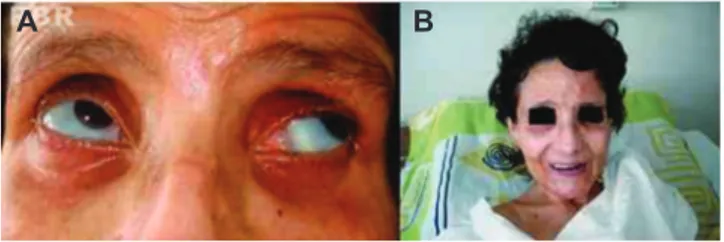
A female patient, 53 years old, born and raised in the city of Rio de Janeiro, Brazil, came to the Rheumatology Service of the Hospital Universitário Clementino Fraga Filho of the Universidade Federal do Rio de Janeiro (HUCFF/UFRJ) for the treatment of osteoporosis associated with a history of recur-rent fractures. Clinical examination revealed blue sclera (Fig. 1A), important limitation in the opening of the mouth (Fig. 1B), and severe deformities of hands and feet (Figs. 2A and 2B). Hormonal investigations discarded osteometabolic dis-eases, such as hypophosphatasia and tumors of the adrenal cortex. The clinical-epidemiological, laboratory, and radio-logical i ndings allowed the diagnoses of congenital syphilis, osteopetrosis, idiopathic juvenile osteoporosis, celiac disease, i brous dysplasia, and bone tumors to be discarded.
Radiographs of the thoracic spine and pelvis showed marked decrease in bone mass with collapse of several dorsal vertebrae, and bone densitometry showed T-scores of -4.7 SD in the lumbar spine (L1-L4) and -4.6 SD in the total femur. A review of the patient’s medical history demonstrated that since age 2 years she had suffered spontaneous fractures, and
that her brother had a similar history of fractures. Neither had been diagnosed in the past with regard to the fractures.
Additionally, the patient had a picture of cumulative sym-metric polyarthritis of large and small joints, which started in childhood, associated with sporadic fever and morning stiff-ness; polyarticular onset JIA was diagnosed at age 15 years. The evaluations at the time of diagnosis showed elevated in-l ammatory markers (ESR = 72 mm/h), presence of rheuma-toid factor (not quantii ed, but coni rmed on other occasions), and irregular use of salicylates and gold salts during periods of arthritis worsening. No other autoantibody was detected.
The diagnosis of JIA associated with OI was clinically con-i rmed, despcon-ite the lack of molecular evcon-idence, and treatment was initiated with an anti-reabsorptive drug (alendronate).
Discussion
JIA was recently classii ed by the International League of As-sociations for Rheumatology (ILAR) into seven subtypes; fe-males are more affected by all of them.3 Polyarticular JIA with
positive rheumatoid factor is the form that resembles the adult disease, evolves with severe joint damage and, in gen-eral, accompanies the patient throughout life, with periods of exacerbation and inactivity.4
JIA patients often have poor oral occlusion due to the effect of the disease on the TMJ.5 The involvement of the TMJ in JIA
is not uncommon and can even occur alone and insidiously. The incidence varies from 17% to 87% of patients, depending on the JIA subtype studied. This condition continues to be one of the most underdiagnosed and undertreated in JIA.1
Patients with TMJ involvement may be asymptomatic or have morning stiffness, reduced capacity of mouth open-ing, rales, trismus, and pain on joint palpation.1 Patients with
more marked alterations are those with longer duration of disease, earlier onset, or those with polyarticular or systemic onset disease.1
The TMJ can be investigated by some complementary tests, such as orthopantomogram, ultrasound, computed tomogra-phy (CI), or magnetic resonance imaging (MRI).2
Ultrasonogra-phy, an examination that yields good joint assessment, depends largely on the experience of the radiologist. The orthopantomo-gram is an affordable and low cost-examination, but has the disadvantage of not differentiating chronic from active lesions.
Computed tomography results in better visualization of the mandibular condyle, but does not detect soft tissue alterations that indicate joint inl ammation. MRI is regarded as the gold standard for diagnosis, since it is able to demonstrate synovial effusion or proliferation (pannus),1 as in the reported case.
OI has a higher prevalence in white women. Most patients have blue sclera and family history of the disease. Its clas-sii cation was described in 19786 and reviewed in 2010,7 but
type 1 remains the mildest and most prevalent form, with a prognosis compatible with life and allowing for ambulation.6
Although there was no molecular verii cation, this patient likely has type 1, in which the deformities are uncommon and there may not be i nal height impairment. The altera-tions observed in extremities are characteristic of JIA without adequate treatment (Figs. 2A and 2B). Additionally, deafness is observed in only 30% of cases.8
Fig. 1 – A, Blueish sclera in patient with osteogenesis imperfecta associated with juvenile idiopathic arthritis. B, Mouth opening limitation due to temporomandibular joint involvement.
Fig. 2 – A, Hand deformities. B, Corresponding X-ray.
A
B
537
R E V B R A S R E U M A T O L . 2 0 1 3 ;5 3 ( 6 ): 5 3 5 – 5 3 7OI patients can have normal teeth, with a moderate change in color, or quite abnormal dentition. Dental frac-tures can occur easily and require extractions.9 The normal
dentin is rich in collagen type 1. JIA patients, however, have signii cantly reduced alveolar bone density.10 These factors
together are enough to interfere with the patient’s dental as-sessment.
During the course of OI, it is possible to observe arthralgia, joint hypermobility, and tendon rupture.11 The disease itself
can lead to deformities in the hands, characterized by swan neck i ngers and reversible contractures. This process is at-tributed to fractures and ligament laxity.12 In this case, it may
even be confused with rheumatoid arthritis and/or JIA. How-ever, OI shows no erosion alterations on radiographs or morn-ing stiffness. The alterations observed in the TMJ and extrem-ities in the present case could be attributed exclusively to JIA.
TMJ involvement is rare in OI. Only 6% of a population of adults with the disease reported having severe TMJ disor-ders.13 This i nding in a patient with a presumptive diagnosis
of hereditary collagen disorder warned u of the possibility of other diagnoses, and the investigation was performed retro-spectively, until the diagnosis of JIA was made.
JIA is a differential diagnosis that should be considered in pediatric patients with spontaneous fractures.14 However,
not only it is a rare form of osteoporosis, but also it does not include blue sclera and is not inl uenced by family history, as in the case described here. OI itself can have a crippling arthropathy form,15 but its clinical course is different from JIA
and does not usually include elevated inl ammatory markers or the presence of rheumatoid factor.
Both OI and JIA cause an increased risk of fractures, with different mechanisms. OI patients have spontaneous frac-tures due to the disease pathogenesis that leads to the bone matrix impairment. In JIA, fractures can occur due to increase in inl ammatory cytokines, reduction of secondary bone mass, physical inactivity, or osteoporosis caused by corticosteroids. As low bone density is found in both diseases, bisphospho-nates are successfully. The efi cacy of treatment and follow-up can be measured by annual bone densitometry.16
The use of bisphosphonates in childhood is well estab-lished, particularly the use of pamidronate in OI and idio-pathic juvenile osteoporosis.17
The authors would like to emphasize that this curious and rare association should be considered when investigating se-vere joint deformities, especially in the TMJ and extremities, associated with joint destruction in patients with a long-term picture of OI.
Conl icts of interest
The authors declare no conl icts of interest.
R E F E R E N C E S
1. Arabshahi B, Cron RQ. Temporomandibular joint arthritis in juvenile idiopathic arthritis: the forgotten joint. Curr Opion Rheumatol. 2006;18(5):490 -5.
2. Santili C, Akkari M, Waisberg, G, Bastos Júnior JOC, Ferreira WM. Avaliação clínica, radiográi ca e laboratorial de pacientes com osteogênese imperfeita. Rev Assoc Med Bras. 2005;51(4):214-20.
3. Ravelli A, Martini A. Juvenile idiopathic arthritis. Lancet. 2007;369(9563):767-78.
4. Gurcay E, Eksioglu E, Yuzer, S, Bal A, Cakci A. Articular damage in adults with juvenile idiopathic arthritis. Rheumatol Int. 2009;29(6):635-40.
5. Barr T, Carmichael NM, Sándor GKB. Juvenile Idiopathic Arthritis: a chronic pediatric musculoskeletal condition with signii cant orofacial manifestations. J Can Dent Assoc. 2008;74(9):813-21.
6. Sillence DO, Rimoin DL. Classii cation of osteogenesis imperfect. Lancet. 1978;1(8072):1041-2.
7. Van Dijk FS, Pals G, Van Rijn RR, Nikkels PG, Cobben JM. Classii cation of Osteogenesis Imperfecta revisited. Eur J Med Genet. 2010;53(1):1-5.
8. Donangelo I, Coelho SM, Farias MLF. Osteogenesis Imperfecta no adulto e resposta ao alendronato: apresentação de caso. Arq Bras Endocrinol Metab 2001;45(3):309-13.
9. Prockop DJ, Kuivaniemi H, Tromp G, Ala-Kokko L. Distúrbios hereditários do tecido conjuntivo. In: Braunwald E, Fauci AS, Kasper DL, Hauser SL, Longo DL, Jameson JL. Harrison - Medicina Interna 15ª ed. Rio de Janeiro: McGrawHill, 2002:2435-2446.
10. Silva TLO, Braga FSFF, Sztajnbok FR, Souza AA, Silva FB, Fischer RG et al.. Redução da densidade óssea alveolar em pacientes com artrite idiopática juvenil. Rev Bras Reumatol. 2012;52(1)33-43.
11. McKiernan FE. Musculoskeletal manifestations of mild osteogenesis imperfecta in adult. Osteoporos Int. 2005;16(12):1698-702.
12. Oz B, Olmez N, Memis A. Osteogenesis imperfecta: a case with hand deformities. Clin Rheumatol. 2005;24(5):565-8. 13. Sæves R, Wekre LL, Ambjørnsen E, Axelsson S, Nordgarden
H, Storhaug K. Oral i ndings in adults with osteogenesis imperfecta. Spec Care Dentist 2009;29(2):102-8.
14. Seda H, Menezes LA, Ghelman SS. Osteoporose idiopatica juvenil. Apresentação de dois casos. Rev bras reumatol.1983;23(2):69-74.
15. Penttinen R, Sipola E, Kouvalainen K, Simila S, Remes M. An arthropathic form of osteogenesis imperfecta. Acta Paediatr Scand. 1980;69(2):263-7.
16. Thornton J, Ashcroft DM, Mughal MZ, Elliott RA, ONeill TW, Symmons D. Systematic review of effectiveness of bisphosphonates in treatment of low bone mineral density and fragility fractures in juvenile idiopathic arthritis. Arch Dis Child. 2006;91(9):753-61.