GAMETE BIOLOGY
Caspase signalling pathways in human spermatogenesis
Carolina Almeida&Sofia Correia&Eduardo Rocha&
Ângela Alves&Luís Ferraz&Joquina Silva&
Mário Sousa&Alberto Barros
Received: 23 November 2012 / Accepted: 20 January 2013 / Published online: 29 January 2013 # Springer Science+Business Media New York 2013
Abstract
Purpose Little is known about the apoptotic mechanisms involved in abnormal spermatogenesis. In order to describe the significance of apoptosis in azoospermia, testicular tis-sue from abnormal spermatogenesis was analysed.
Methods Testicular treatment biopsies were obtained from 27 men. Five presented oligozoospermia, 9 obstructive
azoospermia (4 congenital bilateral absence of the vas deferens; 5 secondary azoospermia) and 13 non-obstructive azoospermia (5 hypospermatogenis; 3 matura-tion arrest; 5 Sertoli-cell-only syndrome). Immunohisto-chemical staining was performed for active caspases-3, −8 and−9. The presence of active caspases in Sertoli cells and germ cells was analyzed using stereological tools.
Results Increased active caspase-3 was found in Sertoli-cell-only syndrome. No significant differences were found in mat-uration arrest. In hypospermatogenesis, primary spermatocytes were the germ cells with higher active caspases. Oligozoosper-mia and secondary obstruction showed significant differences among germ cells for the presence of all active caspases. In oligozoospermia, spermatogonia presented significant in-creased active caspase-9 in relation to active caspase-8. In primary obstruction and hypospermatogenesis, germ cells pre-sented significant increased active caspases-3 and−9. Conclusions Results suggest that increased active caspase-3 might be involved in Sertoli-cell-only syndrome etiology. In cases of hypospermatogenesis, intrinsic lesions at the meiotic stage seem to be related to the pathology. In secondary ob-struction apoptosis is suggested to be initiated due to extrinsic and intrinsic lesions, whereas in primary obstruction only the intrinsic apoptotic pathway seems to be present. Finally, in oligozoospermic patients spermatogonia death by mitochon-drial damage additionally to meiosis malfunctioning, might be on the origin of the decreased sperm output.
Keywords Apoptosis . Caspases . Testis . Obstructive azoospermia . Non-obstructive azoospermia
Introduction
During normal development of an organism a balance be-tween cell proliferation and cell apoptosis is observed.
Capsule Apoptotic mechanisms in testicular tissue from men with abnormal spermatogenesis are described. Immunohistochemical staining performed for active caspases-3, -8 and -9, showed the involvement of apoptosis in oligozoospermia, obstructive azoospermia and non-obtructed azoospermia, except for maturation arrest. C. Almeida (*)
:
A. BarrosDepartment of Genetics, Faculty of Medicine, University of Porto, Porto 4200-319, Portugal e-mail: carola@med.up.pt
S. Correia
Department of Hygiene and Epidemiology, Faculty of Medicine, University of Porto, Porto 4200-319, Portugal
S. Correia
Institute of Public Health, University of Porto, Porto 4050-600, Portugal
E. Rocha
Department of Microscopy, Laboratory of Histology,
Institute of Biomedical Sciences Abel Salazar (ICBAS), UMIB, University of Porto, Porto 4050-313, Portugal
Â. Alves
:
M. SousaDepartment of Microscopy, Laboratory of Cell Biology, ICBAS, UMIB, University of Porto, Porto 4050-313, Portugal
L. Ferraz
Department of Urology, Hospital Centre of Vila Nova de Gaia, Vila Nova de Gaia 4434-502, Portugal
J. Silva
:
M. Sousa:
A. BarrosCentre for Reproductive Genetics Alberto Barros, Porto 4100-009, Portugal
However, under certain stimuli, this balance is disrupted and cell proliferation or cell death by apoptosis deregulation occur, being on the origin of several proliferative and de-generative diseases [11,30].
Caspases are a family of proteins essential in the apo-ptotic mechanism. Upon an apoapo-ptotic stimulus, initiator caspases are activated starting the biochemical apoptotic cascade. The initiator caspases-8 and −9 are responsible for the extrinsic and intrinsic apoptotic pathway activation, respectively, leading to the most important effector cas-pase activation: cascas-pase-3. Cascas-pase-3 activation marks the point of no return in the apoptotic process and is responsible for key proteins cleavage leading to final cell disassembly [5].
During spermatogenesis, there is a requirement of germ cell death by apoptosis in order to maintain normal germ cell development and to achieve a normal sperm output [10]. During this process, damaged and/or exces-sive germ cells are phagocytised by Sertoli cells through a Fas/FasL apoptotic system dependent-manner [25]. Ap-optotic markers like active caspase-3 [1, 20, 32], phos-phatidylserine (PS) externalization [3, 31] and DNA fragmentation [6, 9,20,21] were described to be present in ejaculated sperm from men with normal semen param-eters. However, increased rates of ejaculated sperm apo-ptosis were described in conditions of abnormal sperm output, mainly related to abnormalities of sperm concen-tration, morphology or rapid progressive motility values [1, 3,6, 9, 20, 21, 24, 31–34].
In cases of disrupted spermatogenesis with no ejacu-lated sperm (azoospermia), testicular sperm apoptosis has also been a subject of discussion in the literature. Increased rates of testicular sperm with active caspase-3 and PS externalization were found in cases of azoosper-mia, either in obstructive azoospermia (congenital ab-sence of the vas deferens and secondary obstructive azoospermia) or in hypospermatogenesis when compared to ejaculated sperm [2, 3], suggesting a relationship between testicular sperm apoptosis and testicular failure. Additionally, increased rates of spermatids with PS ex-ternalization were described in men with incomplete spermiogenesis failure [29]. Nuclear and mitochondrial damage in cases of secondary obstructive azoospermia and hypospermatogenesis, respectively, were suggested to be on the origin of testicular sperm apoptosis [2]. However, rates of germ cell apoptosis and possible activation mechanisms in-volved in obstructive and non-obstructive azoospermia are still poorly understood.
Results reported so far show discordant and inconclusive results for the role of apoptosis during testicular failure.
The aims of the present study were to determine, using a stereological approach, the rates of Sertoli cell and germ cells (spermatogonia, primary and secondary spermatocytes and
round spermatids) with active caspase-3 (effector), active caspase-8 (initiator; extrinsic pathway) and active caspase-9 (initiator; intrinsic pathway), in cases with oligozoospermia and in cases with obstructive and non-obstructive azoosper-mia. To search for a possible apoptotic activation mechanism, comparisons between the rates of Sertoli cells and germ cell stages with active caspases-8 and−9 were performed, both per syndrome and between syndromes.
Methods Patients
According to the National Law on Medically Assisted Procreation (PMA, Law 32/2006) and the National Council on Medically Assisted Procreation guidelines, testicular tissues were used after patient informed and written consent.
Testicular tissue was obtained from 27 men undergoing a treatment testicular biopsy during assisted reproduction techniques. Cell characterization for diagnosis was per-formed during the testicular biopsy procedure in an inverted Nikon microscope equipped with Hoffman optics on a heated stage (33 °C).
Five patients had oligozoospermia but normal testicular histology. The other 22 patients were diagnosed as azoo-spermic and subdivided into different groups. Obstructive azoospermia was diagnosed in 9 patients based on con-genital bilateral absence of the vas deferens (CBAVD, n= 4) and secondary obstruction (n=5). At the moment of testicular biopsy, all presented numerous germ cells (all stages) and numerous testicular spermatozoa with in situ and slow progressive motility. Azoospermia with a non-obstructive origin was found in 13 patients based on the presence of [1] reduced number of germ cells and sper-matozoa (hypospermatogenesis, n=5), [2] spermatogenic arrest at the secondary spermatocyte stage (maturation arrest, n=3) and [3] seminiferous tubules with only Sertoli cells (Sertoli-cell-only syndrome, SCOS, n=5) (defined according to [22]). Testicular volumes varied according to pathology: 12–22 ml (oligozoospermia, hypospermato-genesis, SCOS), 22 ml (CBAVD), 15–22 ml (secondary obstruction) and 18–20 ml (maturation arrest). All patients presented normal karyotypes and absence of Y chromo-some microdeletions.
Testicular tissue sampling and preparation for histological analysis
Testicular tissue was obtained during treatment testicular biopsy as previously described [27]. Testicular sample biopsies volume was measured by water displacement
and all presented volumes approximately of 0.016 ml. Samples were fixed in 4 % Paraformaldheide (Merck, Darmstadt, Germany) in PBS (Phosphate Buffered Saline, Sigma, St. Louis, USA). After fixation, samples were washed in PBS, dehydrated in ethanol series, and then embedded in paraffin (Merck).
Histological analysis
With a manual Minot microtome (American Optical, USA), 10 3μm sections were made in each paraffin block and transferred to Vectabound Reagent treated slides (Vector Laboratories, Burlingame, USA). Haematoxylin-eosin (Hemalumen Mayer, Merck; Surgipath Medical Industries, Peterborourg) staining was performed in all samples to confirm testicular diagnosis and tissue integrity.
Immunohistochemistry
Immunostaining for caspase-3 and−9 was performed using Vectastain ABC Kit (Vector Laboratories) according to manu-facturer’s instructions. Briefly, paraffin sections were deparaf-finised in xylol and rehydratated in ethanol series. To suppress endogenous peroxidase activity, tissue sections were treated with 3 % hydrogen peroxide (Hydrogen Peroxide 30 %, Merck) in distilled water. After 1 h of incubation with 5 % of normal horse serum (RT, Vectastain ABC Kit; diluted in 0.1 % Tween20/PBS), primary antibody was incubated overnight at 4 °C: active caspase-3 (Monoclonal Anti-Human/Mouse Cleaved Caspase-3 Antibody, R&D Systems, Minneapolis, USA; 1:150 diluted in 5 % normal horse serum); active caspase-9 (Monoclonal Anti-Caspase-9 Cleavage Site Specif-ic, Sigma; 1:50 diluted in 5 % normal horse serum). Biotiny-lated secondary antibody (Vectastain ABC Kit, 30 min, RT) and ABC reagent (Vectastain ABC Kit, 30 min, RT) were incubated according to manufacturer instructions. The final colorimetric reaction was performed using DAB (3,3′-Dia-minobenzidine; Sigma; final concentration: 0.05 % in 0.015 % H2O2), nuclei counterstained with haematoxylin
(Vector Laboratories) and slides mounted in entellan. Active caspase-8 immunostaining was performed similarly with NovoLink Polymer Detection System (Novocastra Lab-oratories, Newcastle, UK) and a caspase-8 primary antibody (Mouse Monoclonal Antibody Caspase-8, Novocastra; 1:1000 diluted in PBS).
For all immunoreactions a negative control slide was used, with the same treatment described above only omitting primary antibody labelling.
Stereological analysis
Testicular tissue preparation for analysis is usually carried out by paraffin inclusion or other embedding media followed by
3–4 μm sections cuts. This procedure can induce cell distortion in number, size and shape within the section analyzed. In order to overcome possible bias, stereological tools were introduced in histological analysis of testicular tissue [26]. Stereological analysis ensures an uniformed sample where Sertoli cells and germ cells from all stages have the same probability to be sampled and where pos-sible distortions in cell volume and orientation during histological processing will not interfere with the final results [19, 26]. In addition, despite several seminiferous tubules are analyzed, a testicular section only provides estimation on how the entire testis might function. This method provides information relative to the total number of cells in testis in order that final results truly represent the entire testis [19].
Only one testicular section per patient was analysed and, when possible, about 20 seminiferous tubules were evaluated. Samples were analysed under light microscopy (LEITZ DMR—Leica, Germany), with digital camera (DFC480— R2, Leica) and appropriate analysis software (Leica Image Manager 50—IM50, Switzerland). The presence of active caspases-3,−8 and −9 was estimated per germ cells (matogonia—SG, primary—ST1, and secondary—ST2 sper-matocytes, round spermatids—Sa) and Sertoli cells (SC), either per seminiferous tubules or per testicular volume using stereological tools.
The parameter VV represents the relative volume
oc-cupied by a cell relatively to a specific reference space. Considering seminiferous tubules as the reference space, each VVis determined by a manual point counting [23],
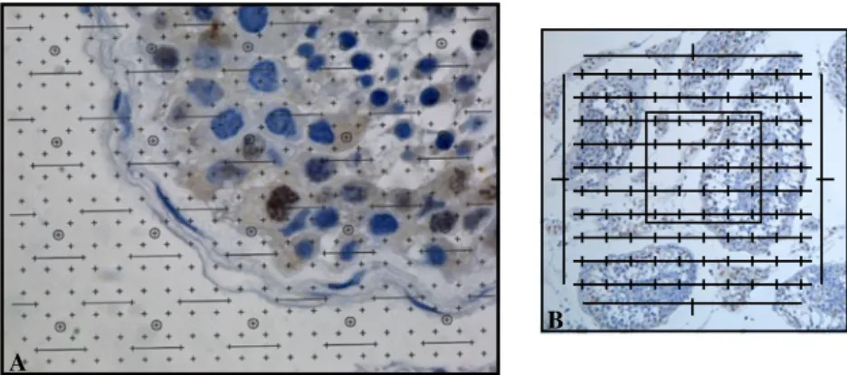
using a multi-system grill placed over the image obtained using an immersion oil objective (100x) (Fig. 1). After counting, results were calculated with the following computation:
VV(cell, seminiferous tubules)=[∑P(cell) x 100] ÷ [R ∑P
(seminiferous tubules)], where“∑P(cell)” corresponds to the total number of points counted over the cell of interest,“∑P (seminiferous tubules)” corresponds to the total number of points over the seminiferous tubules from the testicular section in study, and“R” to the ratio of points considering the counting system used for the analysed cells and for the reference space (seminiferous tubules). This computation gives the estimation of the volume occupied per cell in seminiferous tubules and was calculated per patient in all pathologies. In the seminiferous tubule, results are expressed as relative volume (Vv) occupied per cell with or without active caspases.
In order to calculate the volume per cell occupied in the testis, the following computations were used:
VV (cell, biopsy) = VV (cell, seminiferous tubules) x
VV (seminiferous tubules, biopsy) and V (cell, testis)=VV
(cell, biopsy) x V (biopsy, testis), where the total volume of each biopsy was estimated by fluid displacement before
preparation of samples for paraffin impregnation. The VV
(seminiferous tubules, biopsy) is estimated by point count-ing with a multi-system grill (Integrationsplatte II, 100/25, Carl Zeiss) directly in the microscope ocular and with a 10x objective (Fig. 1). In the entire testis, results are expressed as volume (V) occupied per cell with or without active caspase labelling.
Statistical analysis
Different approaches were applied in the analysis of the relative (Vv) and total (V) volume occupied per cell with active caspases-8,−9 and −3 in both seminiferous tubules and testis, respectively. For each caspase, comparisons were made per group of patients (Oligozoospermia, CBAVD, secondary obstruction, hypospermatogenesis, maturation ar-rest and SCOS) among germ cell types (SC, SG, ST1, ST2, Sa) and also per germ cell type among groups of patients. Kruskal-Wallis test with a 95 % confidence interval (CI) was applied. Multiple comparisons were also performed between cells by the Mann–Whitney U Test (95 % CI) and per group of patients. Ten different comparisons were made: SCvsSG, SCvsST1, SCvsST2, SCvsSa, SGvsST1, SGvsST2, SGvsSa, ST1vsST2, ST1vsSa, ST2vsSa. The Bonferroni method was used to adjust the significance level owing to multiple Mann–Whitney tests. The significance level of 0.05 was maintained using the formula of 0.05O/k inde-pendent hypotheses, with k=10 to reflect the ten planned analyses of interest detailed above. An adjusted signifi-cance level of 0.005 was therefore used to maintain the overall 0.05 level in the context of the multiple compar-isons. Finally, comparisons between active caspase-8 and active caspase-9 relative (Vv) and total volume (V), per group and per cell, were performed using the Mann– Whitney U Test (95 % CI).
In all approaches, medians were used instead of means as no normal distribution was shown by active caspases-8,−9
and −3 Vv and V values, in all groups. All comparisons were performed using Stata (version 9).
Results
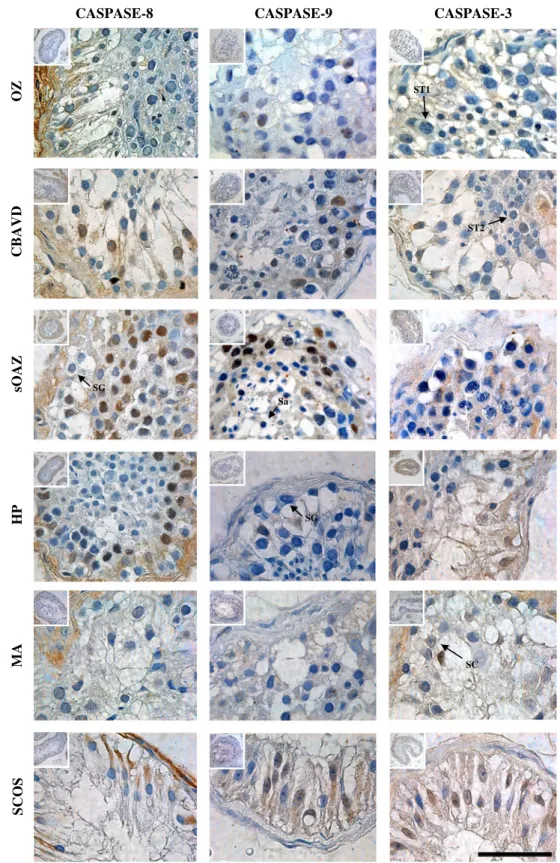
In all groups, active caspases-8,−9 and −3 immunostaining were observed in Sertoli cells’ cytoplasm and germ cells’ cytoplasm and nucleus (Fig.2).
The relative and total volume medians of Sertoli cells and germ cells with active caspases-8,−9 and −3 per testicular groups are shown in Fig.3.
Seminiferous tubules Active caspase-8
In all groups, increased Vv(SC) with active caspase-8 (caspcaspase-8+) was shown to be present in relation to all germ cells types, with no significant differences being found among groups (P=0.632). Comparisons per group revealed significant differences among germ cell types in cases of Oligozoospermia and secondary obstruction (Table 1). Although no significant differences were found by multiple comparisons between germ cell types, in-creased Vv(ST1)casp8+ was observed in both groups. No significant differences were observed in comparison to oligozoospermia and among the other groups, per germ cell type (Table1).
Active caspase-9
Increased Vv(SC) with active caspase-9 (casp9+) was ob-served in all groups in relation to germ cells, but with no significant differences among groups (P=0.554). Comparisons per group showed significant differences among germ cell types (P=0.033, Table 1). Again, increased Vv(ST1)casp9+
A
B
Fig. 1 Grill systems used for the stereological analysis performed. a Howard and Reed multi-scale system placed over a seminiferous tubule image on the computer (100×) in order to estimate the relative volume
(Vv) occupied per cell per seminiferous tubule; b Grill inserted in the left microscope ocular (10×) in order to estimate the volume (V) occupied per cell in the testis (Integrationsplatte II, 100/25, Carl Zeiss)
was found in all groups (Table1). Contrarily to casp8+, com-parisons per germ cell stage among groups, including oligo-zoospermia, showed significant differences (P=0.011) with increased Vv(SG)casp9+ and Vv(ST1)casp9+ in secondary obstruction and increased Vv(ST2)casp9+ and Vv(Sa)casp9+ in CBAVD (Table1).
Active caspase-3
Increased Vv(SC) with active caspase-3 (casp3+) was pres-ent in all groups in relation to germ cells and significant differences were found among groups (P=0.009). Increased values were shown by SCOS and hypospermatogenic
CASPASE-8 CASPASE-9 CASPASE-3
OZ CBAVD sOAZ HP MA SCOS ST1 SC SG ST2 SG Sa Fig. 2 Active caspases-8,−9
and−3 immunohistochemical analysis for the testicular groups analyzed. OZ— Oligozoospermia; CBAVD— congenital bilateral absence of the vas deferens; sOAZ— secondary obstructive azoospermia; HP— hypospermatogenesis; MA— maturation arrest; SCOS— Sertoli-cell-only syndrome. SC—Sertoli-cell; SG— spermatogonia; ST1—primary spermatocyte; ST2—secondary spermatocte; Sa—round spermatid; Bar=50μm
Seminiferous Tubules
Testis
Active Caspase-9 Active Caspase-3
Active Caspase-8
OZ CBAVD sOAZ HP MA SCOS OZ CBAVD sOAZ HP MA SCOS
OZ CBAVD sOAZ HP MA SCOS OZ CBAVD sOAZ HP MA SCOS
OZ CBAVD sOAZ HP MA SCOS
OZ CBAVD sOAZ HP MA SCOS
Fig. 3 Median values for relative volume (Vv) and volume (V) occu-pied by Sertoli cells and germ cells with active caspases-8,−9 and −3, in the seminiferous tubules and testis, respectively.
OZ—Oligozoo-spermia; CBAVD—congenital bilateral absence of the vas deferens; sOAZ—secondary obstructive azoospermia; HP—hypospermatogenesis; MA—maturation arrest; SCOS—Sertoli-cell-only syndrome
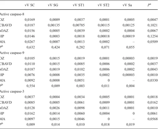
Table 1 Relative volumes of active caspases-8,−9 and −3 per germ cell stage per seminiferous tubules
SC sertoli cell, SG spermatogo-nia, ST1 primary spermatocyte, ST2 secondary spermatocyte, Sa round Spermatid, OZ oligozoo-spermia, CBAVD congenital bilateral absence of the vas deferens, sOAZ secondary obstructive azoospermia, HP hypospermatogenesis, MA maturation arrest, Vv relative volume (expressed in median)
aComparisons per group among
cell stages (except SC);
b
comparisons per cell stage among groups vV SC vV SG vV ST1 vV ST2 vV Sa Pa Active caspase-8 OZ 0,0169 0,0009 0,0037 0,0001 0,0005 0,0047 CBAVD 0,0187 0,00135 0,00785 0,00115 0,00125 0,1021 sOAZ 0,0156 0,0005 0,0039 0,0002 0,0004 0,0067 HP 0,0146 0,0003 0,0018 0,00018 0,00019 0,1254 MA 0,0267 0,0007 0,0013 0,0002 – 0,0509 Pb 0,632 0,424 0,282 0,071 0,055 Active caspase-9 OZ 0,0105 0,0015 0,0019 0,0001 0,00003 0,0019 CBAVD 0,0110 0,0015 0,0085 0,0006 0,0002 0,0037 sOAZ 0,0187 0,0019 0,0102 0,0006 0,0002 0,0043 HP 0,0076 0,0008 0,0035 0,0002 0,00003 0,0010 MA 0,0092 0,0008 0,0051 0 – 0,0330 Pb 0,554 0,009 0,003 0,011 0,004 Active caspase-3 OZ 0,0037 0,0004 0,0034 0,0005 0,0001 0,0018 CBAVD 0,0085 0,0005 0,0061 0,0009 0,0001 0,0162 sOAZ 0,0128 0,0026 0,0098 0,0011 0,0001 0,0010 HP 0,0162 0,0014 0,0060 0,0004 0 0,0061 MA 0,0097 0,0015 0,0046 0 – 0,0568 Pb 0,009 0,014 0,010 0,018 0,019
patients (Table 1). Among germ cell types, all groups, except MA (P=0.057) presented significant differences (Table 1). Although no significant differences were ob-served by multiple comparisons between germ cell types, increased Vv(ST1)casp3+ was found in all testic-ular syndromes. Significant differences were observed when comparisons were made per germ cell stage and among groups (P=0.019). Increased Vv(SG)casp3+, Vv (ST1)casp3+, Vv(ST2)casp3+ and Vv(Sa)casp3+ were all observed for secondary obstruction in relation to the other groups. It should be noted that oligozoospermic men showed the lowest levels of casp3+ in SC and in all germ cells except for Sa where no casp3+ cell was analyzed for hypospermatogenic patients.
Active caspase-8 vs active caspase-9
Comparisons per group showed significant increased Vv (Sa)casp8+ (P= 0.009) and significant increased Vv (SG)casp9+ (P=0.009) in oligozoospermia. Hyposper-matogenic patients showed significant increased Vv (Sa)casp9+ (P=0.028). No significant differences were observed between casp8+ and casp9+ for the other groups (P=0.076).
Testis
Active caspase-8
Results obtained were similar to those obtained at the seminiferous tubule level. Increased V(SC)casp8+ was present in all groups but with no significant differences among them (P=0.706). Significant differences among germ cell types were observed in cases of oligozoo-spermia and secondary obstruction that presented in-creased but not significant V(ST1)casp8+. No significant differences were observed per germ cell stage among groups.
Active caspase-9
Increased V(SC)casp9+ was observed in all groups in rela-tion to germ cell type, but with no significant differences among groups (P=0.102). Comparisons per group showed significant differences for the presence of casp9+ among germ cells (P=0.007) with increased but not significant V (ST1)casp9+ in all groups. An exception was observed within maturation arrest where no significant differences were found (P=0.058). Contrarily to seminiferous tubules, comparisons per germ cell stage among groups showed significant differences only for V(Sa)casp9+ (P=0.048) with increased but not significant V(Sa)casp9+ in secondary obstruction.
Active caspase-3
Increased V(SC)casp3+ was present in all groups in relation to germ cell type and significant differences were found among groups (P=0.009) with increased values shown by SCOS and hypospermatogenic patients. Among germ cell type, all groups, except MA (P=0.058) presented significant differences (P=0.021). Even though multiple comparisons revealed no significant differences between germ cell types, increased V(ST1)casp3+ was found in all testicular syn-dromes. Comparisons per germ cell among groups showed only significant differences for V(Sa)casp3+ (P=0.029) with increased values in CBAVD.
Active caspase-8 vs active caspase-9
Oligozoospermia showed significant increased V(SG)casp9+ (P=0.016) and V(Sa)casp8+ (P=0.009). Hypospermatogene-sis showed significant increased V(Sa)casp8+ (P=0.047).
Discussion
In men with normal sperm output a fine balance between germ cell proliferation and germ cell apoptosis exists [13]. Sertoli cells are responsible for germ cell nurture and sup-port, controlling the number of germ cell production [12]. In the presence of excessive or damaged germ cells, Sertoli cells express FasL inducing germ cell apoptosis by the Fas/FasL system in order to maintain equilibrium, essential to normal spermatogenesis [25]. However, sometimes the adverse conditions are not overcome and this equilibrium is not reached. Increased rates of apoptosis have already been described in cases of azoospermia [17, 18]. However, the apoptotic mechanisms involved are still poorly understood. Increased rates of DNA fragmentation and active caspase-3 were found in testes from patients with SCOS and maturation arrest [16] and decreased rates of TUNEL-positive germ cells were found in hypospermatogenesis, CBAVD and secondary obstruction cases [4]. However, different studies present dis-cordant results [28].
To characterize apoptosis during impaired spermatogen-esis the rates of apoptosis in testicular tissue from fertile men should be assessed. Due to the difficulty in obtaining testicular samples from normozoospermic individuals, we used oligozoospermic patients which although presenting a decreased number of ejaculated sperm have conserved tes-ticular histology with no obstruction [21].
In the present study, the quantification of apoptosis was performed in Sertoli cells and germ cells using stereological analysis. The presence of active caspases−3, −8 and −9 was determined first at the seminiferous tubule level and then results were calculated for the entire testis.
In all cases, Sertoli cells presented higher apoptosis in comparison to germ cells. No significant differences were observed among groups for the presence of active caspase-8 and active caspase-9. During normal spermatogenesis, Sertoli cells and germ cells die by apoptosis in order to maintain equilibrium [15]. In the present study increased levels of Sertoli cells with active caspase-3 were found in SCOS patients suggesting that apoptosis might be an active mechanism in this syndrome. Results are in concordance with previous data where increased TUNEL-positive Sertoli cells and increased rates of active caspase-3 were found in SCOS patients [16].
Contrarily to previous studies where the FasR/FasL system with caspase-3 activation was suggested to be involved in germ cell degeneration in cases of maturation arrest [7, 8, 16], our results suggest no active role for apoptosis in this syndrome. On the contrary, another study showed increased immunostaining of active caspase-3 in spermatocytes or spermatocytes and spermatids in cases of incomplete [4] and complete maturation arrest [14]. These discordant results might be due to differences in MA histology as well as to differences in germ cell quantification and analysis.
In cases of conserved spermatogenesis (secondary ob-struction, CBAVD and hypospermatogenesis), although no significant results were observed, primary spermatocytes showed higher active caspase-3 in comparison to all other germ cell types. Results suggest that a malfunction might exist in the meiotic phase of spermatogenesis, namely in the first meiotic phase, and that might be on the origin of these testicular pathologies. In secondary obstruction significant differences were observed for active caspase-8 and active caspase-9 among germ cells but no differences were found between both. Although the intrinsic and the extrinsic apo-ptotic pathways seemed to be present, results suggest that none of them are likely to prevail. Extrinsic factors on the origin of the obstruction might activate the extrinsic apoptotic pathway with caspase-8 activation but a cross-talk between the intrinsic and the extrinsic pathways, with mitochondrial apoptotic factors release, apparently co-exist, with both mech-anisms contributing to the apoptotic germ cell death observed in secondary obstruction. In cases of CBAVD, no significant differences were found among germ cells for the presence of active caspase-8. On the contrary, significant differences for the presence of germ cells with active caspase-9 and active caspase-3 were observed, with higher levels of labeling appearing at the primary spermatocyte stage, thus suggesting activation of the intrinsic apoptotic pathway. In this mecha-nism, endoplasmic reticulum and mitochondrial lesions might be involved [23].
Previous studies on testicular spermatozoa have shown increased rates of active caspase-3 in secondary obstruction and CBAVD patients, with staining observed mainly in the
sperm nucleus [2]. In the present study, active caspase-3 immunostaining was observed in germ cells cytoplasm and/or nucleus suggesting that external and internal lesions in cases of secondary obstruction and only internal lesions in CBAVD patients might activate the apoptotic pathway inducing caspase-3 activation among germ cells and conse-quent translocation to the nucleus during spermiogenesis.
Like in CBAVD, no significant differences were found for the presence of active caspase-8 among germ cells in hypospermatogenic patients. In these patients significant differences were found for the presence of active caspase-9 with higher levels at the primary spermatocyte stage, sug-gesting that apoptosis through the intrinsic mechanism might be on the origin of the lower numbers of germ cells observed in hypospermatogenic patients. Additionally, in HP, the number of Sertoli cells with active caspase-3 was the highest among all syndromes (exluding SCOS). These results reinforce the results obtained recently where in-creased rates of active caspase-3 were shown to be present in the midpiece of testicular spermatozoa from hyposperma-togenic patients [2].
It should be noted that oligozoospermic men showed the lowest levels of caspase-3 in SC and in all germ cells except for Sa where no caspase-3 positive cell was ana-lysed for hypospermatogenic patients. Significant differ-ences for the presence of active caspase-8 and active caspase-9 were observed among germ cells, with in-creased levels at the primary spermatocyte stage. Compar-isons between both proteins showed significant increased spermatogonia with active caspase-9. Previous results have suggested the presence of a post-testicular apoptotic induction factor as the main cause of decreased ejaculated spermatozoa [2, 3]. The present additional findings sug-gest that germ cell death induced by internal apoptotic factors and meiosis malfunctioning during spermatogene-sis might also be on the origin of decreased sperm production observed in these men.
In conclusion, no significance for apoptosis was found for maturation arrest whereas in SCOS increased levels of active caspase-3 were shown to be present. In all other pathologies (oligozoospermia, secondary obstruction and CBAVD), meiotic germ cell apoptosis seemed to be present. Apoptosis in cases of secondary obstruction is suggested to be initiated by extrinsic factors and intrinsic lesions, whereas in CBAVD only the intrinsic apoptotic pathway seemed to be present. The low numbers of germ cells observed in hypospermatogenesis are suggested to result from Sertoli cell death by apoptosis and mitochon-drial lesions at the primary spermatocyte stage. Finally, in oligozoospermic patients, spermatogonia death by mi-tochondrial damage and meiosis malfunctioning during spermatogenesis might be on the origin of the decreased sperm output.
Acknowledgments From the Centre for Reproductive Genetics Alberto Barros, Porto, we would like to acknowledge Paulo Viana (BSc), Ana Gonçalves (BSc) and Mariana Cunha (BSc) for testicular biopsies processing in the IVF Lab. From the Laboratory of Cell Biology from the Institute of Biomedical Sciences Abel Salazar (ICBAS), Porto, we would like to thank Francisca Reis (BSc) for helping with active caspase-3 immunostainning assays. From the Department of Genetics of the Faculty of Medicine, we would like to acknowledge Dr. Susana Fernandes for helping with manuscript revision.
References
1. Almeida C, Cardoso MF, Sousa M, Viana P, Goncalves A, Silva J, et al. Quantitative study of caspase-3 activity in semen and after swim-up preparation in relation to sperm quality. Hum Reprod. 2005;20(5):1307–13. doi:10.1093/humrep/deh727.
2. Almeida C, Cunha M, Ferraz L, Silva J, Barros A, Sousa M. Caspase-3 detection in human testicular spermatozoa from azoospermic and non-azoospermic patients. Int J Androl. 2011;34:e407–14. 3. Almeida C, Sousa M, Barros A. Phosphatidylserine translocation
in human spermatozoa from impaired spermatogenesis. Reprod Biomed Online. 2009;19:770–7.
4. Bozec A, Amara S, Guarmit B, Selva J, Albert M, Rollet J, et al. Status of the executioner step of apoptosis in human with normal spermatogenesis and azoospermia. Fertil Steril. 2008;90(5):1723–31. 5. Chowdhury I, Tharakan B, Bhat GK. Caspases—an update. Comp
Biochem Physiol B Biochem Mol Biol. 2008;151(1):10–27. 6. Donnelly ET, O’Connell M, McClure N, Lewis SEM. Differences
in nuclear DNA fragmentation and mitochondrial integrity of semen and prepared human spermatozoa. Hum Reprod. 2000;15 (7):1552–61.
7. Eguchi J, Koji T, Nomata K, Yoshii A, Shin M, Kanetake H. Fas-Fas ligand as a possible mediator of spermatogenic cell apoptosis in human maturation-arrested testes. Hum Cell. 2002;15(1):61–8. 8. Francavilla S, D’Abrizio P, Cordeschi G, Pelliccione F, Necozione
S, Ulisse S, et al. Fas expression correlates with human germ cell degeneration in meiotic and post-meiotic arrest of spermatogene-sis. Mol Hum Reprod. 2002;8(3):213–20.
9. Gandini L, Lombardo F, Paoli D, Caponecchia L, Familiari G, Verlengia C, et al. Study of apoptotic DNA fragmentation in human spermatozoa. Hum Reprod. 2000;15(4):830–9.
10. Hikim APS, Swerdloff RS. Hormonal and genetic control of germ cell apoptosis in the testis. Rev Reprod. 1999;4:38–47.
11. Jacobson MD, Weil M, Raff MC. Programmed cell death in animal development. Cell. 1997;88(3):347–54.
12. Johnson L, Thompson Jr DL, Varner DD. Role of Sertoli cell number and function on regulation of spermatogenesis. Anim Reprod Sci. 2008;105(1–2):23–51.
13. Johnson L, Varner DD, Roberts ME, Smith TL, Keillor GE, Scrutchfield WL. Efficiency of spermatogenesis: a comparative approach. Anim Reprod Sci. 2000;60:471–80.
14. Kandirali E, Cayan S, Armagan A, Erol B, Kadioglu A. Does the testicular apoptotic index vary with serum gonadotropins and testic-ular histopathology in infertile men? Urol Int. 2008;83(3):349–53. 15. Kierszenbaum AL. Apoptosis during spermatogenesis: the thrill of
being alive. Mol Reprod Develop. 2001;58(1):1–3.
16. Kim S-K, Yoon Y-D, Park Y-S, Seo JT, Kim J-H. Involvement of the Fas-Fas ligand system and active caspase-3 in abnormal
apoptosis in human testes with maturation arrest and Sertoli cell-only syndrome. Fertil Steril. 2007;87(3):547–53.
17. Lin WW, Lamb DJ, Wheeler TM, Abrams J, Lipshultz LI, Kim ED. Apoptotic frequency is increased in spermatogenic maturation arrest and hypospermatogenic states. J Urol. 1997;158:1791–3. 18. Lin WW, Lamb DJ, Wheeler TM, Lipshultz LI, Kim ED. In situ
end-labeling of human testicular tissue demonstrates increased apoptosis in conditions of abnormal spermatogenesis. Fertil Steril. 1997;68(6):1065–9.
19. Mandarim-de-Lacerda CA. Stereological tools in biomedical re-search. An Acad Bras Cienc. 2003;75:469–86.
20. Marchetti C, Gallego MA, Deffosez A, Formstecher P, Marchetti P. Staining of human sperm with fluorochrome-labeled inhibitor of caspases to detect activated caspases: correlation with apoptosis and sperm parameters. Hum Reprod. 2004;19(5):1127–34. 21. Marchetti C, Obert G, Deffosez A, Formstecher P, Marchetti P.
Study of mitochondrial membrane potential, reactive oxygen spe-cies, DNA fragmentation and cell viability by flow cytometry in human sperm. Hum Reprod. 2002;17(5):1257–65.
22. McLachlan RI, Rajpert-De Meyts E, Hoei-Hansen CE, de Kretser DM, Skakkebaek NE. Histological evaluation of the human testis– approaches to optimizing the clinical value of the assessment: mini review. Hum Reprod. 2007;22(1):2–16. doi:10.1093/humrep/del279. 23. Movassagh M, Foo R. Simplified apoptotic cascades. Hear Fail
Rev. 2008;13(2):111–9. doi:10.1007/s10741-007-9070-x. 24. Oosterhuis GJ, Mulder AB, Kalsbeek-Batenburg E, Lambalk CB,
Schoemaker J, Vermes I. Measuring apoptosis in human spermato-zoa: a biological assay for semen quality? Fertil Steril. 2000;74:245– 50.
25. Pentikainen V, Erkkila K, Dunkel L. Fas regulates germ cell apoptosis in the human testis in vitro. Am J Physiol. 1999;276(2 Pt 1):E310–306.
26. Petersen PM, Giwercman A, Pakkenberg B. Stereological methods as efficient and unbiased tools to quantitate structures in the testis. Scand J Work Environ Health. 1999;25(Suppl1):31–3.
27. Sousa M, Cremades N, Silva J, Oliveira C, Ferraz L, Teixeira da Silva J, et al. Predictive value of testicular histology in secretory azoospermic subgroups and clinical outcome after microinjection of fresh and frozen-thawed sperm and spermatids. Hum Reprod. 2002;17(7):1800–10.
28. Stiblar-Martincic D. Morphometrical evaluation of germ cell apo-ptosis in infertile men. Folia Biol. 2009;55(6):233–7.
29. Tesarik J, Greco E, Cohen-Bacrie P, Mendoza C. Germ cell apoptosis in men with complete and incomplete spermiogenesis failure. Mol Hum Reprod. 1998;4(8):757–62. doi:10.1093/molehr/4.8.757. 30. Thompson C. Apoptosis in the pathogenesis and treatment of
disease. Science. 1995;267(5203):1456–62.
31. Varum S, Bento C, Sousa A, Gomes-Santos CS, Henriques NP, Almeida-Santos T, et al. Characterization of human sperm popu-lations using conventional parameters, surface ubiquitination, and apoptotic markers. Fertil Steril. 2007;87:572–83.
32. Wang X, Sharma RK, Sikka SC, Thomas AJ, Falcone T, Agarwal A. Oxidative stress is associated with increased apoptosis leading to spermatozoa DNA damage in patients with male factor infertil-ity. Fertil Steril. 2003;80(3):531–5.
33. Weng S-L, Taylor SL, Morshedi M, Schuffner A, Duran EH, Beebe S, et al. Caspase activity and apoptotic markers in ejaculated human sperm. Mol Hum Reprod. 2002;8(10):984–91.
34. Zhang H, Chen Z, Ma C, Lu S, Wang L, Li X. Early apoptotic changes in human spermatozoa and their relationships with con-ventional semen parameters and sperm DNA fragmentation. Asian J Androl. 2008;10:227–35.