D e te ctio n o f e arly apo pto sis and ce ll
de ath in T CD 4
+
and CD 8
+
ce lls fro m
le sio ns o f patie nts with lo calize d
cutane o us le ishm aniasis
Laboratório de Imunidade Celular e Humoral em Protozooses, Departamento de Protozoologia, Instituto O swaldo Cruz, FIO CRUZ, Rio de Janeiro, RJ, Brasil
A.L. Bertho, M.A. Santiago, A.M. Da-Cruz and S.G. Coutinho
Abstract
Human localized cutaneous leishmaniasis (LCL), induced by Leish-mania braziliensis, ranges from a clinically mild, self-healing disease
with localized cutaneous lesions to severe forms which can present secondary metastatic lesions. The T cell-mediated immune response is extremely important to define the outcome of the disease; however, the underlying mechanisms involved are not fully understood. A flow cytometric analysis of incorporation of 7-amino actinomycin D and CD4+ or CD8+ T cell surface phenotyping was used to determine whether different frequencies of early apoptosis or accidental cell death occur at different stages of LCL lesions. When all cells obtained from a biopsy sample were analyzed, larger numbers of early apoptotic and dead cells were observed in lesions from patients with active disease (mean = 39.5 ± 2.7%) as compared with lesions undergoing spontaneous healing (mean = 17.8 ± 2.2%). Cells displaying normal viability patterns obtained from active LCL lesions showed higher numbers of early apoptotic events among CD8+ than among CD4+ T cells (mean = 28.5 ± 3.8 and 15.3 ± 3.0%, respectively). The higher frequency of cell death events in CD8+ T cells from patients with LCL may be associated with an active form of the disease. In addition, low frequencies of early apoptotic events among the CD8+ T cells were observed in two patients with self-healing lesions. Although the number of patients in the latter group was small, it is possible to speculate that, during the immune response, differences in apoptotic events in CD4+ and CD8+ T cell subsets could be responsible for controlling the CD4/CD8 ratio, thus leading to healing or maintenance of disease.
Co rre spo nde nce
S.G. Coutinho
Laboratório de Imunidade Celular e Humoral em Protozooses Departamento de Protozoologia Instituto O swaldo Cruz, FIO CRUZ Av. Brasil, 4365
21045-900 Rio de Janeiro, RJ Brasil
Fax: + 55-21-280-1589 E-mail:
coutinho@ gene.dbbm.fiocruz.br
Research supported by the European Economic Community.
Received July 14, 1999 Accepted February 1, 2000
Ke y wo rds
·Apoptosis ·Flow cytometry ·Human leishmaniasis ·Cell death
·T cells
Intro ductio n
Human cutaneous leishmaniasis in Rio de Janeiro, Brazil, is caused mainly by the obligate intracellular protozoan Leishmania braziliensis (1). The parasite replicates in-side the parasitophorous vacuoles of
mul-tiple nodular lesions all over the body (dif-fuse cutaneous leishmaniasis). Few para-sites are detectable in LCL lesions, but tissue destruction and focal points of necrosis sug-gest a cell-mediated hypersensitivity mech-anism of injury.
Several studies utilizing the mouse mo-del suggest that T cell-mediated immunity may be responsible for either a favorable outcome of the disease by activation of mac-rophages and killing of the parasites or ag-gravation of the lesions by inhibition of mac-rophage function (2-4). Cytokines such as IFN-g and TNF-a and -ß, produced by Th1 CD4+ T lymphocytes, play a pivotal role in
this process of macrophage activation and parasite destruction. Alternatively, the mech-anisms for aggravating the disease in mice are related to the effects of cytokines such as IL-4 and TGF-ß, which are primarily pro-duced by Th2 CD4+ T lymphocytes (5-8).
CD8+ T lymphocytes also appear to play an
important role in the immunologic response leading to cure of murine leishmaniasis (9-11); antigen-activated CD8+ T lymphocytes
have been shown to produce IFN-g and may have a cytolytic effect on parasitized macro-phages (11,12).
Research in histology, genetics, and mo-lecular biology during the past 25 years has shown that all cells are genetically pro-grammed to die. Under physiological cir-cumstances, damaged and senescent cells sacrifice themselves through a type of cell death termed apoptosis. This form of pro-grammed cell death is characterized by shrinkage, dense chromatin condensation, DNA fragmentation and formation of apop-totic bodies (13,14), and plays a pivotal role in the development and homeostasis of nor-mal tissues (15), as well as in the pathogen-esis of different diseases (16).
The hallmark for identifying apoptosis is fragmentation of cell DNA via endonu-cleases, and demonstrable by gel electro-phoresis (17). However, flow cytometry has become a method of choice for analysis of
cell death in a variety of cell systems (18-20). A method using 7-amino actinomycin D (7-AAD) and single laser flow cytometry has been described (21-23) which permits discrimination between early apoptotic and live or dead cells (late apoptosis and acci-dental cell death, ACD). The 7-AAD method was used in the present study to measure the frequencies of live, early apoptotic or dead CD4+ and CD8+ T cells from lesions of LCL
patients with active disease or in the sponta-neous healing phase in order to better under-stand the role of cell death in the regulation of the T cell-mediated immune responses in human leishmaniasis.
Mate rial and Me tho ds
Study subje cts
Seventeen adult patients were studied (11 men and 6 women), 15 with active LCL and two with spontaneously healed lesions. After diagnosis, tissue samples were col-lected for examination and the patients were immediately treated with antimony therapy. Since treatment with antimony compounds is usually started immediately, few sponta-neous healed lesions were available for inclusion in the study. All patients were from areas in the regions surrounding Rio de Janeiro where the disease is endemic. The following criteria were used for diagnosis: a) clinical picture and epidemiological evidence for LCL, b) positive Montenegro skin test -delayed-type hypersensitivity reaction to leishmanial antigens, and c) isolation of
Leishmania from lesion biopsy samples af-ter culture in McNeal, Novy and Nicolle medium (24).
Mo no nucle ar ce lls o btaine d fro m le sio ns and
blo o d
ob-tained under local anesthesia from LCL pa-tients. Fat tissue was removed, and the re-maining fragment was placed in a sieve fit-ted with a mesh filter on a Petri dish contain-ing RPMI 1640 medium (Sigma Chemical Co., St. Louis, MO, USA) with 10% fetal calf serum (FCS; Sigma). The tissue was cut into small pieces with a surgical scalpel and extruded through the mesh with a glass rod. The mononuclear cells were separated by centrifugation over a Ficoll-Hypaque gradi-ent (Sigma), adjusted to 5 x 10⁵-1 x 10⁶ cells
per ml and then stained for surface markers and 7-AAD (Sigma) or used in apoptosis-induction experiments.
Blood from healthy adult donors was collected into heparinized tubes and periph-eral blood mononuclear cells (PBMC) were separated by centrifugation over a Ficoll-Hypaque (Sigma) gradient.
Inductio n o f apo pto sis
For induction of apoptosis in LMC- and PBMC-derived cells, 10 µM staurosporine (Stau, Sigma) was added to 1 x 10⁶ cells of
LMC or PBMC cultured in RPMI 1640 supplemented with 1% penicillin/streptomy-cin (Sigma), 10% human AB+ serum (Sigma), 10 mM HEPES, 1.5 mM L-glutamine (Sigma), and 40 µM 2-mercaptoethanol (Sigma) at 37oC in a humidified atmosphere
of 5% CO2 in air for 3 days (21). Cell
cul-tures without Stau treatment were used as control. ACD was induced by placing LMC or PBMC in a water bath at 60oC for 5 min.
D ual-co lo r ce ll surface phe no typing, 7-AAD
staining and flo w cyto me try
For surface antigen staining, 5 µl of each anti-CD4-FITC or anti-CD8-PE monoclonal antibody (Coulter Electronics Inc., Miami, FL, USA) diluted in 90 µl of phosphate-buffered saline (PBS) containing 2% FCS and 0.1% sodium azide (Sigma) (PBSAz)
was added to 5 x 10⁵-1 x 10⁶ LMC followed
by incubation at 4oC for 20 min. After two
washes with 1 ml of PBSAz, the supernatant was removed and the cell pellet was resus-pended in 1 ml of PBSAz with 20 µg of 7-AAD, and then incubated for 20 min at 4oC
protected from light. Samples stained with 7-AAD and surface markers were analyzed with an EPICS 751 flow cytometer (Coulter) equipped with a 488-nm argon-ion laser. Green fluorescence was measured with a 525-nm band pass (BP) filter and orange fluorescence was measured with a 575-nm BP filter. The red fluorescence from 7-AAD was filtered through a 630-nm long pass filter. Electronic compensation among the fluorescence channels was used to remove residual spectral overlap. Fluorescence data were displayed on four-decade log scales. Approximately 30,000 events were analyzed for each sample.
Ce ll staining with PI and flo w cyto me try
Cells were stained with hypotonic citrate solution containing propidium iodide (PI; Sigma) as described elsewhere (26). Briefly, 1 ml of staining solution containing 50 µg of PI, 0.1% sodium citrate (Sigma), and 0.1% Triton X-100 (Sigma) in PBS was added to 1 x 10⁶ cells, kept overnight at 4oC, and then
analyzed with an EPICS 751 flow cytometer. Red PI fluorescence was measured with a 600-nm BP filter and displayed on a four-decade log scale. A low flow rate was set at approximately 400 events/s to improve the coefficient of variation in the DNA histo-grams. A minimum of 15,000 events were analyzed per sample.
Statistical analysis
7
-A
A
D
1000
0.1
R3
R2
R1
7
-A
A
D
1000
0.1
R3
R2
R1 25%
FSC
0 64
FSC
0 64
7
-A
A
D
1000
0.1
R3
R2
FSC
0 64
R1
C
o
u
n
t
291
0
PI
0.1 1000
Sub-G1
21%
A B
C D
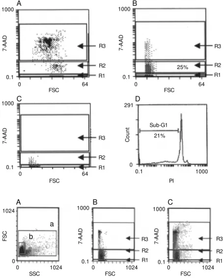
Figure 1 - Tw o-parameter flow cytometry analysis (forw ard scat-ter, FSC vs 7-amino actinomycin D, 7-AAD). PBM C from LCL pa-tients w ere obtained after cen-trifugation over a Ficoll-Hypaque gradient and heated for 1 h at 60oC to induce ACD (A) or cul-tured in the presence of Stau for 3 days to induce early apoptosis (B). Cell cultures w ithout Stau w ere used as control (C). R1 -Live cells (7-AAD-); R2 - early apoptotic cells (7-AADdim); R3 -dead cells (7-AADbright). D, Anal-ysis of Stau-induced apoptosis by hypotonic propidium iodide (PI) staining w as done to con-firm the results. The sub-G1 re-gion encompasses the apoptotic cells.
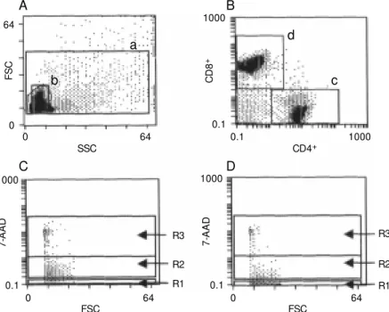
Figure 2 - A representative flow cytometry analysis used to de-termine 7-amino actinomycin D (7-AAD) incorporation based on tw o different gates (A) created in side scatter/forw ard scatter (SSC/FSC) dot plot: a, all cells; b, cells w ith viable patterns; B, cells w ithin gate b; C, cells w ithin gat e a. Region R1 encompasses live cells; R2 - early apoptotic cells; R3 - dead cells (ACD).
F
S
C
1024
0
SSC 0
R3
R2
R1 1024
7
-A
A
D
1000
0.1
7
-A
A
D
1000
0.1 R3
R2
R1
FSC
0 1024
FSC
0 1024
a
b
A B C
Re sults
Mo nito ring the accuracy o f 7-AAD staining
To determine whether the 7-AAD method effectively distinguished between live, early apoptotic and dead cells, LMC and PBMC were heated for 5 min at 60oC to induce
death, or cultured in the presence of Stau for induction of early apoptosis (see Material and Methods).
Figure 1 shows one representative series of 3 individual experiments in which the fluorescence intensity of 7-AAD vs forward
scatter (FSC) dot plot was used to define dead cells induced by heating (Figure 1A), and early apoptotic cells induced by treat-ment with Stau (Figure 1B). Untreated cells were used as controls (Figure 1C). Similar procedures were carried out using mono-nuclear cells obtained from lesions of a cuta-neous leishmaniasis patient (data not shown). Negligible 7-AAD incorporation was seen in live cells (region R1), while 7-AADdim
was observed in early apoptotic cells (R2) and 7-AADbright in dead cells (R3). For a
fol-lowed by agarose gel electrophoresis where the formation of characteristic DNA ladders was observed (data not shown).
Cells undergoing induced apoptosis or ACD were detectable in side vs forward light scatter plots. Treatment with Stau reduced FSC and increased side scatter (SSC), which is a well-established morphological feature of apoptosis; on the other hand, thermal injury at 60oC increased both FSC and SSC,
which is a characteristic of cells undergoing ACD (data not shown).
The 7-AAD and hypotonic PI staining techniques were also compared. PBMC or LMC were cultured for 3 days in medium containing Stau, or heated for 5 min at 60oC
and then stained in parallel with 7-AAD or with a hypotonic solution containing PI. In the 7-AAD dot plots (R2) we observed 25% of cells within the early apoptosis gate. These data closely matched the percentage of lym-phocytes in the sub-G1 region where the DNA fragmented content was detected in the PI histogram (21%) (Figure 1B and D, respectively).
Ce ll de ath analysis o f LMC o btaine d fro m LCL
patie nts
The 7-AAD method was used to evaluate the cell death events occurring in the lesions of LCL patients with active disease or spon-taneous healing. Figure 2 shows a represen-tative experiment in which a flow cytometry protocol was used to quantitatively deter-mine the frequencies of live, early apoptotic or dead cells in these groups of patients. 7-AAD incorporation (Figure 2B and C) was evaluated by two different gates created in the SSC/FSC dot plot: gate a, surrounding all analyzed cells including those supposed to be dead according to the light scatter pat-terns and deeply stained with PI, and gate b, constructed around cells with apparently normal morphology (unmodified SSC/FSC) and considered as viable cells by their ability to exclude vital dyes such as PI
(Fig-Figure 3 - Quantitative analysis of early apoptotic and dead cells obtained from lesions of LCL patients w ithin gates a and b ac-cording to Figure 2. A, Analysis w ithin gate a; B, cells analyzed w ithin gate b. Black bars - M ean frequencies ± SEM of early ap-optosis in cells from patients during active disease (N = 15) and during spontaneous healing (N = 2). Gray bars - mean fre-quencies ± SEM of dead cells in cells from patients during active disease and during spontaneous healing. * P<0.0001 compared to % dead cells (M ann-Whitney U-test).
ure 2A). Analysis within gate a (Figure 3A) showed higher numbers of early apoptotic cells in lesions of patients with active dis-ease (mean = 39.5 ± 2.7%) as compared to patients with spontaneous healing (mean = 17.8 ± 2.2%). Regarding dead cells, no sig-nificant difference was observed between the two groups of patients (16.3 ± 2.0 and 12.6 ± 4.0%, respectively, P = 0.49). It is interesting to note that even in gate b, cells supposed to be viable according to their scatter profiles showed important frequen-cies of early apoptotic events. Patients with active disease showed high numbers of early apoptotic cells (mean = 28.5 ± 3.8%) when compared to patients with spontaneously healing lesions (mean = 15.3 ± 3.0%) (P = 0.0001). As expected, Figure 3B shows small numbers of dead cells within gate b (as de-scribed in Figure 2), regardless of the group of patients analyzed (active disease = 5.1 ± 2.0%; healing lesions = 2.0 ± 1.0%).
Analysis o f ce ll de ath in CD 4+ o r CD 8+ T
ce lls fro m le sio ns o f LCL patie nts
Live, early apoptotic or dead CD4+ or
CD8+ T cells were evaluated in the LMC
obtained from lesions of LCL patients by the 7-AAD method and by dual-color cell sur-face staining for phenotypic analysis. The
%
o
f
c
e
lls
50
Spontaneous healing 40
30
20
10
0
*
*
Active disease
*
*
%
o
f
c
e
lls
40
30
20
10
0
Spontaneous healing Active disease
A
Figure 4 - Representative flow cytometric analysis carried out t o det erm ine live (R1), early apoptotic (R2) or dead (ACD) (R3) CD8+ or CD4+ T cells obtained from lesions of an LCL patient.
A, Scatter histogram (side/for-w ard, SSC/FSC) in (side/for-w hich gates a and b w ere created follow ing scatter patterns according to Fig-ure 2. B, Dual-color dot plot of CD8+-PE and CD4+-FITC T cells based on gate b according to Fig-ure 2. C, FSC vs 7-amino actino-mycin D (7-AAD) gated on CD8+ T cells w ithin gate d; D, FSC vs
7-AAD gated on CD4+ T cells w ithin gate c.
quantitation of the frequency of cell death events in cells within gate a was difficult because cells in advanced stages of apopto-sis and necroapopto-sis can lose their surface antigen expression. In this respect, early apoptosis and phenotypic determinations were per-formed only on gate b, created as described in Figure 2. Two other gates were also con-structed around the CD4+ (gate c) or CD8+
(gate d) T cells. Double positive or double negative cells were not considered relevant in this analysis. The 7-AAD incorporation in each subset was measured in two 7-AAD vs
FSC dot plots which combined definitions for gate b + c or gate b + d. These dot plots provided simultaneous determinations of live (region R1), early apoptotic (region R2) or dead (region R3) CD4+ or CD8+ T
lympho-cytes (Figure 4).
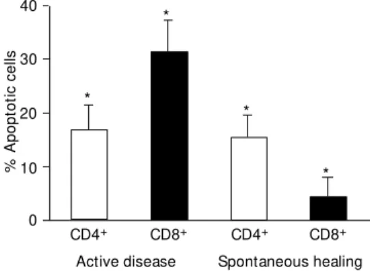
In patients with active disease, CD8+ and
CD4+ T cells exhibited high frequencies of
apoptosis, with significantly more apoptosis among CD8+ T cells than CD4+ T cells (means:
CD8+ = 31.3 ± 5.9%; CD4+ = 16.7 ± 4.7%; P =
0.00003). Conversely, patients with spontane-ous healing showed a general reduction in the frequency of apoptotic cells. Moreover, the numbers of apoptotic events was significantly
lower among CD8+ T cells than CD4+ T cells
(mean CD8+ = 4.3 ± 3.6%; mean CD4+ = 15.2
± 4.1%, P= 0.0004). Additionally, the num-bers of apoptotic CD4+ T cells in
spontane-ously healed lesions remained similar to those observed in patients with active disease, whereas the number of apoptotic CD8+ T cells
decreased in patients showing spontaneous healing (P = 0.016) (Figure 5).
D iscussio n
Apoptosis has been described as essen-tial for normal organogenesis and tissue de-velopment. This phenomenon plays an im-portant role in the immunopathogenesis of several parasitic diseases (27,28). Studies on cell death in protozoonoses have also fo-cused on its possible role in the immunopa-thology of experimental Chagas disease (29-31). Infection with the intracellular proto-zoan Toxoplasma gondii induces apoptosis of host CD4+ T lymphocytes, which may
involve a cooperative effect of IFN-g on Fas-mediated cell death (32). In leishmaniasis there is evidence suggesting that the sensi-tivity of Leishmania promastigotes to pro-grammed cell death is induced by heat shock
F
S
C
64
0
SSC 0
R3
R2
R1 64
C
D
8
+
1000
a
b A
7
-A
A
D
1000
0.1
0.1
CD4+
0.1 1000
B
c d
R3
R2
R1
FSC
0 64
7
-A
A
D
1000
0.1
C D
FSC
and modulated by calcium (33). It has also been demonstrated that intracellular infec-tion by Leishmania donovani inhibited mac-rophage apoptosis (34).
The importance of apoptotic events in the modulation of the immune response oc-curring in lesions of LCL patients is still unclear. To quantify cell death events in T cell subsets after they were obtained ex vivo
from lesions of LCL patients, we utilized the 3-color staining method which permitted us to measure live, early apoptotic or dead T cell subsets.
As expected, by analyzing all LMC from active LCL patients, we detected high num-bers of early apoptotic and dead cells. These data are in accordance with some histologi-cal findings which show the presence of fibrinoid necrosis in active LCL (35). Ex-pressive numbers of early apoptotic cells were detected in another analysis based on cells which exhibited a normal, unmodified scatter profile. This means that, despite the apparently normal morphological patterns, cells were undergoing early apoptosis.
When cell death events were evaluated in CD4+ or CD8+ T lymphocytes we observed
that active LCL patients had larger numbers of apoptotic CD8+ T cells than apoptotic
CD4+ T cells. These data suggest that during
active disease a large number of CD8+
lym-phocytes underwent early apoptosis and thereby had their immunologic functions compromised, leading to low cytokine pro-duction (mainly IFN-g) and probably impair-ing a favorable effect on the course of the disease.
Regarding patients with a tendency to self-healing, the small number of patients studied here did not allow us to reach con-sistent conclusions, although some interest-ing data were obtained. A lower rate of cell death was observed in these patients, a fact probably related to the clearance of infec-tion. In this situation, preserved T cells may maintain their efficacy, producing cytokines and activating macrophages to kill the
para-sites. In the case of apoptotic CD4+ and
CD8+ T cell analysis, these patients with
spontaneously healed lesions showed small numbers of apoptotic CD8+ T cells although
the frequencies of apoptotic CD4+ T cells
were similar to those observed in patients with active disease. Despite the small num-ber of patients studied, we can speculate that the low percentage of apoptosis in CD8+ T
cells of spontaneous healing patients may point to a significant role of this cell sub-population in the mechanism of cure of LCL. This possibility is in accordance with previ-ous results from our group (36-39) showing that CD8+ T cells are associated with cure
and protection in LCL.
Some hypotheses can be raised in order to explain the role of apoptosis mediating the immune responses in active or healing le-sions. One possibility refers to a T cell hy-persensitivity to leishmanial antigens, which is apparently the main immunopathological component in this disease, leading to activa-tion-induced cell death (AICD). The phe-nomenon of AICD is thought to operate in situations in which there is an excess of antigen, and may be a mechanism that pre-vents immunopathology resulting from over-activation of the immune system (40). This AICD is mediated by interaction of Fas (CD95) with its ligand (FasL), which is tran-siently expressed on activated T cells. In fact, such affirmation is in agreement with our preliminary observations showing that there was a high expression of CD95 in cells
Figure 5 - Early apoptosis in CD4+ or CD8+ T cells obtained from lesions of patients w ith ac-tive disease (N = 15) or sponta-neous healing (N = 2) as meas-ured by flow cytometry. Solid bars represent early apoptotic CD8+ T cells, and open bars, early apoptotic CD4+ T cells. Data are reported as mean ± SEM . * P<0.0004 - early apopto-tic CD8+ T cells compared to early apoptotic CD4+ T cells (M ann-Whitney U-test).
%
A
p
o
p
to
ti
c
c
e
lls
Spontaneous healing 40
30
20
10
0
Active disease
CD4+ CD8+ CD4+ CD8+
*
*
*
from lesions of patients with active disease (data not shown). Apoptosis may also be associated with modulation of the immune response leading to a beneficial effect or with cell and tissue destruction leading to aggravation of lesions. In active disease an important number of CD8+ and CD4+ T cells
undergo early apoptosis and therefore have their functional characteristics altered.
Re fe re nce s
1. Grimaldi Jr G, Tesh RD & M cM ahon-Pratt D (1989). A review of the geographic dis-tribution and epidemiology of leishmania-sis in the New World. American Journal of Tropical M edicine and Hygiene, 41: 687-725.
2. Coutinho SG, Louis JA, M auel J & Engers HD (1984). Induction by specific T lym-phocytes of intracellular destruction of
Leishmania major in infected murine mac-rophages. Parasite Immunology,6: 157-161.
3. Liew FY, M illott S, Li Y, Lelchuk R, Chan WL & Ziltener H (1989). M acrophage acti-vation by interferon-gamma from host-protective T cells is inhibited by interleu-kin (IL) 3 and IL4 produced by disease-promoting T cells in leishmaniasis. Euro-pean Journal of Immunology, 19: 1227-1232.
4. Titus RG, Kelso A & Louis JA (1984). Intra-cellular destruction of Leishmania tropica
by macrophages activated w ith macro-phage activating factor/interferon. Clinical and Experimental Immunology, 55: 157-165.
5. Barral A, Barral-Net t o M , Yong EC, Brow nell CE, Tw ardzik DR & Reed SG (1993). Transforming grow th factor ß as a virulence mechanism for Leishmania bra-ziliensis. Proceedings of the National A-cademy of Sciences,USA, 90: 3442-3446. 6. Liew FY & O’Donnell CA (1993). Immu-nology of leishmaniasis. Advances in Para-sitology, 32: 161-259.
7. M oll HR, Scollay R & M itchell GF (1988). Resistance to cutaneous leishmaniasis in nude mice injected w ith L3T4+ T cells but not w ith Ly-2+ T cells. Immunology and Cell Biology, 66: 57-65.
8. Reiner SL, Wang ZE, Hatam F, Scott P & Locksley RM (1993). Th1 and Th2 cell antigen receptors in experimental leish-maniasis. Science, 259: 1457-1460. 9. Bertho AL, Santiago M A & Coutinho SG
(1994). An experimental model of the pro-duction of metastases in murine cutane-ous leishmaniasis. Journal of Parasitology, 80: 93-99.
10. M üller I, Fruth U & Louis JA (1992). Im-munobiology of experimental leishmania-sis. M edical M icrobiology and Immunol-ogy, 181: 1-12.
11. M üller I, Kropf P, Etges RJ & Louis JA (1993). Gamma interferon response in secondary Leishmania major infection: role of CD8+ T cells. Infection and Immu-nity, 61: 3730-3738.
12. Conceição-Silva F, Perlaza BL, Louis JA & Romero P (1994). Leishmania major infec-tion in mice primes for specific major his-tocompatibility complex class I-restricted CD8+ cytotoxic T cell responses. European Journal of Immunology, 24: 2813-2817. 13. Kerr JFR, Wyllie AH & Currie AR (1972).
Apoptosis: A basic biological phenome-non w ith w ide-ranging implications in tis-sue kinetics. British Journal of Cancer, 26: 239-257.
14. M ajno G & Joris I (1995). Apoptosis, oncosis, and necrosis. An overview of cell death. American Journal of Pathology, 146: 3-15.
15. White E (1996). Life, death and the pur-suit of apoptosis. Genes and Develop-ment, 10: 1-15.
16. Thompson CB (1995). Apoptosis in the pathogenesis and treatment of diseases.
Science, 267: 1456-1462.
17. Telford WG, King LE & Fraker PJ (1994). Rapid quantitation of apoptosis in pure and heterogeneous cell populations using flow cytometry. Journal of Immunological M ethods, 172: 1-16.
18. Darzynkiew icz Z, Bruno S, Del Bino G, Gorczyca W , Hotz M A, Lassota P & Traganos F (1992). Features of apoptotic cells measured by flow cytometry. Cy-tometry, 13: 795-808.
19. Darzynkiew icz Z, Juan G, Li X, Gorczyca
W, M urakami T & Traganos F (1997). Cy-tometry in cell necrobiology: analysis of apoptosis and accidental cell death (ne-crosis). Cytometry, 27: 1-20.
20. Sw at W, Ignatow icz L & Kisielow P (1991). Det ect ion of apopt osis of im m at ure CD4+8+ thymocytes by flow cytometry.
Journal of Immunological M ethods, 137: 79-87.
21. Schmid I, Krall WJ, Uittenbogaart CH, Braun J & Giorgi JV (1992). Dead cell dis-crimination w ith 7-amino-actinomycin D in combination w ith dual color immuno-fluorescence in single laser flow cytom-etry. Cytometry, 13: 204-208.
22. Schmid I, Uittenbogaart CH, Keld B & Giorgi JV (1994). A rapid method for meas-uring apoptosis and dual-color immuno-fluorescence by single laser flow cytom-etry. Journal of Immunological M ethods, 170: 145-157.
23. Schmid I, Uittenbogaart CH & Giorgi JV (1994). Sensitive method for measuring apoptosis and cell surface phenotype in human thymocytes by flow cytometry.
Cytometry, 15: 12-20.
24. Nicolle CH (1908). Culture du parasite de bouton d’Oriente. Comptes Rendus de L’Académie des Sciences, 146: 842-843. 25. M odlin RL, Kato K, M ehra V, Nelson EE, Xue-Dong F, Rea TH, Pattengale PK & Bloom BR (1986). Genetically restricted suppressor T-cell clones derived from le-promatous leprosy lesions. Nature, 322: 459-461.
26. Nicoletti I, M igliorati G, Pagliacci M C, Grignani F & Riccardi C (1991). A rapid and single method for measuring thy-mocyte apoptosis by propidium iodide staining and flow cytometry. Journal of Immunological M ethods, 139: 271-279. 27. Barcinski M A & DosReis GA (1999).
zilian Journal of M edical and Biological Research, 32: 395-401.
28. Neves Jr I, Bert ho AL, Veloso VG, Nascimento DV, Campos-M ello DLA & M orgado M G (1998). Improvement of the lym phoproliferative im m une response and apoptosis inhibition upon in vitro treat-ment w ith zinc of peripheral blood mono-nuclear cells (PBM C) from HIV+ individu-als. Clinical and Experimental Immunol-ogy, 111: 264-268.
29. Lopes M F, da Veiga VF, Santos AR, Fonseca M E & DosReis GA (1995). Acti-vation-induced CD4+ T cell death by ap-optosis in experimental Chagas’ disease.
Journal of Immunology, 154: 744-752. 30. Lopes M F & DosReis GA (1995).
Apopto-sis as a cause of T-cell unresponsiveness in experimental Chagas’ disease. Brazil-ian Journal of M edical and Biological Re-search, 28: 913-918.
31. Lopes M F & DosReis GA (1996). Trypano-soma cruzi-induced immunosuppression: selective triggering of CD4+ T-cell death by the T-cell receptor-CD3 pathw ay and not by the CD69 or Ly-6 activation path-w ay. Infection and Immunity, 64:
1559-1564.
32. Liesenfeld O, Kosek JC & Suzuki Y (1997). Gamma interferon induces Fas-depend-ent apoptosis of Peyer’s patch T cells in m ice follow ing peroral infection w ith
Toxoplasma gondii. Infection and Immu-nity, 65: 4682-4689.
33. M oreira M EC, Del Portillo HA, M ilder RV, Balanco JM F & Barcinski M A (1996). Heat shock induction of apoptosis in promasti-gotes of the unicellular organism Leish-mania (LeishLeish-mania) amazonensis. Journal of Cellular Physiology, 167: 305-313. 34. M oore RJ & M atlashew ski G (1994).
Intra-cellular infection by Leishmania donovani
inhibits macrophage apoptosis. Journal of Immunology, 152: 2930-2936.
35. Bittencourt AL & Andrade ZA (1967). As-pectos imunopatológicos na leishmaniose cutâneo-mucosa. Hospital,71: 975-978. 36. Da-Cruz AM , Conceição-Silva F, Bertho
AL & Coutinho SG (1994). Leishmania -reactive CD4+ and CD8+ T cells associ-ated w ith cure of human cutaneous leish-maniasis. Infection and Immunity, 62: 2614-2618.
37. Coutinho SG, Oliveira M P, Da-Cruz AM ,
De Luca PM , M endonça SCF, Bertho AL, Soong L & M cM ahon-Pratt D (1996). T-cell response of American cutaneous leishmaniasis patients to purified Leish-mania pifanoi amastigote antigens and
Leishmania braziliensis promastigote an-tigens: immunologic patterns associated w ith cure. Experimental Parasitology, 84: 144-155.
38. Coutinho SG, Da-Cruz AM , Bertho AL, Santiago M A & De-Luca P (1998). Immu-nologic patterns associated w ith cure in human American cutaneous leishmania-sis. Brazilian Journal of M edical and Bio-logical Research, 31: 139-142.
39. M endonça SCF, De Luca PM , M ayrink W, Restom TG, Conceição-Silva F, Da-Cruz AM , Bertho AL, Costa CA, Genaro O, To-ledo VPCP & Coutinho SG (1995). Charac-terization of human T lymphocyte-medi-ated immune responses induced by a vac-cine against American tegumentary leish-maniasis. American Journal of Tropical M edicine and Hygiene, 53: 195-201. 40. Abbas AK (1996). Die and let live: