INSTITUTO DE INVESTIGAÇÃO E FORMAÇÃO AVANÇADA
ÉVORA, JUNHO DE 2017
ORIENTADORES:Professora Doutora Joana Margarida Ferreira da Costa Reis Professor Doutor José Alberto Caeiro Potes
Tese apresentada à Universidade de Évora para obtenção do Grau de Doutor em Ciências Veterinárias
Maria Teresa Carvalho Oliveira
Development of a large animal model for
percutaneous vertebroplasty for in vivo
evaluation of a new injectable cement
Para ser grande, sê inteiro: nada Teu exagera ou exclui.
Sê todo em cada coisa. Põe quanto és No mínimo que fazes.
Assim em cada lago a lua toda Brilha, porque alta vive.
À memória dos meus avós Aos meus amores
v
O meu mais profundo agradecimento à minha orientadora, Professora Doutora Joana Reis, pela confiança que depositou continuamente em mim, até quando a mesma em mim teimava em falhar. Pela motivação e orientação constantes ao longo destes anos, dando-me sempre asas para voar.
Ao meu orientador, Professor Doutor José Potes, por toda a generosa e filantropa transmissão de conhecimentos, quer no âmbito deste projeto, quer no restante da minha actividade profissional, enquanto médica veterinária.
O meu agradecimento à Universidade de Évora, que apoiou financeiramente o meu trabalho através de uma bolsa individual de doutoramento, por sua vez suportada pela Comissão Europeia, ao abrigo do Sétimo Programa-Quadro, através do projeto RESTORATION – RESORBABLE CERAMIC BIOCOMPOSITES FOR ORTHOPAEDIC AND MAXILLOFACIAL APPLICATIONS–, ao abrigo da ação “SME-targeted Collaborative project”, grant agreement NMP.2011.2.1-1.
Agradeço à Professora Doutora Maria Cristina Queiroga a ajuda preciosa nos trabalhos decorrentes do projeto, quer fosse nas cirurgias, quer no tratamento das ovelhas internadas no Hospital Veterinário. Mais agradeço todo o auxílio prestado na correção dos artigos publicados, uma e outra vez.
Ao Professor Doutor José Lopes de Castro agradeço toda a inestimável amizade e companheirismo demonstrados desde os primeiros dias de convivência. Muito obrigada pela sua entusiástica disponibilidade para todas as tarefas que lhe propúnhamos, mesmo quando se
vi
e transmissão de conhecimentos no tratamento e bem-querer das nossas “amigas”, ao longo de todo este projeto. Não tenho qualquer dúvida que o seu trabalho, conjuntamente com o trabalho do Professor Lopes de Castro, foi fundamental para o sucesso inolvidável deste projeto.
Ao Professor Doutor Kenneth Dalgarno, da Universidade de Newcastle, e à Doutora Sarrawat Rehman, da JRI Orthopaedics, pela pronta disponibilidade que demonstraram para nos auxiliar nos testes mecânicos efetuados no decorrer do estudo ex vivo, tendo que, para isso, abandonar o conforto dos seus lares e famílias e vir até Portugal.
À Professora Doutora Chiara Vitale-Brovarone, do Politécnico de Turim, e ao Doutor Antonio Manca, da Unidade Radiológica do Instituto Oncológico Candiolo de Turim, pela amabilidade que tiveram em corrigir em tempo recorde o artigo “Novel mesoporous bioactive glass/calcium sulphate cement for percutaneous vertebroplasty and kyphoplasty: in vivo study”, só assim tornando possível a entrega desta tese “a tempo e horas”.
Ao Professor Doutor António Ramos, na representação do TEMA, da Universidade de Aveiro, por se ter disponibilizado para nos auxiliar na execução dos testes mecânicos das vértebras, abrindo-nos a porta da sua “casa” e acompanhando-me nesta minha aprendizagem.
Ao Professor Doutor António Completo, o meu agradecimento pelo convite dirigido à Professora Doutora Joana Reis e, posteriormente, estendido à minha pessoa, para a submissão do capítulo “Bone: functions, structure and physiology”, inserido num livro, neste momento em fase de edição, intitulado de “The bone tissue computational mechanics - Biologic behaviour, remodelling algorithms and numerical applications”.
vii
aventura.
Aos colegas do Hospital Veterinário da Universidade de Évora e alunos que se encontravam em atividades hospitalares/ complementares, o meu muito obrigada por todo o auxílio prestado nas cirurgias e tratamentos dos animais.
À Leonor Pinho e à Margarida Costa, o meu eterno obrigada. Nunca me esquecerei dos nossos anos no HVUÉ, das nossas estórias, das nossas alegrias e tristezas, convivências e divergências, e todos os ingredientes que constroem relações duradouras.
O meu desmedido agradecimento ao João Fragoso e à Ana Bota por toda a amizade, companheirismo, motivação e ajuda disponibilizados desde sempre, quer no âmbito do projeto, quer fora dele. Vocês fazem parte da minha família de Évora.
O meu eterno obrigada às minhas amigas de coração, que mesmo longe estão perto, sempre à distância de um telefonema, e que são sempre as primeiras a quem recorro nos momentos mais difíceis. À Susana, minha companheira de viagem, obrigada pela paciência para ouvires os meus dilemas quando por vezes os teus se afiguram gigantes. À Ana Isabel, minha amiga desde sempre, que admiro enquanto mãe e profissional, e que continua a ser uma das minhas referências quando volto à minha terra-mãe. À minha prima Marta, pelo exemplo de força de mulher que é e dá. Todas lutamos e almejamos por vidas melhores, mais justas e plenas.
À minha família, de sangue e de coração, que em tempos de necessidade são os primeiros a acudir.
viii
À minha amada irmã, por toda a paciência e ajuda na correção de boa parte destes escritos.
Aos meus amores pequeninos, que sem quererem ou pedirem, se viram privados da mãe, mais do que, por vezes, a própria desejava. Amo-vos daqui até à Lua e voltar.
ix
Nota: O conteúdo da presente tese foi parcialmente publicado ou submetido para publicação. Como tal, os capítulos 2, 3, 4, 5 e 6 encontram-se formatados de acordo com as normas de formatação e de referenciação bibliográfica das respetivas publicações. A indexação das secções, figuras e tabelas dos capítulos supracitados aparece de acordo com a ordem original, precedida pelo número do capítulo correspondente.
xi Abbreviations xxi Abstract xxv Sumário xxvii Chapter 1 1 1. Introduction 3
1.1. Context and Objectives 3
1.2. References 8
Chapter 2 11
2. Bone: functions, structure and physiology 13
2.1. Introduction 13
2.2. And yet it moves 14
2.2.1. Bone functions 14
2.2.2. Regulations of bone metabolism (modelling/ remodelling)
16 2.2.2.1. Parathormone (PTH), Vitamin D and
calcitonin
16 2.2.2.2. Growth hormone (GH) 17 2.2.2.3. Sexual hormones and steroids (oestrogen and testosterone)
18 2.2.2.4. Thyroid hormones 18 2.2.2.5. Leptin (“satiety” hormone) 19 2.2.2.6. Bone Morphogenetic Proteins (BMPs) 19 2.2.2.7. Insulin and insulin-like growth factors
(IGF-1 and IGF-2)
19 2.2.3. Bone structure and mechanical properties 20
2.2.4. The bone matrix 24
2.2.5. Bone cell population 26 2.2.5.1. Osteoblasts 26
2.2.5.2. Osteocytes 26
2.2.5.3. Osteoclasts 28 2.2.6. Bone remodelling and cell interplay 30 2.2.7. Bone mechanotransduction 31
2.2.7.1. The membrane elements, ECM-cell and cell-cell adhesions
31 2.2.7.2. Primary cilia 34 2.2.7.3. The cytoskeleton 35 2.2.8. Mechanotransduction mechanisms 37 2.2.8.1. Strain, frequency and loading duration 37 2.2.8.2. Bone piezoelectricity 38
xii 3.3. Experimental 64 3.4. Discussão 65 3.5. Conclusões 66 3.6. Agradecimentos 66 3.7. Referências 66 Chapter 4 67
4. Ex vivo Model For Percutaneous Vertebroplasty 69
4.1. Abstract 69
4.2. Introduction 69
4.3. Experimental 70
4.4. Results and Discussion 71
4.5. Conclusions 73
4.6. Acknowledgements 73
4.7.References 74
4.8.DOI References 75
Chapter 5 77
5. Percutaneous vertebroplasty: a new animal model 79
5.1. Abstract 79
5.2. Introduction 80
5.3. Materials and methods 80 5.3.1. Ex vivo model development 80 5.3.1.1. Animal Model 80 5.3.1.2. Micro-CT assessment 80 5.3.1.3. Bone defect creation 80 5.3.1.4. Cement injection 81 5.3.1.5. Mechanical testing 81 5.3.1.6. Statistical analysis 81 5.3.2. In vivo model application 81 5.3.2.1. Animal model 81 5.3.2.2. Anesthetic protocol 81
5.3.2.3. Surgery 82
5.3.2.4. Biomaterial injection 82 5.3.2.5. Post-surgery 82 5.4. Results and discussion 83
5.4.1. Ex vivo model 83 5.4.2. In vivo model 85 5.5. Conclusions 85 5.6. Acknowledgments 87 5.7. Supplementary material 87 5.8. References 87
xiii
6.3. Materials and methods 95 6.3.1. Cement development and characterization 95 6.3.2. In vivo large model development 95 6.3.3. Assessment of tissue regeneration 96 6.3.3.1. Macrocospic inspection 96 6.3.3.2. Micro-CT assessment 97 6.3.3.3. Histology 98 6.3.3.3.1. Undecalcified histology 98 6.3.3.3.2. Decalcified histology 99 6.3.4. Statistical analysis 100 6.4. Results 100
6.4.1. Cement development and characterization 100 6.4.2. In vivo findings 101 6.4.3. Assessment of tissue regeneration 102 6.4.3.1. Macroscopic 102 6.4.3.2. Micro-CT assessment 103 6.4.3.3. Histology 105 6.4.3.3.1. Undecalcified histology 105 6.4.3.3.2. Decalcified histology 108 6.5. Discussiom 110 6.6. Conclusions 113 6.7. Acknowledgements 113 6.8. Conflicts of interest 113 6.9. References 114 Chapter 7 119 7. Discussion 121
7.1. Analgesia in animal models 121 7.2. Ex vivo model development 124
7.3. In vivo study 127
7.4. References 134
Chapter 8 145
8. Conclusions and Future Perspectives 147
8.1. Conclusions 147
8.2. Future Perspectives 148
xv
Figure 2.2. Microphotograph of cortical bone in vertebrae (undecalcified bone section of sheep vertebra, Giemsa-Eosin, 20x magnification; slide digitalized using Nanozoomer SQ, Hamamatsu Photonics, Portugal). Haversian systems are evident, along with organized fibrils. Osteocytes are visible in their lacunae, in between lamellae.
22
Figure 2.3. Image of vertebral trabecular bone (undecalcified bone section of sheep lumbar vertebra, Giemsa-Eosin, 1.25x magnification; slide digitalized using Nanozoomer SQ, Hamamatsu Photonics, Portugal). The picture illustrates the sponge-like structure of cancellous bone.
23
Figure 2.4. Osteoblasts are round cells that when actively deposing matrix on bone surfaces show prominent Golgi complexes (on the left, microphotograph, undecalcified bone section, Giemsa Eosin, magnification 100x). When quiescent, osteoblasts appear as flat bone lining cells.
26
Figure 2.5. Microphotograph of undecalcified bone section of sheep vertebra, Giemsa-Eosin, on the left, showing osteocytes (Giemsa-Eosin, 24x magnification; slide digitalized using Nanozoomer SQ, Hamamatsu Photonics, Portugal). Some of the canaliculi where cell processes run are evident. The image on the right illustrates the resulting three-dimensional syncytium.
27
Figure 2.6. On top, microphotograph of TRAP positive osteoclasts in cutting cone; on bottom, a schematic detail of the ruffled border membrane in direct contact with bone. This is the resorbing organelle; along its enlarged ruffled contact surface, proton pumps lower the local pH, dissolving hydroxyapatite.
29
Figure 3.1. Vertebroplastia percutânea com fluoroscopia. 64 Figure 3.2. Vertebroplastia em ovino com monitorização anestésica adequada. 65 Figure 3.3. Ovinos intervencionados em pasto. 65 Figure 4.1. 3D partial reconstructed L4 showing major anatomical landmarks
and PVP access point, with the advocated access angle (large arrow).
71
Figure 4.2. Scan image of a ovine lumbar vertebra showing the wide nutritional foramen (white arrow).
72
Figure 4.3. 3D partial reconstructed L4 showing the interconnected defect (white arrow).
72
Figure 4.4. MicroCT scan image of a L4 showing the disrupted vertebral foramen (white arrow)
xvi
instruments’ orientation regarding a transverse plane.
Figure 5.2. Three-dimensional (3D) partially reconstructed L4 showing the instruments’ orientation regarding a frontal plane.
81
Figure 5.3. Anesthetized sheep, with the surgical location (over the lumbar vertebrae) already clipped and sheep’s position supported with foam wedges.
82
Figure 5.4. Anesthetic monitor providing data from the sheep. 82 Figure 5.5. C-Arm image showing an L4 with an interconnected defect, in a
dorsoventral projection.
82
Figure 5.6. C-Arm image showing Cerament filling the interconnected defects (in vivo study), in a dorsoventral projection.
83
Figure 5.7. Scan image of an ovine lumbar vertebra showing the wide nutritional foramen (white arrow).
83
Figure 5.8. 3D partial reconstructed L4 showing the interconnected defect (white arrow).
83
Figure 5.9. Defect volume of interest (VOI)’s values distribution shown in a histogram with normal curve (SPSS 22 graph).
84
Figure 5.10. Micro-computed tomography (micro-CT) scan image of an L4 showing the disrupted vertebral foramen (white arrow).
84
Figure 5.11. C-Arm image showing an L4 with defect injected with Cerament in a dorsoventral projection.
84
Figure 5.12. Micro-computed tomography (micro-CT) scan image of an L4 showing the artefact caused by Cerament.
85
Figure 5.13. (Top) Compression testing of vertebra with Cerament-filled defect. (Middle) Vertebrae stiffness (Groups A-D) (SPSS 22 graph). (Bottom) Vertebrae normalized stiffness (Groups A-D) (SPSS 22 graph).
86
Figure 5.14. Micro-Computed tomography (micro-CT) scan image injected with Cerament, from the in vivo study, showing cement resorption, new bone formation, and no artefact.
87
Figure 5.15. Micro-Computed tomography (micro-CT) scan image of an L4 injected with Cerament, from the in vivo study, showing a potential cortical disruption of the vertebral foramen (white arrow).
xvii
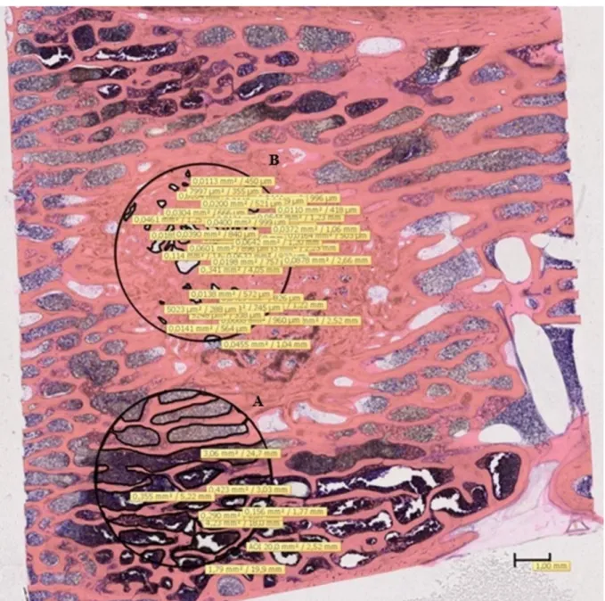
Figure 6.1. Histomorphometric study. Giemsa staining, 0.55x magnification; the circles are limiting the two areas of interest: A) newly formed trabecular bone within the defect; B) mature trabecular bone outside the defect. Scale bar on image.
99
Figure 6.2. Cement characterization. A) Syringe after the injection of Spine-Ghost, coupled with a 13-gauge vertebroplasty needle and Spine-Ghost cement extruded on a paper sheet to prove its injectability; B) Spine-Ghost radiopacity assessment C) FE-SEM micrograph of precipitated HAp on Spine-Ghost after 7 days of immersion in SBF and relative EDS spectrum.
101
Figure 6.3. Macroscopic assessment. A) Instrumentation entry point with a pink discoloration pointed by the cannula; B) hemivertebra, after sagittal cut, with cement still evident (large white arrow) and adjoining newly formed bone (small white arrow); C) macroscopic evidence of cortical disruption of the vertebral canal.
102
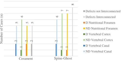
Figure 6.4. Clustered stacked chart presenting micro-CT qualitative evaluation. Data obtained from the injected vertebrae: group A (n=7) – Cerament™ –, and group B (n=8) – Spine-Ghost. Legend: ND – not disrupted; D – disrupted.
103
Figure 6.5. Micro-CT post-mortem assessment. Here it can be seen the reconstructed cross-section images and the 3D rendered models of 2 injected vertebrae – one from each group –, explanted from the sheep closest to the mean, when it comes to the trabecular bone mineral densities of the intact caudal hemivertebrae (BMDCHv): 1) vertebra injected with Spine-Ghost, with a
BMDCHv of 0.45 gcm-3; 2) vertebra injected with Cerament™, with a BMDCHv
of 0.50 gcm-3; a) cross-section image of CHv; b) 3D rendered image of 30 cross-sections of VOICHv; c) cross-section image of the defect; d) 3D rendered
image of 30 cross-sections of VOIDefect.
105
Figure 6.6. Undecalcified Technovitt 9100 sections of two vertebrae. A) Cerament™ augmented vertebra section (magnification 0.55x) with areas of lighter pink stain where bone was more recently mineralized (black arrows); B) Spine-Ghost augmented vertebra section (magnification 0.54x) showing the affinity of Giemsa to the biomaterial, which stains in shades of blue (white arrowheads). Both defect areas are fulfilled with an intricate network of trabeculae, with multiple directions, surrounding and penetrating the remains of the cements. In contrast, is evidenced the trabecular structure of intact tissue, mostly parallel to the long axis of the vertebrae. C) Cerament™ augmented vertebrae section (magnification 0.55x) where empty areas with some cement present may be seen; it’s also visible the disruption of the cortex of the vertebral canal (black arrow). This section belongs to same vertebra shown above in Figure 3. D) Spine-Ghost augmented vertebra section (magnification 0.55x) where an empty area with some cement present may be seen. This was the only
xviii
marking showing different patterns of bone apposition and bone remodeling, with alizarin complexone (small arrow) encircling a trabecula. Scale bar on images.
Figure 6.8. Sections of decalcified histology with Mallory and Masson trichromes staining. A) Spine-Ghost augmented vertebra demineralized section (Masson Trichrome with aniline blue), 0.5x magnification, showing the intricated net of trabecular bone within the defect area; B ) and D) Spine-Ghost and Cerament™ augmented vertebrae demineralized sections, respectively (Mallory’s trichrome), 10x magnification, showing biomaterial integration into the trabecular bone structure (arrowheads), blue staining of collagen fibres (small arrows); C) Cerament™ augmented vertebra demineralized section (Masson Trichrome with aniline blue), 0.58x magnification. Scale bar on images.
109
Figure 6.9. Sections of immunohistochemistry of Spine-Ghost augmented vertebra demineralized sections. A) 100x magnification, anti-osteopontin antibody, showing DAB stained biomaterial (large arrow); osteocytes, bone lining cells and cells within bone marrow are also positive (arrowheads). B) 1000x magnification, anti-TRAP antibody. The image shows an area of cement/bone interface. Scale bar on images.
xix
Table 6.1. Descriptive analysis of the 3D structural parameters 104 Table 6.2. Descriptive analysis of the Histomorphometric Parameters 107
xxi
AOI Area of interest
ASBMR American Society for Bone and Mineral Research
BAr Bone area
BCIS Bone cement implantation syndrome BMC Bone mineral content
BMD Bone mineral density
BMP Bone morphogenetic protein BMU Basic multicellular unit BRU Bone remodelling unit BSP Bone sialoprotein BS/BV Specific surface BTM Bone turnover marker BV/TV Relative bone volume Ca2+ Calcium ions
CaS Calcium sulphate CHv Caudal hemivertebra CNS Central nervous system COX-2 Cyclooxygenase-2
CSR Calcium-sensing receptor
CSH α-Calcium sulphate hemihydrate CTGF Connective tissue growth factor DAB Diaminobenzidine
DGAV Direcção Geral de Alimentação e Veterinária ECG Electrocardiograma
ECM Extracellular matrix
EISA Evaporation-induced self-assembly
FELASA Federation of European Laboratory Animal Science Associations
FGF23 Fibroblast Growth Factor 23 GABA Gamma-aminobutyric acid
xxii
KP Kyphoplasty
MAR Mineral apposition rate MBG Mesoporous bioactive glass
M-CSF-1 Macrophage colony stimulating factor-1 Micro-CT Micro-computed tomography
MSC Mesenchymal stem cells
NSAID Non-steroidal anti-inflamatory drug
NO Nitric oxide
ODF Osteoclast differentiation factor OPG Osteoprotegerin
OPN Osteopontin
PFF Pulsating fluid flow
PG Proteoglycan PGE2 Prostaglandin E2 Pi Phosphorus ions PMMA Polymethylmethacrylate PKP Percutaneous kyphoplasty PTH Parathormone PVP Percutaneous vertebroplasty
RANK Receptor activator of nuclear factor-κB RANKL Receptor activator of nuclear factor κB ligand
RESTORATION Resorbable Ceramic Biocomposites for Orthopaedic and Maxillofacial Applications
ROI Region of interest
Rt-PCR Reverse transcription-polymerase chain reaction Runx2 Runt-related transcription factor 2
SBF Simulated body fluid SMC Smooth muscle cell
SPARC Secreted protein acidic and rich in cysteine TbN Trabecular number
xxiii
TIVA Total intravenous anaesthesia TRAP Tartrate-resistant acid phosphatase TSH Thyroid stimulating hormone
TV Tissue volume
T3 Triiodothyronine
T4 Thyroxine
VBH Vertebral body height
VCF Vertebral compression fracture VOI Volume of interest
[1,25(OH)2D] 1,25 dihydroxyvitamin D3 [1,25(OH)2D3]
2D Two-dimensional
xxv
percutaneous bone interventions are based on a polymeric non-resorbable matrix. However, they can present some complications. Calcium suphate-based cements are effective bone substitutes. Disadvantages include their limited shear and compressive strength.
To go beyond the state of the art, a new bioactive calcium sulphate-based cement was developed – Spine-Ghost. To test the suitability of the injectable cement for percutaneous vertebroplasty, a preclinical study was mandatory.
A new large animal model for percutaneous vertebroplasty was developed in sheep. In the ex vivo model, bone defects were created in the cranial hemivertebrae through a bilateral modified parapedicular approach, and mechanical tests were performed. The ex vivo model is reproducible, and safe under physiological loads.
In the in vivo study, two groups of Merino sheep were defined (n=8): a) the control group, injected with a commercial ceramic cement; and b) the experimental group, injected with Spine-Ghost. Of the first interventioned animals, two presented cardiorespiratory distress during the cement injection, and one had mild neurologic deficits in the hindlimbs. All sheep survived and completed the 6-month implantation period.
After sacrifice, the samples were assessed by micro-computed tomography, histological, histomorphometric, and immunohistological studies. There was no evidence of cement leakage into the vertebral foramen. No signs of infection or inflammation were observed. Most importantly, there was cement resorption and new trabecular bone formation in the bone defects of all sheep.
The model of percutaneous vertebroplasty is considered suitable for preclinical in vivo studies, mimicking clinical application.
Spine-Ghost proved to be an adequate material for percutaneous vertebroplasty, with a biological response identical, if not superior, to the one elicited by the available commercial control.
xxvii Sumário
O trabalho aqui apresentado teve por objetivo a avaliação in vivo de um novo biomaterial injetável para vertebroplastia a cifoplastia percutâneas.
As fraturas de compressão vertebral com indicação cirúrgica são tratadas com recurso a técnicas minimamente invasivas. Presentemente, a maioria dos cimentos utilizados baseiam-se numa matriz polimérica não reabsorvível. Podem, no entanto, causar algumas complicações. Os cimentos à base de sulfato de cálcio são substitutos ósseos eficazes, cujas desvantagens incluem resistência limitada a esforços de corte e compressão.
Um novo cimento bioativo de sulfato de cálcio – Spine-Ghost – foi desenvolvido. Para testar a sua aplicabilidade na vertebroplastia percutânea, tornou-se imperativo um estudo pré-clínico.
Para o efeito, um novo modelo animal para vertebroplastia percutânea foi desenvolvido em ovinos e sujeito a ensaios mecânicos. No modelo ex vivo, foram criados bilateralmente dois defeitos ósseos interligados, nas hemivértebras craniais, através de uma abordagem parapedicular modificada. O modelo ex vivo é reprodutível e seguro sob cargas fisiológicas.
No estudo in vivo, definiram-se dois grupos de ovelhas Merino (n=8): a) grupo controlo, injetado com cimento comercial de base cerâmica; b) grupo experimental, injetado com Spine-Ghost. Nos primeiros animais intervencionados, dois apresentaram alterações cardiorrespiratórias durante a injeção de cimento, e um défices neurológicos ligeiros nos membros pélvicos. Todos os animais sobreviveram e completaram o período de implantação de 6 meses.
Após a ocisão, as amostras foram avaliadas por microtomografia computorizada, histologia, histomorfometria e imunohistoquímica. Não se observou derrame de cimento para o canal vertebral, nem sinais de infeção ou inflamação. Ademais, verificou-se a reabsorção do cimento e a neoformação de tecido ósseo trabecular no interior dos defeitos ósseos, em todos os animais.
O modelo de vertebroplastia percutânea é considerado adequado para estudos pré-clínicos, mimetizando a aplicação clínica.
3
transdisciplinary way. The contemporary “One medicine – One health” concept implies that multidisciplinary teams of medical doctors, veterinarians, engineers, biologists, among other experts (Cook & Bal, 2014), share the responsibilities and synchronise local and global activities to address health problems at the animal-human-environment interfaces, either these problems concern the whole population or the individual, since a better understanding and a more adequate control of animal health (often through environmental management) potentially lead to less human risks (Kaplan et al., 2008). The collaboration between the specialists from the different scientific areas is the vital key that will result in a better health for humans and animals in a near future.
The improvement of the living conditions and the aging of the world population, along with the evolution of medicine, led to the necessity of new approaches to pathologies and the development of novel therapeutics. Likewise, the evolution of animal healthcare and welfare and the subsequent raise of the life span of our pets and domestic animals triggered the increase of disorders commonly associated with geriatrics. These diseases occasionally can be compared with some human conditions, considering the interspecies similarities. Conditions like cancer, diabetes, asthma, orthopaedic disorders – like osteoarthritis and osteoporosis –, cardiovascular diseases, and neurologic diseases – like dementia – are thoroughly studied in companion animals, as well as in other species. These studies can be of benefit to both animal and human health, in the sense that they can generate longer, healthier lives for all species.
The testing of innovative biomaterials – for clinical application in orthopaedic surgery and other areas – in living and sentient animal models in direct benefit to the human race, must be the subject of careful planning and consideration, given the ethical conflict that arises. There are several currents of thought within the international scientific community, either for or against the use of animals. For example, the "Medical Research Modernization Committee" believes that animal testing is mainly driven by economic interests. Moreover, it says that animal testing should not be considered a valid method for medical research due to the anatomical, physiological and pathological differences between human and non-human (Anderegg, 2002). Bearing this in mind, in order to deepen the knowledge in bone structure and physiology, and responding to an invitation made by Professor Joana Reis, Chapter 2 was written. Nonetheless, numerous evidences show that animal testing is inexorable and an
4
In accordance to the previous insights, the present doctoral research work was integrated in a multidisciplinary European project – Resorbable Ceramic Biocomposites for Orthopaedic and Maxillofacial Applications [RESTORATION] – led by the University of Newcastle Upon Tyne, United Kingdom. This project involved several consortium partners, each with different tasks – from the development of the biomaterials, through the in vitro testing and the in vivo testing in the small animal model, until the in vivo testing in the large animal model.
The project’s research team from the University of Évora, coordinated by Professor Joana Reis, was responsible for the development and application of the large animal models and ulterior biomaterials testing. Encompassing the different models, a total of 40 Merino sheep were intervened. All animals were daily assessed and taken care by members of the research team, for over 6 months. After the sacrifice of the animals, the samples were collected and processed for ulterior analysis. This work is the result of multiple tasks, such as sheep handling and care, surgery planning and performance, postoperative animal care, and bone samples processing and evaluation. For confidentiality reasons imposed on the Consortium partners, in this thesis the only shown data are those relative to vertebroplasty, even though this was just one of the models developed. Nevertheless, other surgical models were implemented and biomaterials assessed.
The entire project was designed and developed respecting the prevailing European legislation for the protection of animals used for scientific purposes – Directive 2010/63/EU (Official Journal of the European Union, 2010) –, and the Federation of European Laboratory Animal Science Associations [FELASA] guidelines (Mähler et al., 2014), which follows the 3R’s recommendations of replacement, reducing and refinement of animals. Consortium partners’ local laws were also respected; hence, the work regarding the large animal models was developed under the Portuguese law decree – Decreto-Lei nº 113/2013 (Diário da República, 2013).
Accordingly, researchers are advised to minimize the use of animals through Replacement – e.g. with computational models, in vitro studies, studies in invertebrates –; Reducing – e.g. with proper study’s experimental design, pilot tests, ex vivo studies, and the use of non-invasive techniques of diagnostic, like ultrasonography, fluoroscopy, radiology and
5
knowledge as imperative requisites to be able to recognize the distinct behaviours of each chosen experimental species and to prevent and minimize the negative impact over the individuals, not only acting when the distress or pain are installed, but also assuming a proactive attitude and avoiding unnecessary noxious stimulus or manipulations.
In consideration of the foregoing, it is within the 3R’s concepts that the veterinarian role assumes utmost importance in basic science’s research, through the development of new better models and other refinement techniques, in a synergy with the other researchers – e.g. physicians, engineers, and biologists –, which help to minimize the animal usage. This way, in addition to contribute to the progress of medicine, the veterinarian guarantees the best health care to the experimental animals. Therefore, it is of utter importance the investment in veterinary science as a mean to a general medical benefit, diminishing the gap between animal models and clinical trials and profiting every species (Cook et al., 2010; Vainio, 2012; Baird et
al., 2013).
Finally, the ultimate purpose of the project was to evaluate, in a controlled in vivo study, the bone response to a novel resorbable calcium sulphate injectable bone cement, developed by one of the consortium partners for application in percutaneous vertebroplasty. As in the clinical context, the biomaterial was to be injected in a vertebral body defect through a minimally invasive procedure. Considering the difficulties and limitations found in previous techniques and studies (Zhu et al., 2011; Galovich et al., 2011; Benneker et al., 2012; Verron et al., 2014), the development of a new reproducible and feasible preclinical model for percutaneous vertebroplasty in sheep was mandatory. The use of sheep is largely accepted due to its translational features regarding the human species, besides its availability, low cost, easy handling and good homogeneity when selected for age, race and gender. In addition, sheep are considered a good model for orthopaedic research due to its anatomical similarities when compared to the human model, when it comes to the size, weight, skeletal structure, bone remodelling and biomechanical behaviour of the bone, also consenting the use of some of the same prosthetic material (Alini et al., 2008; Li et al., 2015; Wancket et al., 2015). Chapter 4 presents an article describing the development of the ex vivo model and chapter 5 the in vivo application of the previously developed model for the novel biomaterial testing.
6
Up to date, most of the cements used in orthopaedic surgery are based on a polymeric matrix – polymethylmethacrylate (PMMA) (Magnan, 2013; Puoci, 2015). These cements have been thoroughly used and investigated over the years, mainly for its immediate effect and safety, but they also present some limitations, such as high polymerization temperatures, low bioactivity, bioinertia, absence of resorbability, and high elastic modulus. These properties are related to the – relatively rare – complications that have been documented with the use of PMMA. For instance, the high polymerization temperatures potentially induce inflammation and/or necrosis of the neighbour structures; likewise, the formation of new healthy tissue is hindered by the cement’s the low bioactivity. Another syndrome, known as Bone Cement Implantation Syndrome (BCIS), is described as a possible complication of PMMA injection during total hip arthroplasty and vertebroplasty/kyphoplasty. It occurs around the cement injection, secondary to medullary fat embolism, and is characterized by hypoxia and/ or hypotension, with or without unexpected loss of consciousness and ultimately death. Finally, subsequent fractures of the contiguous vertebrae are also described as complications, due to the cement’s high elastic modulus (Becker et al., 2014; Puoci, 2015).
Synthetic ceramics are widely known and are proved to be safe and effective in bone substitution, since they are highly biocompatible, resorbable, and osteoconductive, displaying mechanical properties similar to those of the cancellous bone, with reduced Young’s Modulus, and a low risk of infection or donor site morbidity (Campana et al., 2014). They also present low setting temperatures and short setting times. Some disadvantages include their limited shear and compressive strength, when compared to those of the normal bone (Campana et al., 2014; Gupta et al., 2015). To overcome these limitations, ceramics are most of the times combined with other composites (Campana et al., 2014). Numerous different formulations are commercially available, like different-sized granules, blocks, and injectable pastes. Furthermore, currently there is an investment in developing new composites that can act as delivery vehicles for cells, growth factors and antibiotics into fractures (Waselau et al., 2007; Larsson et al., 2011; Campana et al., 2014).
Calcium sulphate-based injectable ceramics present themselves as good alternatives to PMMA, concerning vertebroplasty/kyphoplasty. Calcium sulphate offers an effective, low cost gap filler, by allowing vascular ingrowth and by being resorbable, thus allowing new bone
7
biomaterials.
To progress beyond the state of the art in terms of available calcium sulphate cements for percutaneous vertebroplasty, a novel bioactive injectable cement for percutaneous vertebroplasty was developed – Spine-Ghost (Vitale-Brovarone et al., 2015). Spine-Ghost was implanted into a sheep vertebral defect model. The performance was compared to a commercial biphasic cement – Cerament™|Spine Support. Cerament™’s application for vertebral compression fractures (VCFs) has been well documented (Marcia et al., 2014). Spine-Ghost supported new bone formation into the vertebral body defect, while slowly biodegraded, suggesting it allows a safe and unpainful medical recovery, thus reducing pain and morbidity. Chapter 6 presents the final in vivo study and the biological response to the biomaterial.
8
Baird, A. W., Rathbone, M. J., & Brayden, D. J. (2013). Human: Veterinary Technology Cross Over. In Long Acting Animal Health Drug Products (pp. 359-375). Springer US.
Benneker, L. M., Gisep, A., Krebs, J., Boger, A., Heini, P. F., & Boner, V. (2012). Development of an in vivo experimental model for percutaneous vertebroplasty in sheep. Veterinary
and Comparative. Orthopaedics and Traumatololy V.C.O.T., 25 (3), 173-177.
Cassidy, C., Jupiter, J. B., Cohen, M., Delli-Santi, M., Fennell, C., Leinberry, C., Husband, J., Ladd, A., Seitz, W. R., & Constanz, B. (2003). Norian SRS cement compared with conventional fixation in distal radial fractures. A randomized study. Journal of Bone and
Joint Surgery. American volume, 85(11), 2127–2137.
Cook, J. L., Kuroki, K., Visco, D., Pelletier, J. P., Schulz, L., & Lafeber, F. P. J. G. (2010). The OARSI histopathology initiative–recommendations for histological assessments of osteoarthritis in the dog. Osteoarthritis and cartilage, 18, S66-S79.
Cook, J. L., & Bal, B. S. (2014). The Clinical Biomedical Research Advances Achievable Utilizing One Health Principles. In Confronting Emerging Zoonoses (pp. 233-239). Springer Japan.
Decreto-Lei nº 113/2013, de 7 de Agosto. Diário da República, 1.ª série — N.º 151. Ministério da Agricultura, do mar, do ambiente e do ordenamento do território, Lisboa.
Directive 2010/63/EU. (2010). Official Journal of the European Union, L276/33-79, ISSN 1725-2601.
Galovich, L. A., Perez-Higueras, A., Altonaga, J. R., Gonzalo Orden, J. M., Barba, M. M., & Morillo, M. C. (2011). Biomechanical, histological and histomorphometric analyses of calcium phosphate cement compared to PMMA for vertebral augmentation in a validated animal model. European Spine Journal, 20(3), 376-382.
Goodman, S. B., Bauer, T. W., Carter, D., Casteleyn, P. P., Goldstein, S. A., Kyle, R. F., Larsson, S., Stankewich, C. J., Swiontkowski, M. F., Tencer, A. F., Yetkinler, D. N., &
9
(2015). Bone graft substitutes for spine fusion: A brief review, World Journal of
Orthopaedics, 6(6), 449-456.
Jubel, A., Andermahr, J., Mairhofer, J., Prokop, A., Hahn, U., & Rehm, K. E. (2004). Use of the injectable bone cement Norian SRS for tibial plateau fractures. Results of a prospective 30-month follow-up study. Der Orthopäde, 33(8), 919–927.
Kaplan, B., Kahn, L. H., & Monath, T. P. (2008). The brewing storm. Veterinaria italiana,
45(1), 9-18.
Kopylov, P., Jonsson, K., Thorngren, K. G., & Aspenberg, P. (1996). Injectable calcium phosphate in the treatment of distal radial fractures. The Journal of hand surgery (British
and European Volume), 21(6):768–771.
Larsson, S., & Hannink, G. (2011). Injectable bone-graft substitutes: current products, their characteristics and indications, and new developments. Injury, 42, S30-S34.
Li, Y., Chen, S. K., Li, L., Qin, L., Wang, X. L., & Lai, Y. X. (2015). Bone defect animal models for testing efficacy of bone substitute biomaterials. Journal of Orthopaedic
Translation, 3(3), 95-104.
Kumar, C. Y., Nalini, K. B., Jagdish Menon, D. K. P., & Banerji, B. H. (2013). Calcium sulfate as bone graft substitute in the treatment of osseous bone defects, a prospective study.
Journal of clinical and diagnostic research: JCDR, 7(12), 2926-2928.
Magnan, B., Bondi, M., Maluta, T., Samaila, E., Schirru, L., & Dall’Oca, C. (2013). Acrylic bone cement: current concept review. Musculoskeletal surgery, 97(2), 93-100.
Mähler, M., Berard, M., Feinstein, R., Gallagher, A., Illgen-Wilcke, B, Pritchett-Corning K, Raspa M (2014). FELASA recommendations for the health monitoring of mouse, rat, hamster, guinea pig and rabbit colonies in breeding and experimental units. Laboratory
10
Paul, E.F., Paul, J. (eds.) (2001). “Why Animal Experimentation Matters: The Use of Animals in Medical Research”, Transaction Publishers.
Vainio, O. (2012). Translational animal models using veterinary patients–An example of canine osteoarthritis (OA). Scandinavian Journal of Pain, 3(2), 84-89.
Verron, E., Pissonnier, M. L., Lesoeur, J., Schnitzler, V., Fellah, B. H., Pascal-Moussellard, H., Pilet, P., Gauthier, O., & Bouler, J. M. (2014). Vertebroplasty using bisphosphonate-loaded calcium phosphate cement in a standardized vertebral body bone defect in an osteoporotic sheep model. Acta biomaterialia, 10(11), 4887-4895.
Vitale-Brovarone C., Pontiroli L., Novajra G., Tcacencu I., Reis J., Manca A. (2015). Spine-Ghost: a new bioactive Cement for Vertebroplasty. Key Eng Mat, 631, 43-47.
Wancket, L. M. (2015). Animal Models for Evaluation of Bone Implants and Devices Comparative Bone Structure and Common Model Uses. Veterinary pathology, 52(5), 842-850.
Waselau, M., Samii, V. F., Weisbrode, S. E., Litsky, A. S., & Bertone, A. L. (2007). Effects of a magnesium adhesive cement on bone stability and healing following a metatarsal osteotomy in horses. American Journal of Veterinary Research, 68(4), 370-378.
Wolfe, S. W., Pike, L., Slade, J. F. III, & Katz, L. D. (1999). Augmentation of distal radius fracture fixation with coralline hydroxyapatite bone graft substitute. The Journal of hand
surgery, 24(4):816–827.
Zhu, X.S., Zhang, Z.M., Mao, H.Q., Geng, D.C., Zou, J., Wang, G. L., Zhang, Z. G., Wang, J. H., Chen, L., & Yang, H. L. (2011). A novel sheep vertebral bone defect model for injectable bioactive vertebral augmentation materials. Journal of Materials Science:
Chapter 2
Bone: functions, structure and physiology
Accepted for publication in July 2016 as a chapter of the book “The bone tissue computational mechanics - Biologic behaviour, remodelling algorithms and numerical applications”, in the book series entitled: Lecture Notes in Computational Vision and Biomechanics, Springer.
13
Bone: functions, structure and physiology
Maria Teresa Oliveira1 and Joana da Costa Reis2
Abstract The bone is reviewed regarding its functions, regulation, morphological structure and physiology; addressing how complex, how responsive to external and internal stimuli, and how intimately intertwined with other organs it is. From embryogenesis to endocrine regulation and bone remodelling, an overall view is presented. Special emphasis is given to how cell structure and tissue organization contribute to the response to mechanical stimuli.
1. Introduction
Bones are dynamic structures. They vary in shape, size and number, and are divided in axial and appendicular skeleton. Through life they are subjected to loads and strains that induce their shape, with old matrices being replaced by newly formed ones. This process is important for maintaining bone volume and strength. In the case of fractures, bones are capable of healing, as long as stability and alignment are assured.
It is the entanglement of environment, cell-to-cell interactions and cell-extracellular matrix interactions that direct and model osteogenesis, bone repair and remodelling. Mechanical forces are essential in early embryonic development. There is evidence that morphogenesis is regulated through fluid flow mechanisms and by cellular contractility. Cells generate tensional forces through contraction of actin-myosin cytoskeleton filaments, which are transmitted through cadherin-mediated adhesion sites to neighbour structures, these being either cells or extracellular matrix. Cohesivity determines morula contraction and the definition of multiple layers - with the development of endoderm, mesoderm and ectoderm in the blastula - and thus early embryo shaping (Oster et al., 1983; Takeichi, 1988; de Vries et al., 2004;
1 Maria Teresa Oliveira
Universidade de Évora, Largo dos Colegiais, Évora, email: [email protected] 2 Joana da Costa Reis
14
Ingber, 2006). The stress-dependent changes in cytoskeletal conformation and cell shape act locally to regulate cell phenotype, of utter importance are interactions with the extracellular matrix (Ingber, 2006). Unidirectional fluid flow, dependent on the specialized motor protein complex dynein, determines organ lateralization and asymmetry, by causing differences in key molecules expression (such as the TGF-family signalling molecules) (Collignon et al., 1996, Okada et al., 1999; Cartwright et al., 2004, Nakamura et al., 2006). Lateralization may also depend on fluid shear, in the embryo, by acting on a group of non-motile cilia, coupled to calcium channels; fluid shear may cause the intracellular calcium concentrations to rise and initiate the cascade of events responsible for lateralization (McGrath et al., 2003). The mechanical environment is also determinant for vasculogenesis, angiogenesis (Schmidt et al., 2007; le Noble et al., 2008; Patwari and Lee, 2008), and neuronal development (Bray, 1979; Dennerll et al., 1989; Anava et al., 2009).
The embryo mesoderm is constituted by spindle or star-shaped cells called mesenchymal stem cells (MSCs). MSCs are the most pluripotential cells in the organism, giving rise to different tissues such as the connective tissue, muscle, cardiovascular tissue and the entire skeletal system. Bone, cartilage, tendons and ligaments develop through mechanisms of proliferation, migration and differentiation, but also apoptosis (Carter and Beaupré, 2001).
For a long time, bone was generally regarded as a less interesting organ, but we are only now starting to address how complex it is in its functions, its responsiveness to external and internal stimuli, and its intimate intertwining with other organs.
2.
And yet it moves2.1 Bone functions
Bone or osseous tissue is the most rigid and resilient tissue of the body. Constituted by dense connective tissue, it’s the primary tissue of the skeleton, thus providing structure, support, and protection to vital organs, like the brain (skull), the spinal cord (vertebrae), and the heart and lungs (ribs and sternum). Moreover, the vertebrae participate in the spine shock absorbance – providing adequate load cushioning for the intervertebral disks (fibrocartilaginous joints) –, whilst long bones, along with the joints, enable body movement – providing levers for the muscles.
Additionally, bones act as the major source of blood, since haematopoiesis occurs in their medullary cavity. In infants, the bone marrow of all long bones is capable of this synthesis. As a person gets older, part of the red marrow turns into yellow fatty marrow, no longer capable of haematopoiesis. Functional red marrow in adults is restricted to the vertebrae and the extremities of femur and tibia.
15
Bones also have an important role as:
• Mineral storage: mostly calcium (Ca2+), phosphate (Pi), and magnesium; it plays an important metabolic role, regulating mineral homeostasis (Bélanger et al., 1968; Zallone et al., 1983; Teti & Zallone, 2009), a process mediated by many hormones.
• Growth factor storage: insulin like growth factors 1 and 2 (1 and IGF-2), transforming growth factor-beta (TGF-β), acidic and basic fibroblast growth factor, platelet-derived growth factor, and bone morphogenetic proteins have been isolated from bone matrix (Mohan & Baylink, 1991). Osteoblasts have been shown to produce many of these growth factors and their production is regulated by systemic hormones and local mechanical stress (Baylink et al., 1993).
• Adipose tissue storage (yellow bone marrow as a fatty acid/ energy reserve) (Rosen et al., 2009; Krings et al., 2012; Suchacki et al., 2016). • Acid-base balance, as it buffers the blood against excessive pH changes by
absorbing or releasing alkaline salts (Green & Kleeman, 1991; Arnett et al., 2003; Bushinsky & Krieger, 2015).
• Heavy metal and other foreign elements storage, after detoxification from the blood, that are, later on, excreted (Roelofs-Iverson et al., 1984; Sharma et al., 2014).
• Endocrine organ, as it produces two known circulating hormones: a. Fibroblast Growth Factor 23 (FGF23): produced mainly by
osteocytes (Rhee et al., 2011), but also by osteoblasts (Masuyama et al., 2006), acts on the kidney to inhibit 1α-hydroxylation of vitamin D and promote phosphorus excretion in urine (Shimada et al., 2004; Fukumoto & Martin, 2009; Haussler et al., 2012). FGF23 also inhibits phosphorus absorption in the intestine, thus regulating inorganic phosphate metabolism and mineralization (Feng et al., 2006). It is now acknowledged that the serum calcium concentration regulates FGF23 production (David et al., 2013), thus making FGF23 into a calcium-phosphorus regulatory hormone (Lopez et al., 2011; Rodriguez-Ortiz et al., 2012). Hence, FGF23 excess or deficiency results in several abnormalities of phosphate metabolism. Excess FGF23 inhibits renal phosphate reabsorption and 1,25 dihydroxyvitamin D3 [1,25(OH)2D] production, leading to hypophosphatemia and suppression of circulating 1,25(OH)2D levels and, ultimately, rachitic changes in bone (Fukumoto & Yamashita, 2007), such as happens in autosomal dominant hypophosphatemic rickets and osteomalacia (ADHR) and in the paraneoplastic syndrome called tumour-induced osteomalacia (TIO). In contrast, reductions in FGF23 cause the syndrome of tumoral calcinosis (Shimada et al., 2001), characterized by hyperphosphatemia, increased 1,25(OH)2D and soft tissue calcifications (Lyles et al., 1988; Fukumoto & Yamashita, 2007). An obligate FGF23 coreceptor was identified – Klotho– (Urakawa et al., 2006). Klotho is required to activate FGF
16
receptors and their signalling molecules. Secreted Klotho suppresses, by itself, activity of insulin, insulin-like growth factor-1 (IGF-1) (Kurosu et al., 2005), Wnt (Liu et al., 2007), and TGF-β1 (Doi et al., 2011) by interacting with these growth factors or their receptors. The resulting FGF23-Klotho axis represents a specialized system responsible for the external and internal Ca2+ e Pi balance in the bone, intestine and kidney. FGF23-Klotho axis works under parathormone regulation, since parathormone directly promotes FGF23 expression by osteocytes (Quarles et al., 2012), whereas balance is sought by FGF23 inhibiting action on parathyroid glands (Ben-Dov et al., 2007; Krajisnik et al., 2007).
b. Osteocalcin: a protein produced by osteoblasts in bone, major regulator of insulin secretion by direct action over the pancreatic β-cell, and increasing sensitivity of peripheral tissues, e.g. muscles and liver, enhancing glucose uptake and energy expenditure, thus intervening in glycaemia regulation (Lee & Karsenty, 2008; Ferron et al., 2008; Ferron et al., 2010; Fulzele et al., 2010); it also acts on adipocytes to increase adiponectin, thus reducing fat deposition (Ribot et al., 1987; Reid et al., 1992).Furthermore, studies on clinical diabetes have shown that blood osteocalcin levels are significantly lower in diabetics, when compared to non-diabetic controls, and that these levels are inversely related to fat mass and blood glucose (Kindblom et al., 2009; Pittas et al., 2009). Lastly, osteocalcin shows some influence in male fertility, by enhancing testosterone production by Leydig cells in the testes (Oury et al., 2011).
2.2 Regulation of bone metabolism (modelling/ remodelling)
Bone functions, like (re)modelling and fracture repair, are accomplished by four types of cells: osteoblasts, bone lining cells, osteocytes and osteoclasts. These processes are regulated locally by cytokines and growth factors, and systemically by hormones, neuropeptides and other mediators (Harada and Rodan, 2003; Karsenty et al., 2009).
2.2.1 Parathormone (PTH), Vitamin D and calcitonin:
The regulation of the bone mineral metabolism (calcium and phosphorus) results from the interplay between parathormone (PTH), calcitonin, FGF23 and vitamin D. PTH is released from the parathyroid glands in response to low levels of extracellular ionized calcium, through the presence of specific cell-surface calcium-sensing receptor (CSR) on the glands. High levels of PTH cause the increase of the number of osteoclasts, and resorption of bone matrix, with consequent release of calcium phosphate and increasing calcaemia. Inversely, low levels of PTH cause the elevation of osteoblast numbers. It also acts over osteoblasts’ receptors, thus stimulating osteoblasts to stop synthetizing collagen, and to inhibit the secretion of stimulating factor by osteoclasts (Calvi et al., 2003). At renal level PTH, stimulated
17
by low plasma calcium, inhibits phosphate reabsorption and accelerates its excretion, stimulates calcium reabsorption, and upregulates a hydroxylase enzyme (CYP27B1), thus stimulating the final step of 1,25(OH)2 vitamin D3 synthesis (Murayama et al., 1998).
Circulating hormonal metabolite, 1α,25-dihydroxyvitamin D3 (1,25(OH)2D3) originates an activated complex that enhances several physiological functions, including intestinal calcium and phosphate absorption, bone phosphate and calcium resorption, and renal calcium and phosphate reabsorption, which results in a rise in the blood calcium and phosphate, required for bone passive mineralization of unmineralized bone matrix to occur (Haussler et al., 1998; Saini et al., 2013). Additionally, 1,25(OH)2D3 stimulates differentiation of osteoblasts and the expression of several bone proteins, like bone-specific alkaline phosphatase, osteocalcin, osteonectin, osteoprotegerin, and other cytokines; and influences the proliferation and apoptosis of other skeletal cells, including hypertrophic chondrocytes (Clarke, 2008).
Calcitonin is produced by parafollicular cells of the thyroid, in direct relationship to extracellular calcium, through the same sensor that regulates the production of PTH. It inhibits matrix resorption, promotes calcium and phosphate excretion, thus reducing calcium and phosphate serum levels; calcitonin has an inhibiting effect over osteoclast mobility and over the secretion of proteolytic enzymes through its receptor on osteoclasts (Boissy et al., 2002; Hadjidakis & Androulakis, 2006).
2.2.2 Growth hormone (GH):
Growth hormone, or somatotropin, is produced in the anterior hypophysis, stimulates growth in general, particularly over the epiphyseal cartilage (Isaksson et al., 1982). It stimulates certain organs, as the liver and the skeleton, to synthetize somatomedins that have effect over growth, like insulin-like growth factor 1 (IGF-1) and 2 (IGF-2). Thus, GH stimulates bone formation in two ways: (IGF-1) via a direct interaction with GH receptors on osteoblasts and 2) via an induction of endocrine and autocrine/paracrine IGF-1 (Ohlsson et al., 1998).
According to Ohlsson et al. (1998), GH action in bone remodelling follows a “biphasic model”: initially it increases bone resorption with a concomitant bone loss, which is followed by a phase of increased bone formation. When bone formation is stimulated more than bone resorption (transition point), bone mass is increased. A net increase of bone mass will be seen after 12–18 months of GH treatment in GH deficient adults.
Moreover, a recent study developed in rats showed that GH increased bone growth, by increasing both periosteal and endocortical bone formation, bone mineral content (BMC) and bone mineral density (BMD), and that the administration of GH along with PTH increases bone growth and bone formation, decreases bone resorption, and has a synergistic effect on increasing bone density and bone mass (Guevarra et al., 2010).
18
2.2.3 Sexual hormones and steroids (oestrogen and testosterone):
Oestrogen is an important regulator of skeletal development and homeostasis, both via direct and indirect effects over the skeleton (Turner et al., 1994; Prince et al., 1994). Indirectly it influences, for example, the calcium intestinal absorption (Liel et al., 1999; ten Bolscher et al., 1999) and secretion (Draper et al., 1997), and the calcium renal excretion; oestrogen also influences the secretion of PTH (Väänänen et al., 2005; Robinson et al., 2009). On the other hand, the mechanisms by which oestrogen acts directly on bone tissue are not completely understood. Nevertheless, it’s now acknowledged that oestrogen maintains bone homeostasis by inhibiting osteoblast and osteocyte apoptosis (Tomkinson et al., 1997; Kousteni et al., 2002; Emerton et al., 2010). Moreover, oestrogen inhibits the osteoclast formation and activity as well as induces osteoclast apoptosis, thus indirectly preventing excessive bone resorption (Hughes et al., 1996; Rodan & Martin, 2000; Faloni et al., 2007; Faloni et al., 2012; Khosla et al., 2012).
Androgens are also important to bone homeostasis, in both sexes. It has been shown that they stimulate bone formation in the periosteum, through several effects on osteoblasts and osteoclasts, and by influencing the differentiation of pluripotent stem cells toward distinct lineages (Wiren & Marcus, 2010). Similarly, to the loss of oestrogen, the loss of androgen influences the rate of bone remodelling by removing restraining effects on osteoblastogenesis and osteoclastogenesis, and creates an imbalance between resorption and formation, as it prolongs the lifespan of osteoclasts and shortens the lifespan of osteoblasts. Likewise, androgens maintain cancellous bone mass and integrity, regardless of age or gender (Compston et al., 2001; Vanderschueren et al., 2004).
2.2.4 Thyroid hormones:
Thyroid hormones (T3 and T4) levels influence bone growth during early development as well as adult bone turnover and maintenance. They act both indirectly, by enhancing the effects of growth hormone over tissues, and directly, by stimulating bone resorption and formation. Hypothyroidism causes impaired bone formation and growth retardation; inversely, thyrotoxicosis is an established cause of secondary osteoporosis. Osteoclastic receptors for thyroid hormone have been demonstrated (Abu et al., 1997). Thus, bone turnover is increased by thyroid hormones, which is confirmed by increased biochemical markers of bone turnover, such as osteocalcin and bone-specific alkaline phosphatase (Harvey et al., 1991; El Hadidy et al., 2011; Waring et al., 2013), and therefore bone loss can occur (Britto et al., 1994; Hadjidakis & Androulakis, 2006). Recent studies also suggest a potential direct effect of thyroid stimulating hormone (TSH) on bone (Abe et al., 2003) and TSH receptors have been found on osteoblasts and osteoclasts. Furthermore, recombinant TSH showed antiresorptive effects in ovariectomized rats (Abe et al., 2003; Sun et al., 2008) and lower TSH levels – with no apparent association with free T4 levels – have been related with hip fracture risk, supporting the idea that TSH effect on the skeleton may be independent from free T4 (Waring et al., 2013). Finally, abnormal thyroid hormone signalling has been recognized as an osteoarthritis’ risk factor (Bassett & Williams, 2016).
19
2.2.5 Leptin (“satiety” hormone):
Leptin is produced mainly in adipose tissue, influences bone metabolism via direct signalling from the central nervous system (CNS). It inhibits osteoclast generation (Holloway et al., 2002), promotes the decrease in cancellous bone and increase in cortical bone, thus enhancing bone enlargement (Ducy et al., 2000; Elefteriou et al., 2004; Hamrick & Ferrari, 2008); it also increases osteoblast number and activity, acting primarily through the peripheral pathways (Turner et al., 2013). Another study showed that leptin increases bone mineral content and density, especially at the lumbar spine (Mantzoros et al., 2011). Leptin also increases the expression of IGF-1 receptor and IGF-1 receptor messenger RNA (mRNA) within the chondrocytes and the progenitor cell population, thus acting as a skeletal growth factor with a direct peripheral effect on skeletal growth centre; it also induces chondrocytes proliferation and differentiation through specific leptin binding sites (Maor et al., 2002). Leptin is also a key up regulator of FGF23 secretion (Tsuji et al., 2010) and it has recently been described as a direct enhancer of parathormone secretion (Lopez et al., 2016).
2.2.6 Bone Morphogenetic Proteins (BMPs):
BMPs are group of 15 growth factors also known by cytokines, which belong to the transforming growth factor β (TGF-β) superfamily,with the ability to induce the formation of bone (Urist et al., 1965) and cartilage (Kobayashi et al., 2005). Bone formation is a very complex process wherein BMPs play the major role in the regulation of osteoblast lineage-specific differentiation and later bone formation (Beederman et al., 2013). Alterations in BMPs activity are often associated to a great variety of clinical pathologies, like skeletal and extra-skeletal anomalies, autoimmune, cancer, and cardiovascular diseases (Rahman et al., 2015). BMPs crosstalk with several other major signalling pathways, e.g. Wnt, Akt/mTOR, miRNA, among others, having Runx2 as a key integrator (Lin & Hankenson, 2011; Rahman et al., 2015). Among all BMPs, several authors refer to BMP9 as one of the most potent BMPs in inducing osteogenic differentiation of MSCs, both in vitro and in vivo (Kang et al., 2004; Kang et al., 2007; Beederman et al., 2013); moreover, TGF-β and GH are known to act synergistically with BMP9 to enhance bone formation (Li et al., 2012; Huang et al., 2012; Rahman et al., 2015). In addition to BMP9, other BMPs also have shown the ability to induce osteogenesis in vivo, such as BMP2, BMP6 and BMP7 (Franceschi et al., 2000; Jane et al., 2002; Cheng et al., 2003), with recombinant human-BMP2 and -BMP7 already being commercialized with the purpose of enhancing bone healing (Carreira et al., 2014). Contrariwise, BMP3 is known to be a negative regulator of bone formation (Kang et al., 2004).
2.2.7 Insulin and insulin-like growth factors (IGF-1 and IGF-2):
IGF-1 stimulates chondrocyte proliferation in the growth plate, thus playing a crucial role in longitudinal bone growth (Lupu et al., 2001). It is also involved in the formation of trabecular bone, which is essential to bone mineralization (Zhang et al., 2002). Insulin and IGF-1 have shown to be anabolic agents in osteoblast and bone development, mainly through the activation of Akt and ERK signalling
20
pathways; also, IGF-1, but not insulin, was capable of inducing osteoblasts in vivo proliferation (Zhang et al., 2012). IGF-1 inhibits the gene expression of osteocalcin, a marker for differentiating osteoblasts, whilst insulin enhances it. Furthermore, insulin indirectly enhances Runx2 expression, which is a regulator of osteoblast differentiation, by inhibiting the expression of Runx2 inhibitor Twist2 (Fulzele et al., 2010; Zhang et al., 2012). A study with insulin-deficient type I diabetic mice showed that these mice presented a decreased expression of Runx2 and the Runx2-regulated genes, like osteocalcin and collagen type I, and a secondary decrease in bone formation. Bone loss was restored after insulin treatment, which increased Runx2 expression and the expression of related genes (Fowlkes et al., 2008). Likewise, IGF-2 potentiates BMP-9-induced osteogenic differentiation and bone formation (Chen et al., 2010) through PI3K/AKT signalling. Moreover, a recent study in mice aortas showed that IGF-2 induces the expression of miR-30e, in a feedback loop, which is a major down-regulator of osteogenic differentiation in MSCs and smooth muscle cells (SMCs) (Ding et al., 2015).
2.3 Bone structure and mechanical properties
Bone mechanical properties depend on mineralization degree, porosity, composition and organization of solid matrix. Therefore, the mechanical behaviour of a whole bone is highly dependent on its properties at a microscale (Rho et al., 1998).
Bone is composed of 70% inorganic component (of which 95% is hydroxyapatite and 5% are impurities impregnated in hydroxyapatite), 22% to 25% of organic component (of which 94-98% is mainly collagen type I and other non-collagen proteins and 2%-6% are cells) and 5 to 8% is water (Sommerfeldt & Rubin, 2001). Mature, lamellar bone is morphologically classified in two different types: cortical or compact and cancellous bone. Cortical and cancellous bone types differ in structure and functional properties.
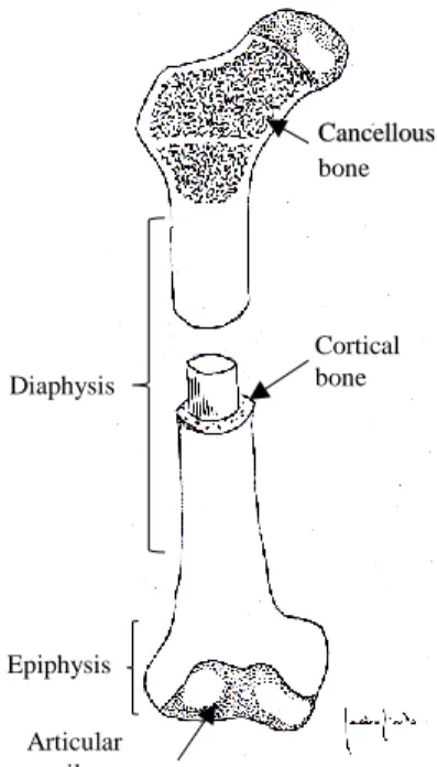
The typical structure of a long bone such as the femur or the humerus comprises the shaft, or diaphysis, and the extremities, or epiphysis (Fig. 1). The diaphysis consists of a cylinder of compact bone surrounding the central medullary cavity, lined by a thin layer of connective tissue, the endosteum.
21
Fig. 1 Illustration of a long bone structure, showing
the distribution of two different types of lamellar bone: cancellous and cortical compact bone.
The epiphysis consists of cancellous bone surrounded by a layer of cortical bone. Within the porous chambers of cancellous bone lays red bone marrow. A layer of dense connective tissue called periosteum covers the outer surface of the bone, except for the areas of articulation, covered with hyaline cartilage. The periosteum is highly vascular and is responsible for appositional bone growth (Van De Graaff, 2001).
Cortical bone (Fig. 2) is a porous mineralized tissue and accounts for approximately 80% of the skeletal mass. It is formed by tightly packed collagen fibrils, forming concentric lamellae. Each lamella is 2 – 3 μm thick and is arranged in several discrete layers of parallel fibrils, each layer having a different orientation of fibrils (Weiner et al., 1999). Apatite crystals (mainly carbonated apatite) are deposited within and around these fibrils. The lamellae form cylinders containing a hollow central tube wherein blood vessels and nerves run. The ensemble is called Haversian system or osteon and it is the microstructural unit of cortical bone. Blood vessels form a three-dimensional network, from the centre of the osteon (Haversian canals) and penetrating the cortical bone layer perpendicularly (Volkman’s channels) (Meyer & Wiesmann, 2006). In between the osteons are remnants of incomplete osteons, known as interstitial systems or interstitial bone.
Articular cartilage Cortical bone Diaphysis Epiphysis Cancellous bone
22
Fig. 2 Microphotograph of cortical bone in vertebrae (undecalcified bone section
of sheep vertebra, Giemsa-Eosin, 20x magnification; slide digitalized using Nanozoomer SQ, Hamamatsu Photonics, Portugal). Haversian systems are evident, along with organized fibrils. Osteocytes are visible in their lacunae, in between lamellae.
Cancellous (or trabecular) bone has a more loosely organized structure and higher porosity. The lamellae are organized in a parallel manner, forming trabeculae in a flattened and spongy-like network (Fig. 3). Trabeculae are covered by osteoblasts and bone-lining cells. Osteoblasts actively depose extracellular matrix (ECM) and bone-lining cells are in an inactive state.
The metabolic rate of trabecular bone is higher than that of cortical bone, and so are the remodelling phenomena. The trabecular network is a light structure, of uttermost importance for load transfer through the bone. This can be clearly seen in epiphyses and metaphysis of long bones, but also in vertebrae and ribs; trabeculae are orientated according to routine load bearing direction (Carter & Beaupré, 2001; Currey, 2003).