UNIVERSIDADE DE LISBOA
FACULDADE DE CIÊNCIAS
(DEPARTAMENTO DE BIOLOGIA VEGETAL)
On the trail of Mycobacterium avium subsp. paratuberculosis and
Mycobacterium bovis in mainland Portugal: a microbiological and
molecular survey in wildlife
Mestrado em Biologia Molecular e Genética
Ana Cristina Lopes dos Reis
Dissertação orientada por:
Professora Doutora Mónica Vieira Cunha
ii
Agradecimentos
Na base da concretização deste trabalho, duas palavras-chave, esforço e empenho, que só surgiram com o contributo e apoio incondicional de muitas pessoas e é a essas que manifesto o meu eterno agradecimento.Dirijo o meu primeiro agradecimento ao Instituto Nacional Investigação Agrária e Veterinária (INIAV), instituição que me acolheu, e cujas instalações tornaram possível a execução prática deste trabalho. Dirijo o meu agradecimento em particular à UEISPSA e ao Laboratório de Bacteriologia e Micologia.
À minha orientadora, Professora Doutora Mónica Cunha, agradeço por me ter acolhido no INIAV e por me ter proporcionado todas as condições físicas e imateriais para o desenvolvimento deste trabalho. Agradeço-lhe igualmente o suporte financeiro para a participação no curso “Applied methods in Biogeography”, ministrado pelo CE3C (Centre for Ecology, Evolution and Environmental Changes).
À Dra. Célia Leão, agradeço toda a disponibilidade manifestada para me auxiliar na execução do trabalho respeitante à pesquisa de Mycobacterium avium subsp.
paratuberculosis.
À Dra. Ana Canto agradeço toda a ajuda disponibilizada na realização da técnica de
spoligotyping.
Ao Professor Doutor Carlos Fonseca e Mestre Victor Bandeira, do Departamento de Biologia & CESAM da Universidade de Aveiro, agradeço a disponibilização de amostras de carnívoros, veados e javalis, bem como a informação sobre a localização geográfica dos animais.
Ao Doutor Luís Miguel Rosalino, do CE3C, agradeço igualmente a cedência de amostras, e informação geográfica das mesmas.
À Estradas de Portugal IP, aos caçadores, produtores e gestores de caça, à Dra. Paula Simões e Fencaça, ao Clube Português de Monteiros, e à Confederação Nacional dos Caçadores Portugueses, pela generosa contribuição de amostras para o banco de tecidos que esteve na base do presente trabalho.
iii À Fundação para a Ciência e a Tecnologia (FCT). O trabalho presente nesta dissertação foi parcialmente financiado pela FCT, no âmbito do projeto nacional com a referência PTDC/CVT/117794/2010, com enquadramento no Projeto 3599 – Promover a Produção Científica e Desenvolvimento Tecnológico e a Constituição de Redes Temáticas.
À Doutora Ana Botelho e à Dra. Teresa Albuquerque, pela cedência de isolados de M. bovis e M. caprae para a realização da técnica de spoligotyping, e de dados sobre a origem geográfica dos mesmos.
Aos meus colegas de gabinete, Marta Vaz, Ana Prata e Tiago Baeta, agradeço não só toda a ajuda disponibilizada, mas também a boa disposição e o companheirismo demonstrados ao longo deste ano de realização de trabalho experimental.
Aos meus pais, agradeço todo o apoio que sempre demonstraram para com todas as minhas decisões, mas em especial a todo o apoio ao longo do meu percurso académico, e sem o qual não teria sido possível a realização deste Mestrado.
Ao meu irmão, agradeço ter estado sempre presente com a sua forma peculiar de apoio e motivação, o que me deu muita força para terminar esta dissertação.
Por fim, a todos os meus familiares e amigos, agradeço toda a motivação para a conclusão desta dissertação.
iv
Abstract
Paratuberculosis and bovine tuberculosis (bTB) are important infectious diseases of cattle caused by pathogenic mycobacteria, whose control present a challenge to livestock producers and veterinary authorities. These diseases represent a major problem for animal health and cause substantial economic losses associated with decreased productivity, limitations on animal trade and transactions of animal products, and slaughtering of infected animals in the case of bTB. Moreover, due to the zoonotic potential of these mycobacteria, and although the association of the paratuberculosis agent and Crohn's disease in humans is still ambiguous, it is necessary to strengthen the knowledge of the epidemiology of bTB and paratuberculosis, particularly the genetic signatures of the pathogens, their hosts, their routes of transmission and pathogenesis.Mycobacterium avium subsp. paratuberculosis (MAP) is responsible for causing
paratuberculosis or Johne's disease, a chronic infection of the gastrointestinal tract that affects ruminants worldwide. Ruminants appear to be the preferred or natural host, but MAP has already been isolated from other animal species, both domestic and wild.
Mycobacterium bovis (M. bovis) is the causative agent of bovine tuberculosis. Besides cattle, M. bovis can infect other domestic and wild species. In Iberian Peninsula, red deer and wild
boar are known to act as maintenance hosts for M. bovis in hotspot areas.
In this work, the occurrence of MAP and M. bovis in wild carnivores, wild boar and red deer was assessed based on the molecular screening and microbiological culture of tissue samples opportunistically obtained from road-killed or hunted animals in mainland Portugal.
MAP detection was performed in feces and spleen samples from 225 animals, belonging to
seven different species, namely Egyptian mongoose (n=149), red fox (n=40), common genet (n=5), stone marten (n=4), European badger (n=4), red deer (n=21) and wild boar (n=2), collected between 2005 and 2015. Samples were tested by culture in Herrold‟s egg yolk solid medium, supplemented with and without mycobactin J, and by nested real-time PCR to amplify IS900 MAP-specific sequence.
The occurrence of MAP-positive animals, detected exclusively by PCR, was 8,4% (95%CI: 5,47-12,8%), which included nine Egyptian mongoose, four red fox, one stone marten, one common genet, one wild boar and three red deer, from several geographic locations. The percentages of MAP-positive Egyptian mongoose and red fox were 6,0% (95%CI: 3,2-11%) and 10% (95%CI: 4,0-23%), respectively.
The detection of MAP-positive red deer suggests an environmental source and indirect transmission, possibly indicating the presence of MAP-contamined water/pasture; while the identification of MAP-positive carnivores and wild boar also suggest contamination of the environment and/or ingestion of infected prey.
v To our knowledge, MAP circulation in genets is reported for the first time. The presence of
MAP-positive animals in several geographic locations, from north to the south of Portugal,
also represents new data, since all the information published so far on wildlife is limited to animals from the center and northeast regions of mainland Portugal.
MAP DNA was detected in both biological matrices under study, being this survey the first
report of MAP DNA in feces from wildlife. These results suggest contamination of the surrounding environment promoting the continuation of the infectious cycle.
The statistical analysis to identify risk factors for MAP exposure showed a significant association (p<0,05) between MAP-positive animals and their geographical location, being Faro, Aveiro and Viana do Castelo, the districts with increased risk. Other variables like species, gender, age class and type of organic matrix, were also analyzed, but no statistically significant differences were obtained.
Genotyping of MAP-positive samples was performed through MIRU-VNTR analysis, and, although several optimization approaches were attempted, it was not possible to obtain a complete allelic profile for any of the positive samples. Despite this fact, some alleles are reported for the first time, and multiple alleles were detected in VNTR X3, 7, 10, 25 and 292, which suggest co-infection with more than one MAP strain and/or mutation in these particular
loci. Moreover, the occurrence of four alleles in VNTR X3 was registered for the first time.
The exposure to M. bovis was tested in feces and liver samples of 121 animals from five different species, namely Egyptian mongoose (n=93), red fox (n=4), stone marten (n=1), wild boar (n=2), and red deer (n=21) collected in 2012, 2013 and 2015, by culture in Lowenstein-Jensen pyruvate and Stonebrink solid media and using a semi-nested real-time PCR approach targeting IS6110.
M. bovis was isolated from one red deer from Castelo Branco, a geographic area enclosed in
the epidemiological risk area for bovine tuberculosis in big game animals. None of the other samples under analysis was positive by culture or PCR.
The molecular characterization of M. bovis and M. caprae isolates from the bio-bank of INIAV was performed, by spoligotyping, disclosing the genetic diversity of 117 M. bovis and 10 M.
caprae strains from domestic (cattle, sheep and pig) and wild animals (wild boar and red
deer) isolated between 2011 and 2015. Spoligotyping exhibited a good discriminatory power (D=0,91) for the strains, revealing 27 different patterns. SB1174 was the most common spoligotype, accounting for 23% of the M. bovis isolates; and SB0157 was the only pattern obtained for M. caprae.
Four new spoligotypes (SB2354-SB2357) were identified and deposited in the international
M. bovis database (http://www.Mbovis.org). The patterns SB1269, SB1265, SB1375,
vi Eight patterns (SB1174, SB1264, SB0122, SB0265, SB1266, SB1195, SB0295 and SB1483) were common to wild boar and red deer and, with the exception of SB1483, the animals share the same geographical location, suggesting a common source of infection in those areas.
Despite the constraints related to our opportunistic sample, calculation of the diversity indices Shannon-Wiener, Simpson and Berger-Parker, combining information about species richness and abundance, and of the non-parametric estimators of species richness, chao 1 and chao 2, evidenced that, although sampling efforts could be reinforced, the panel of sampled species and number of specimens included was adequate for the purposes of this study, enabling inference of the epidemiological situation for the pathogens surveyed, particularly in the case of MAP.
Results from this work thus confirm that MAP and M. bovis circulate in livestock and widely distributed wildlife species from specific geographic regions of mainland Portugal and suggest the possibility of environmental contamination, reinforcing the need for increased surveillance and adjustment of control measures in order to enable successful eradication of these relevant diseases.
Key-words: Mycobacterium avium subsp. paratuberculosis, Mycobacterium bovis, wildlife, epidemiology, real-time PCR
vii
Resumo
A paratuberculose e a tuberculose bovina são duas doenças infeciosas causadas por micobactérias patogénicas, com uma elevada importância ao nível da saúde e bem-estar animal e que provocam perdas económicas substanciais associadas à diminuição de produtividade, limitações ao comércio de animais e de produtos de origem animal e o abate sanitário de animais infetados, no caso da tuberculose. Acresce que, devido ao potencial zoonótico dos respetivos agentes etiológicos e apesar de estar ainda por demonstrar inequivocamente a associação do agente da paratuberculose à doença de Crohn em humanos, é necessário reforçar o conhecimento sobre a epidemiologia destas doenças, nomeadamente através de uma melhor caraterização dos agentes patogénicos, os seus hospedeiros, as vias de transmissão e a patogenia.Mycobacterium avium subsp. paratuberculosis (MAP) é o microorganismo responsável pela
paratuberculose ou doença de Johne, uma infeção crónica do trato gastrointestinal que afeta ruminantes em todo o mundo. Os ruminantes parecem ser o hospedeiro preferencial de
MAP, contudo esta micobactéria já foi isolada a partir de amostras biológicas de um elevado
número de outras espécies animais, domésticas e selvagens.
Mycobacterium bovis (M. bovis) é o agente etiológico da tuberculose bovina. À semelhança
de MAP, também o isolamento de M. bovis já foi reportado num elevado número de hospedeiros, incluindo animais domésticos e selvagens. Na Península Ibérica, o veado e o javali estão identificados como reservatórios de M. bovis em determinadas áreas geográficas de elevada prevalência, tendo um importante papel no ciclo epidemiológico da doença.
Neste trabalho, pretendeu-se contribuir para o estudo da epidemiologia da paratuberculose e da tuberculose animal em Portugal, através da vigilância molecular e microbiológica de populações silvestres rastreadas de forma oportunística. Realizou-se a pesquisa de MAP e
M. bovis em amostras biológicas de carnívoros, veados e javalis, doadas para fins científicos
por várias entidades, e provenientes de atropelamento acidental, ações cinegéticas recreativas ou de correção de densidade de predadores devidamente autorizadas em Portugal continental.
Cento e trinta e quatro amostras de fezes e 149 amostras de baço, provenientes de 225 animais silvestres, foram rastreadas para a presença de MAP. Os espécimes testados pertenciam a sete espécies diferentes, nomeadamente, sacarrabos (n=149), raposa (n=40), geneta (n=5), fuinha (n=4), texugo (n=4), javali (n=2) e veado (n=21). As matrizes biológicas utilizadas foram processadas para realização de cultura em meio Herrold‟s com e sem micobactina J, e para extração de DNA. O DNA extraído foi testado para a presença da sequência de inserção IS900, específica de MAP, através de nested PCR em tempo real.
viii A presença da sequência IS900 foi detetada em amostras provenientes de 19 dos animais testados (8,4%; 95%CI: 5,47-12,8%), evidenciando a sua exposição àquele agente. Os animais positivos incluem nove sacarrabos, quatro raposas, uma fuinha, uma geneta, um javali e três veados, originários de várias localizações geográficas. A percentagem de animais positivos das espécies sacarrabos e raposa foi de 6,0% (95%IC: 3,2-11%) e 10% (95%IC: 4,0-23%), respetivamente.
A deteção de MAP em veados sugere a existência de uma fonte ambiental e transmissão indireta, possivelmente através da existência de água e/ou vegetação contaminada com
MAP, sendo que a identificação do microorganismo em amostras de carnívoros e javalis
também sugere a possibilidade de contaminação do ambiente ou das presas, nas localizações geográficas de onde eram oriundos os animais positivos.
Tendo em consideração a informação disponível na literatura, este é o primeiro estudo onde é detetada a presença de DNA de MAP em genetas. A deteção de DNA de MAP em animais provenientes de norte a sul de Portugal, também representa uma nova informação, uma vez que todos os dados publicados até ao momento, se reportam a animais selvagens oriundos da região centro e nordeste do país.
Registou-se a deteção do DNA de MAP em ambas as matrizes biológicas, tratando-se este do primeiro estudo em que é reportada a deteção de MAP em fezes de animais selvagens. A deteção do microrganismo nas fezes sugere a sua excreção, que na forma viável poderá promover a continuação do ciclo infecioso.
A análise estatística dos resultados evidenciou uma associação significativa (p<0,05) entre os animais positivos para a presença de MAP e a sua localização geográfica, sendo Faro, Aveiro e Viana do Castelo, os distritos com maior risco. Por outro lado, para as variáveis espécie, género, classe etária e tipo de matriz biológica analisada, não foram obtidas diferenças estatisticamente significativas.
A caracterização molecular das amostras MAP-positivas foi realizada através da análise de MIRU-VNTR. Apesar de ter sido realizada uma otimização do protocolo aplicado, não foi possível obter um perfil alélico completo para nenhuma das amostras em análise.
Contudo, alguns alelos foram observados pela primeira vez, e alelos múltiplos foram detetados para o VNTR X3, 7, 10, 25 e 292, o que pode ser explicado pela presença de uma co-infeção e/ou ser devido a uma mutação nos referidos loci. Registou-se ainda pela primeira vez a ocorrência de quatro alelos para o VNTR X3.
Relativamente à pesquisa de M. bovis, foram analisadas 23 amostras de fezes e 98 amostras de fígado, provenientes de 121 animais. Os animais utilizados pertenciam a cinco espécies diferentes, especificamente sacarrabos (n=93), raposa (n=4), fuinha (n=1), javali (n=2) e veado (n=21). A metodologia seguida foi semelhante à aplicada para a pesquisa de
ix realização de cultura em meio Lowenstein-Jensen com piruvato e Stonebrink, e o DNA extraído foi testado através de uma abordagem de PCR em tempo real semi-nested para a presença da sequência de inserção IS6110, a qual é específica para os membros do complexo Mycobacterium tuberculosis.
M. bovis foi isolada a partir de uma amostra de fezes de um veado, proveniente do distrito
de Castelo Branco, uma área geográfica pertencente à zona epidemiológica de risco definida pelas autoridades Portuguesas para a tuberculose em caça maior. Nenhuma das restantes amostras em estudo demostrou ser positiva por cultura ou PCR.
O presente estudo vem também alargar o conhecimento sobre os genótipos de M. bovis e
M. caprae que circulam em Portugal, e das relações epidemiológicas entre as diferentes
espécies de hospedeiros, através da caraterização molecular de 127 isolados por
spoligotyping. Os isolados utilizados (117 de M. bovis e 10 de M. caprae) encontravam-se
depositados no biobanco do INIAV, e são provenientes de animais selvagens (veado e javali) e domésticos (bovinos, caprinos e suínos). A elevada diversidade de estirpes existente em Portugal é confirmada, tendo sido obtidos 27 perfis distintos e um bom poder de discriminação do método (D=0,91). O spoligotipo mais frequente entre os isolados de M.
bovis foi SB1174 (23%), enquanto SB0157 foi o único perfil obtido para todos os isolados de M. caprae.
Quatro novos spoligotipos (SB2354-SB2357) foram identificados neste trabalho e adicionados à base de dados internacional (http://www.Mbovis.org). Os spoligotipos SB1269, SB1265, SB1375, SB0948 e SB1060 foram obtidos pela primeira vez em Portugal. Oito dos spoligotipos identificados (SB1174, SB1264, SB0122, SB0265, SB1266, SB1195, SB0295 e SB1483) são comuns a javalis e veados, e à exceção do SB1483, os animais compartilham a mesma origem geográfica, sugerindo uma fonte comum de infeção nestas áreas.
Apesar das limitações inerentes a uma amostragem oportunística, o cálculo dos índices de diversidade Shannon-Wiener, Simpson e Berger-Parker, que combinam Informação sobre a ruiqueza e abundância de espécies, e dos estimadores não paramétricos de riqueza de espécies, chao 1 e chao 2, evidenciaram que, apesar do esforço de amostragem poder ser melhorado, o painel de espécies e número de espécimes incluídos neste estudo são adequados aos objetivos delineados, possibilitando inferências epidemiológicas acerca dos agentes patogénicos rastreados, particularmente no caso de MAP.
Concluindo, a paratuberculose e tuberculose bovina são doenças com elevado impacto ao nível da saúde animal e da economia. As micobactérias patogénicas responsáveis por estas doenças circulam em animais domésticos e selvagens em Portugal continental, reforçando assim a necessidade de uma maior vigilância e revisão das medidas de controlo, de modo a favorecer a erradicação das mesmas.
x Palavras-chave: Mycobacterium avium subsp. avium, Mycobacterium bovis, vida selvagem, epidemiologia, PCR em tempo real
xi
Index
Agradecimentos ... ii Abstract ... iv Resumo ... vii Index ... xiFigures index ... xiv
Table index ... xvi
Abbreviations and Acronyms ... xvii
1. Introduction ... 1
1.1 The genus Mycobacterium ... 1
1.2 Mycobacterium avium subspecies paratuberculosis ... 1
1.2.1 MAP strain types ... 1
1.2.2 Paratuberculosis or Johne‟s disease ... 2
1.2.2.1 MAP host species ... 2
1.2.2.2 Paratuberculosis progression and MAP transmission ... 3
1.2.2.3 Potential sources of MAP infection ... 3
1.2.2.4 Paratuberculosis worldwide ... 3
1.2.2.5 Paratuberculosis in Portugal ... 4
1.2.2.6 Diagnosis methods of paratuberculosis ... 4
1.2.2.7 Control and eradication strategies for paratuberculosis ... 5
1.3 Mycobacterium bovis ... 5
1.3.1 M. bovis genotyping ... 5
1.3.2 Animal Tuberculosis ... 6
1.3.2.1 M. bovis host species ... 6
1.3.2.2 Tuberculosis progression and transmission ... 6
1.3.2.3 Animal tuberculosis worldwide ... 7
1.3.2.4 Animal tuberculosis in Portugal ... 7
1.3.2.5 Diagnosis methods ... 8
xii
1.4. Objectives of the present work... 9
2. Methods ...10
2.1 Animal samples used in this study ...10
2.2 Samples used for Mycobacterium avium subsp. paratuberculosis survey ...10
2.2.1 DNA extraction from feces and tissue suspension ...11
2.2.2 Nested real-time PCR for the detection of IS900 insertion sequence ...11
2.2.3 Genotyping of MAP-positive samples by MIRU-VNTR technique ...11
2.2.4 Strain type differentiation of MAP- positive samples ...12
2.3 Samples used for Mycobacterium bovis survey ...12
2.3.1 DNA extraction from feces and tissue suspension ...13
2.3.2 DNA extraction from culture ...13
2.3.3 Semi-Nested real-time PCR for the detection of IS6110 insertion sequence ...13
2.3.4 Species identification of bacterial isolates by gyrB PCR-REA ...13
2.4 Spoligotyping of M. bovis and M. caprae isolates ...13
2.5 Statistical analyses of the results ...14
2.6 Diversity analyses of the samples under study ...14
3. Results and Discussion ...15
3.1 Detection of MAP in feces and spleen of wild species ...15
3.1.1 Demographic characteristics of the samples under study ...15
3.1.2 Diversity analyses of the samples under study ...15
3.1.3 Detection of MAP-positive animals ...17
3.1.4 MAP-positive samples genotyping by MIRU-VNTR ...21
3.1.5 Strain differentiation of MAP-positive samples ...23
3.2 Detection of Mycobacterium bovis in liver and feces of wild species ...23
3.2.1 Demographic characteristics of the samples under study ...23
3.2.2 Detection of M. bovis-positive animals ...25
3.3 Spoligotyping ...26
3.3.1 Spoligotype distribution by geographical location...27
xiii
4. Final Discussion and Perspectives ...28
5. Bibliographic References ...30
6. Supplementary Tables ... xix
xiv
Figures index
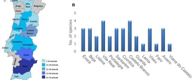
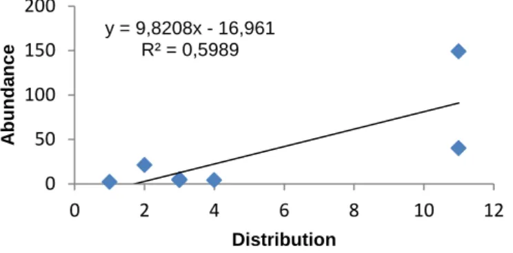
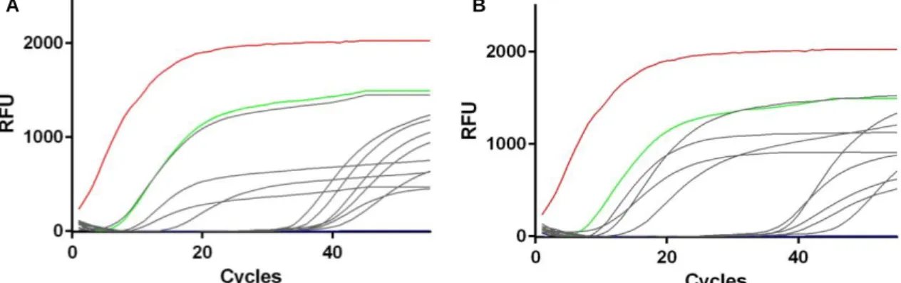
Fig. 1.1 - World map distribution of paratuberculosis, for both domestic and wild species, based on data related to last semester of 2014 reported by worldwide countries to OIE (accessed on October 2015). ... 4 Fig. 1.2 - Summary of MAP diagnosis methods. AFB - Acid Fast Bacillus, IHQ - Immunohistochemistry. ... 4 Fig. 1.3 - Summary of the most common typing techniques used for M. bovis. Spoligotyping - Spacer Oligonucleotide Typing, DR - Direct Repeat. ... 6 Fig. 1.4 - World map distribution of animal tuberculosis, for both domestic and wild species, based on data related to last semester of 2014 reported by worldwide countries to OIE (accessed on October 2015). ... 7 Fig. 1.5 - (A) Trend of bovine tuberculosis epidemiological indicators in mainland Portugal for the period 2007-2014. Adapted from DGAV (2014) [108]. (B) Epidemiological risk area for big game species. Adapted from DGAV (2014) [110]. ... 8 Fig. 1.6 - General laboratory algorithm used for bovine tuberculosis diagnosis. ... 9Fig. 3.1 - (A) Geographic distribution of the 225 animals under analysis across the districts of mainland Portugal. (B) Number of species analyzed per sampled district. ...15 Fig. 3.2 - Relationship between abundance of animals and their distribution across sampled districts. ...16 Fig. 3.3 - Real-time PCR amplification curves targeting IS900 in fecal (A) and spleen (B) samples. Positive controls (DNA extracted from MAP ATCC19698 (red line) and from a previously positive fecal sample (green line)). Positive samples analyzed in this study are depicted in grey lines and the negative control (water instead of DNA) is represented by the blue line. RFU - Relative Fluorescence Units. ...17 Fig. 3.4 - (A) Percentage of DNA-positive animals within each species (HI - Herpestes
ichneumon; VV - Vulpes vulpes; MF - Martes foina; GG - Genetta genetta; CE - Cervus elaphus; and SS - Sus scrofa). (B) Geographical distribution of MAP-positive animals. The
pie charts show the proportion of MAP-positive animals within each district; red=MAP-positive animals, blue= MAP-negative animals. ...19 Fig. 3.5 - Distribution of the Cq values obtained by semi-nested real-time IS900 PCR for feces and spleen samples (A); and for Egyptian mongoose and red foxes samples (B). HI -
Herpestes ichneumon and VV - Vulpes vulpes. ...19
Fig. 3.6 - Real time PCR amplification curves of TR 47 (A) and TR 10 (B) loci. Positive control (DNA extracted from MAP ATCC 19698) is represented by red line, sample F16 by
xv green line, sample F20 by blue line, sample F7 (only TR 10) by purple line and negative control by orange line. ...22 Fig. 3.7 - Agarose gel (2%) electrophoresis of the amplification products of the PCR targeting TR 47 (A), TR 10 (B, C) and TR 292 (D) loci. (B) Results of MAP-positive samples in different dilutions. Results for positive culture control (MAP ATCC 19698, C+) and MAP-positive samples are depicted in the image (molecular size marker – Nzytech ladder VI)...22 Fig. 3.8 - (A) Geographic distribution of the 121 animals under analysis across the districts of mainland Portugal. (B) Number of species analyzed per sampled district. ...24 Fig. 3.9 - Relationship between abundance of animals and their distribution across sampled districts. ...24 Fig. 3.10 - (A) Agarose gel (2%) electrophoresis of IS6110 PCR for positive controls (M.
tuberculosis H37RV (C+) and positive sample (S+)) and DNA from fecal samples under
analysis (S) (molecular size marker – Nzytech ladder VI). (B) IS6110 targeted real-time PCR amplification curve obtained from DNA of the red deer positive fecal sample culture. Positive controls (DNA extracted from M. tuberculosis H37RV culture (red line) and from positive spleen sample (green line)). Positive sample is depicted in grey and negative control (water instead of DNA) is represented by the blue line. ...25
xvi
Table index
Table 1.1 - Summary of the major phenotypic and epidemiological characteristics of MAPstrain types. ... 2
Table 1.2 - Nomenclature of MAP strain types determined by different typing procedures and their relationship to the designated Type S and Type C strains. ... 2
Table 2.1 - Family, species, number and type of animal specimens processed for MAP detection. ...10
Table 2.2 - Family, species, number and type of animal specimens processed for M. bovis detection. ...12
Table 2.3 - Number of bacterial isolates, per year and animal species, spoligotyped. ...14
Table 3.1 - Diversity indices values by sampled district and NUTS (Statistical terrestrial units). ...16
Table 3.2 - Characteristics of MAP DNA positive animals. ...18
Table 3.3 - MIRU-VNTR profile for the positive samples under analysis. ...21
Table 3. 4 - Diversity index values by sampled districts and NUTS. ...25
Table 3.5 - Spoligotyping patterns, hosts and geographic location of M. bovis and M. caprae isolates. ...26
xvii
Abbreviations and Acronyms
AFB: Acid Fast BacillusbTB: Bovine Tuberculosis CE: Cervus elaphus CD: Crohn‟s Disease
DGAV: Direção Geral de Alimentação e Veterinária DR: Direct Repeat
EU: European Union
ELISA: Enzyme-Linked Immunosorbent Assay GG: Genetta genetta
HEYM: Herrold‟s Egg Yolk Medium HI: Herpestes ichneumon
HPC: Hexadecylpyridinium Chloride Monohydrate IHQ: Immunohistochemistry
INIAV: Instituto Nacional de Investigação Agrária e Veterinária, IP LJ: Lowenstein-Jensen
LPSN: List of prokaryotic names with standing in nomenclature
M. bovis: Mycobacterium bovis
MAC: Mycobacterium avium complex
MAP: Mycobacterium avium subspecies paratuberculosis
MF: Martes foina
MIRU: Mycobacterial Interspersed Repetitive Units MM: Meles meles
MTBC: Mycobacterium tuberculosis complex NaOH: Sodium hydroxide
NUTS: Statistical terrestrial units
OIE: World Organization for Animal Health OMS: World Health Organization
OTF: Officially Tuberculosis Free PCR: Polymerase Chain Reaction PFGE: Pulsed-Field Gel Electrophoresis REA: Restriction Endonuclease Analysis
RFLP: Restriction Fragment Length Polymorphism RFU: Relative Fluorescence Units
SNP: Single Nucleotide Polymorphism
xviii SS: Sus scrofa
SSR: Short Sequence Repeats TR: Tandem Repeat
USA: United States of America
VNTR: Variable Number Tandem Repeats VV: Vulpes vulpes
1
1. Introduction
1.1 The genus MycobacteriumThe genus Mycobacterium is part of the Mycobacteriaceae family, presently encompassing 170 species, as enlisted in LPSN (list of prokaryotic names with standing in nomenclature). In general, it includes rod shaped, aerobic, alcohol-acid resistant (due to the waxy mycolic acid cell wall), non-motile organisms with an optimal growth temperature ranging from 25 to 45 °C, a high G+C content (61-71%) and several pathogens known to cause disease in numerous hosts, including man [1–3].
Mycobacteria are widespread microorganisms, and can be found in soil, water, vegetation and food; however, some species appear to be obligate parasites. According to growth in culture media, mycobacteria can be grouped as fast and slow growers, and the colonies formed can be non-pigmented or have a yellow to red color [1,2].
1.2 Mycobacterium avium subspecies paratuberculosis
Mycobacterium avium subspecies paratuberculosis (MAP) is a slow-growing intracellular
obligate parasite and the causative agent of paratuberculosis or Johne‟s disease, a chronic infection of the gastrointestinal tract that affects ruminants worldwide.
MAP is a member of the Mycobacterium avium complex (MAC), a group of environmental
slow-growing mycobacteria, also composed by Mycobacterium avium subspecies avium,
Mycobacterium avium subspecies silvaticum, Mycobacterium avium subspecies hominissuis, Mycobacterium intracellulare, Mycobacterium colombiense, Mycobacterium chimaera, Mycobacterium vulneris, Mycobacterium arosiense, Mycobacterium bouchedurhonense, Mycobacterium marseillense and Mycobacterium timonense [4–11].
MAP genome is a circular chromosome with approximately 69% of G+C content, containing
several insertion sequences like IS900 (14 to 18 copies), ISMav2 (three copies), ISMap02 (six copies) and F57 element (one copy), which are MAP exclusive [12–15].
1.2.1 MAP strain types
In a general way, MAP can be divided into two major strain groups (or strain types) designated Type I/S/sheep and Type II/C/cattle (Table 1.1 and Table 1.2). Initially, strain types were named according to the species from which they were first isolated, however the designation of „sheep‟ and „cattle‟ is not entirely correct, since interspecies transmission of prototypical strains has been detected [16–18].
The Type III/intermediate as a subtype of Type I/S strains and Type B/Bison, as a subtype of Type II/C strains, were recently described [19–21].
2
Table 1.1 - Summary of the major phenotypic and epidemiological characteristics of MAP strain types. Characteristic Type I/Sheep Type II/Cattle
Growth rate [17] Very slow-growing isolates Slow-growing isolates
Colony morphology[17] Smooth and uniform colonies Rough and non-uniform colonies
Pigmentation [17,19] Mostly yellow colonies Mostly non-pigmented colonies
Recommended solid medium [22]
LJ, Middlebrook 7H10 or 7H11 agar media with mycobactin J
HEYM with mycobactin J
Components of the medium [22]
Highly sensitive to certain antibiotics Addition of sodium pyruvate enhances bacterial recovery
Typical incubation time for primary growth on solid media [23]
4-12 months 2-4 months
Decontamination [24] Highly sensitive to decontamination
Host range preference
[16,25,26]
Preference for sheep and goats Very broad host range, including ruminants and non-ruminants Legend: LJ - Lowenstein Jensen, HEYM - Herrold‟s Egg Yolk Medium;
Using a polyphasic approach, the most common genotyping methods provide basic information on strain type, and sub-typing tools can be applied in order to further discriminate
MAP isolates, and in that way, obtain a higher discrimination power [27] (Table 1.2).
Table 1.2 - Nomenclature of MAP strain types determined by different typing procedures and their relationship to the designated Type S and Type C strains.
Typing methods Characteristics Type S Type C
IS900-RFLP [18,20] IS900-based RFLP using PstI, BstEII and/or PvuII as restriction endonucleases. Together with PFGE, these are the most widely used typing methods.
I/S III II/C
PFGE [17,18] Whole genome restriction using SnaBI and SpeI as restriction endonucleases, followed by electrophoresis with continuous changes in the electric field.
I/S III II/C
IS13111 PCR-REA
[21,28]
IS1311 amplification followed by restriction with HinfI and MseI. I/S II/C B
Sub-typing methods
SSR analysis [29] Analysis of G mononucleotide repeats and GGT trinucleotide repeats loci.
VNTR analysis
[30–32]
PCR method based on the polymorphism analysis of VNTR loci, namely the determination of the number of repetitions. MIRUs are a specific type of VNTR applicable to mycobacteria, and can be used in this analysis.
Legend: RFLP (Restriction Fragment Length Polymorphism), PFGE (Pulsed-Field Gel Electrophoresis), PCR - Polymerase Chain Reaction, REA - Restriction Endonuclease Analysis, SSR - Short Sequence Repeats, VNTR (Variable Number Tandem Repeat) and MIRU - Mycobacterial Interspersed Repetitive Units.
1.2.2 Paratuberculosis or Johne’s disease 1.2.2.1 MAP host species
Ruminants appear to be the preferred or natural host for MAP, primarily domestic ruminants as cattle, sheep and goat, but the disease in wild ruminants is also well documented including a large number of hosts, as red deer (Cervus elaphus), roe-deer (Capreolus
capreolus), fallow deer (Dama dama), white-tailed deer (Odocoileus virginianus), alpine ibex
(Capra ibex) and riverine buffalo (Bubalus bubalis) [33–38].
In non-ruminant animals, MAP was already detected in a very broad host range, that includes wild rabbit (Oryctolagus cuniculus), raccoon (Procyon lotor), coyote (Canis latrans),
3 stoat (Mustela erminea), weasel (Mustela nivalis), brown bear (Ursus arctos), and jackdaw (Corvus monedula) [34,39–45].
1.2.2.2 Paratuberculosis progression and MAP transmission
Disease progression and clinical signs are best characterized in domestic ruminants. MAP typically enters the host by the oral route and then, eventually, will cause a chronic inflammatory state with the presence of granulomatous lesions in the small and large intestine, leading to progressive weight lost, diarrhea and decreased milk production [46]. The animals can remain asymptomatic for two to five years, but during this period shedding at different levels may occur [47–49]. MAP excretion occurs, preferentially, in the feces but in an advanced-stage of infection, the pathogen may also be found in colostrum and milk, saliva, uterine fluid and semen, being also possible for fetal infections to occur in utero [46,50–52].
The main route of intra- and inter-species transmission is fecal-oral, by direct ingestion of
MAP-contaminated feces, colustrum and milk or, indirectly, via MAP-fecal-contaminated
water and/or feed and preys [53,54].
1.2.2.3 Potential sources of MAP infection
Despite the fact that MAP is only able to replicate inside host cell, it can be found in water, vegetation and soil, as well as in animal-derived products, which contributes to animal and/or human exposure to the pathogen. The human exposure raises public health concern, since the causal association between MAP and Crohn‟s disease in humans is still not clear.
In environment, MAP was cultured for up to 55 weeks from soil, for up to 24 weeks from the aerial parts of grasses and for up to 48 weeks from water, in a controlled environment in Australia [55,56].
In food, MAP has been detected by PCR and/or culture in raw and pasteurized milk from cows, sheep and goats; powdered infant milk; and cheeses [57–60]. Moreover, MAP has been detected by PCR in drinking water in the United States of America (USA) [61].
1.2.2.4 Paratuberculosis worldwide
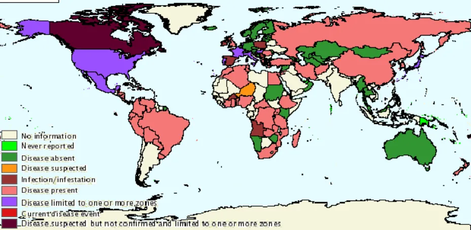
Notification of paratuberculosis to OIE (World Organization for Animal Health) is mandatory and, according to the information on WAHID interface (Worldwide Animal Health Information Database), 54 out of 191 countries reported the presence of MAP infection and/or clinical disease during last year (Fig. 1.1). The worldwide herd prevalence of paratuberculosis is estimated to be 7 to 40%, based on serological monitoring tests [62].
In Europe, it is estimated that more than 50% of the dairy cattle is infected with MAP, however paratuberculosis was not included in the European Union (EU) programs of eradication, control and surveillance of animal diseases and zoonoses for 2015-2017 [63].
4
Fig. 1.1 - World map distribution of paratuberculosis, for both domestic and wild species, based on data related to last semester of 2014 reported by worldwide countries to OIE (accessed on October 2015).
1.2.2.5 Paratuberculosis in Portugal
So far, only a few geographically-limited studies have tried to assess the prevalence of paratuberculosis in Portugal, mainly in small ruminants [64–69], which suggest that probably this disease is under-diagnosed.
In the most recent data, the serological analysis by ELISA (Enzyme-Linked Immunosorbent Assay) of blood samples from 5370 sheep and goats and 2562 sheep has evidenced a prevalence of 27% and 9,1% in Lisboa and Serra da Estrela, respectively [66,67]. Moreover, a survey in sheep‟s from the Northeast of the country was performed, and 3,7% of the 3900 blood samples were seropositive by ELISA, and 18,7% of the pooled samples from a set of 1500 blood samples (in pools of five) were positive by IS900 PCR approach [68,69].
Paratuberculosis cases have to be reported to the Portuguese authorities since 2009. However, in this work it was not possible to find official information regarding the numbers of cases. Furthermore, in the reports sent by Portugal to OIE, in the last five years, and available at WAHID, disease report is limited to the domestic population.
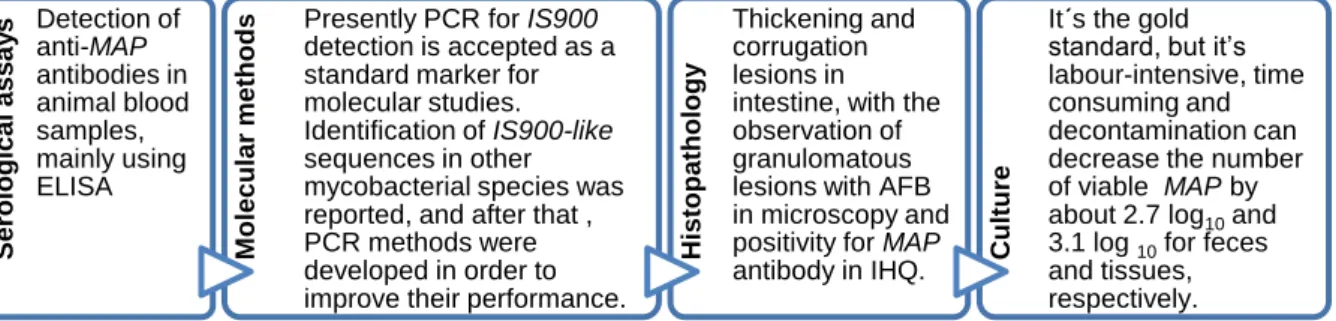
1.2.2.6 Diagnosis methods of paratuberculosis
Diagnostic methods include bacteriological culture of clinical samples, mainly from feces and intestine tissues, and serological/molecular tests (Fig. 1.2) [22,24,62,70–77].
Fig. 1.2 - Summary of MAP diagnosis methods. AFB - Acid Fast Bacillus, IHQ - Immunohistochemistry.
Serologic al as sa y s Detection of anti-MAP antibodies in animal blood samples, mainly using ELISA M ol e c ul a r m e tho
ds Presently PCR for IS900 detection is accepted as a
standard marker for molecular studies. Identification of IS900-like sequences in other mycobacterial species was reported, and after that , PCR methods were developed in order to improve their performance.
His top a tho lo gy Thickening and corrugation lesions in intestine, with the observation of granulomatous lesions with AFB in microscopy and positivity for MAP antibody in IHQ. Cultu
re
It´s the gold standard, but it‟s labour-intensive, time consuming and decontamination can decrease the number of viable MAP by about 2.7 log10 and 3.1 log 10 for feces and tissues, respectively.
5 Decontamination with oxalic acid and NaOH and culture in Lowenstein-Jensen (LJ) medium, or decontamination with HPC (hexadecylpyridinium chloride monohydrate) and culture in Herrold‟s Egg Yolk Medium (HEYM) or Middlebrook medium, are the common protocols followed for MAP culture. The culture media needs to be supplemented with an iron chelator, named mycobactin, being this dependence characteristic, but not MAP exclusive [78].
1.2.2.7 Control and eradication strategies for paratuberculosis
There are still no ideal, cost-effective, methods for the control of paratuberculosis. Control schemes based on a test-and-cull approach depend on the availability of suitable diagnostic assays to detect infected animals as early as possible, but the available serological methods are most reliable during the later stages of infection [23,71].
Vaccination has been used in some European countries, generally in small ruminants, and with good results (the number of clinical and subclinical animals was reduced), so is a possible control measure [23].
1.3 Mycobacterium bovis
Mycobacterium bovis (M. bovis), together with Mycobacterium tuberculosis, Mycobacterium africanum, Mycobacterium microti, Mycobacterium canettii, Mycobacterium pinnipedii and Mycobacterium caprae, constitute the Mycobacterium tuberculosis complex (MTBC) [79–82].
MTBC members, and the causative agents of tuberculosis in numerous species, can be best understood as a series of host-adapted ecotypes, with each ecotype having distinct host tropisms, phenotypes and pathogenicity, and being marked by molecular differences [83–86].
M. bovis, the causative agent of bovine TB (bTB), is an aerobic slow-growing bacteria with a
genome formed by a circular chromosome with approximately 66% G+C content [87].
1.3.1 M. bovis genotyping
The molecular typing techniques most commonly used for M. bovis genotyping can also be applicable to all members of MTBC and are described in Fig. 1.3 [88–92].
• PCR and reverse hybridization method based on the polymorphisms of DR locus, which is comprised of conserved direct repeats of 36 bp interspersed with unique spacer sequences. The simplicity of the method, that detects the presence/absence of these spacer sequences, allowed the establishment of international, open-source, spoligotype databases, such as M.bovis.org. Spoligotyping
• Several VNTR loci, including MIRUs, were already used for genotyping M. bovis. The common approach determines the number of repetitions of 12, 15 or 24 locus per analysis and the final result can be introduced in international databases, such as MIRU-VNTR plus database.
VNTR analysis
• IS6110 is a multi-copy sequence (1-25 copies) found within MTBC, although M. bovis normally contains only one copy. IS6110-RFLP uses PvuII as restriction endonuclease. This method has low discriminatory power, especially for strains with low-copy number of IS6110.
6
Fig. 1.3 - Summary of the most common typing techniques used for M. bovis. Spoligotyping - Spacer Oligonucleotide Typing, DR - Direct Repeat.
1.3.2 Animal Tuberculosis
M. bovis is the main etiological agent of bTB but occasionally M. caprae is also isolated from
suspected tuberculosis lesions. The disease caused by both ecotypes is not substantially different and the same diagnostic methods can be applied [77].
1.3.2.1 M. bovis host species
Besides cattle, M. bovis is also able to infect other animal species, including domestic animals as sheep, goat, pig, dogs and cats [93,94], but also several wildlife species worldwide, causing animal TB. Some of the wildlife species are known to act as maintenance hosts and source of infection to other animals, namely the African buffalo(Syncerus caffer) in South Africa, lechwe antelope (Kobus leche) in Zambia, brushtail possums (Trichosurus
vulpecula) in New Zealand, white-tailed deer (Odocoileus virginianus) in EUA, Canadian
bison (Bison bison) in Canada, badger (Meles meles) in United Kingdom and Republic of Ireland, and red deer (Cervus elaphus) and wild boar (Sus scrofa) in Iberian Peninsula [95– 98].
Surveys in other wildlife species were already performed and the presence of M. bovis was detected in a very broad host range that includes coyote (Canis latrans), red fox (Vulpes
vulpes), black bear (Ursus americanus), raccoon (Procyon lotor), fallow deer (Dama dama)
and Iberian lynx (Lynx pardinus) [99–101].
1.3.2.2 Tuberculosis progression and transmission
Bovine TB is an infectious, chronic, but progressive, disease characterized by the formation of typical granulomatous lesions in several different organs. Clinical signs include weakness, anorexia, emaciation, dyspnoea, and cough, particularly in the advanced stages [77].
The excretion of virulent and viable bacilli is dependent on the localization of the infection. Thus, the excretion of M. bovis occurs, essentially, through aerosols and respiratory secretions, feces and urine. In advanced stages of the disease, the pathogen can also be found in milk, saliva, vaginal and uterine discharges, and purulent material of cutaneous abscesses or open lesions of peripheral lymph nodes [97,102–104].
The anatomical localization of the lesions provides some information about the route of transmission: animals with lesions restricted to the thoracic cavity are presumed to have been infected by the inhalation of aerosols through sharing of the same habitat (food/water spots); while those with lesions in abdominal organs are thought to have acquired the infection by ingestion of infected animals or contaminated pastures/water [102–104].
In a controlled environment in the USA,M. bovis persisted up to 88 days in soil and 58 days
7 cattle and/or wildlife [105]. Moreover, in Portugal, the presence of M. bovis/ caprae was already detected in the environment in areas where bTB is highly prevalent in wildlife [106].
1.3.2.3 Animal tuberculosis worldwide
Bovine TB is notifiable to OIE and, according to the information on WAHID interface, 71 out of the 191 countries reported the presence of M. bovis infection and/or clinical disease during the last year (Fig. 1.4).
Concerning Europe, several countries and specific regions, including the Algarve in Portugal, achieved the OTF (Officially Tuberculosis Free) status in 2012, in accordance with EU legislation (Decision 2012/204/EU) [107]. In the EU non-OTF regions, infection with M. bovis was reported in 18,256 (1,33%) cattle herds in 2013 [107]. Bovine TB is one of the diseases included in the EU programs of eradication, control and surveillance of animal diseases and zoonoses for 2015-2017 [63].
Fig. 1.4-World map distribution of animal tuberculosis, for both domestic and wild species, based on data related to last semester of 2014 reported by worldwide countries to OIE (accessed on October 2015).
1.3.2.4 Animal tuberculosis in Portugal
All bTB cases detected in Portugal have to be reported to the Portuguese authorities since 1953, and also to the European authorities (EU Comission) and to OIE.
The prevalence of M.bovis infected cattle in mainland Portugal has remained low and has been decreasing, with animal and herd prevalence being 0,03% and 0,11%, respectively, in 2008 (Fig. 1.5 A) [108]. However, in 2009 the number of animals almost doubled, reaching in 2010 the values of 0,26% prevalence in animals and 0,9% prevalence in herds [108].
This increase of bTB indicators in Portugal may be a combination of genuine disease increase with higher vigilance efficacy [109]. The reorganization of the veterinary services that was performed in 2009, and the reorganization of the existent farms might also have contributed to the escalation of epidemiological indicators [108].
Since 2012, the values have remained constant and low (0,06% and 0,37% of animal and herd prevalence, respectively, in 2014).
8
Fig. 1.5 - (A) Trend of bovine tuberculosis epidemiological indicators in mainland Portugal for the period 2007-2014. Adapted from DGAV (2014) [108]. (B) Epidemiological risk area for big game species. Adapted from DGAV (2014) [110].
The eradication program of bTB in Portugal is based on the detection of positive live animals by the single intradermal comparative tuberculin test and interferon- test; routine surveillance at slaughterhouse; compulsory slaughter of positive animals; compensation to owners of slaughtered animals; and restriction of movements and pre-movement testing [108]. Only the isolation of M. bovis during culture or the observation of typical TB lesions during histopathological analysis confirms the animal as being TB infected, being INIAV the reference laboratory for diagnosis [109].
Overpopulation of big game animals, namely red deer and wild boar, occurs in certain regions of mainland Portugal, increasing intra and interspecific transmission. After the publication of several studies evidencing the circulation of M. bovis in wild populations [111– 113], the Portuguese authorities defined in April 2011 an epidemiological risk area for TB in big game (Fig. 1.5 B). This implies that big game hunting activities may be accompanied by a credentialed veterinarian to perform a post-mortem examination of hunted animals and eventually collect samples from suspected tuberculosis lesions for laboratorial confirmation; and to ensure proper disposal of hunting by-products [110].
In mainland Portugal, the isolation of M. bovis from red deer and wild boar is well documented and genotyping studies of circulating isolates obtained from domestic and wild animals, from different geographic locations have been performed over the years [111–115].
1.3.2.5 Diagnosis methods
Bovine tuberculosis infection in cattle is usually diagnosed in living animals on the basis of delayed hypersensitivity reactions and, after death, with the use of histopathological, bacteriological and molecular techniques (Fig. 1.6) [88,90,91,116]
0,00% 0,20% 0,40% 0,60% 0,80% 1,00% 2007 2008 2009 2010 2011 2012 2013 2014
Herd prevalence Herd incidence Animal prevalence
A
9 For the microbiological culture, decontamination with detergent (0,375-0,75% HPC), alkali (2-4% NaOH) or acid (5% oxalic acid), and incubation in LJ pyruvate, Stonebrink and Coletsos base are the common approaches. Moreover, the culture media needs to be supplemented with pyruvate, since M. bovis has an alteration in genes involved in the pyruvate metabolism [117].
Fig. 1.6 - General laboratory algorithm used for bovine tuberculosis diagnosis.
1.3.3 Impact of M. bovis in human health
In countries with programs of control and eradication of bTB, M. bovis can especially affect people that work directly with infected animals by airborne transmission, such as veterinarians, zoo workers, hunters, slaughterhouse workers and farmers. In countries without surveillance, the contact might happen through the consume of unpasteurized dairy products or raw/undercooked meat from infected animals [118–121]. There are evidences of human-to-human transmission through the airborne via, especially among immunossupressed people [122,123].
1.4. Objectives of the present work
MAP and M. bovis are pathogenic mycobacteria that can infect livestock and wildlife, causing
paratuberculosis and animal TB, respectively. These diseases may have a huge impact in economy and animal health throughout the world, and, due to their zoonotic potential, raising concerns for public health. In that way, their control and eradication can only be achieved through the understanding of the transmission routes of the pathogens and the ascertainment of the importance of wildlife species in the epidemiological cycle.
As such, the main objective of this work was to contribute to the disclosure of wild hosts that may potentially be exposed to MAP or M. bovis in mainland Portugal. For that purpose, geographically widely distributed animal specimens from the Mustelidae, Herpestidae, Viverridae, Canidae, Suidae and Cervidae families were surveyed for the presence of these bacteria using a polyphasic diagnostic approach based on microbiological and molecular
• Official test: Intradermal comparative tuberculine test • Complementary test: interferon- assay Ante mortem diagnostic tools
• Sample collection from seropositive animals and
from unsuspected animals with tuberculosis-like lesions Post mortem observation in slautherhouse • Histopathology • Microbiological culture • Molecular confirmation • Genotyping Laboratory approach towards diagnosis
10 methods. The clarification of epidemiological links between wildlife and livestock animals was also envisaged based on genotyping techniques applied to isolates of both origins and using the limited epidemiological information available.
Samples for M. bovis survey were selected based on previous knowledge, meaning that the majority of the samples used are from animals killed in the epidemiological risk area.
2. Methods
All the experimental work described below was developed at Instituto Nacional de Investigação Agrária e Veterinária (INIAV, IP; UEISPSA strategic unit) in Lisboa, Portugal.2.1 Animal samples used in this study
The biological matrices used in this study resulted from animals found dead on the road and from legal hunting actions, and were donated for scientific purposes, by the hunting associations and under a protocol established by Dr. Mónica Cunha with CE3C, FCUL, University of Aveiro and Fencaça. All tissue and fecal samples were collected during necropsy, and conserved at -20ºC until being processed in agreement with OIE and OMS (World Health Organization) guidelines.
2.2 Samples used for Mycobacterium avium subsp. paratuberculosis survey
The circulation of MAP in wildlife was investigated in biological specimens of wild carnivores, wild ruminants (red deer) and wild boar, collected between 2005 and 2015, in mainland Portugal (Supplementary Table 1).
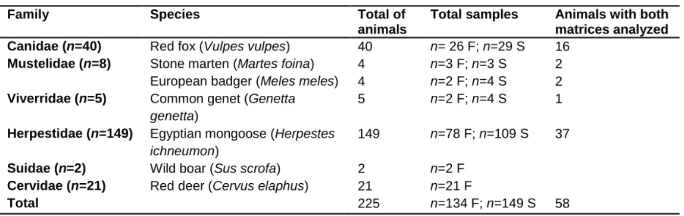
Feces (n=134) and spleen (n=149) samples from 225 animals, 105 males and 101 females, representing seven different species, were the biological matrices screened for MAP (Table 2.1).
Table 2.1 - Family, species, number and type of animal specimens processed for MAP detection.
Family Species Total of
animals
Total samples Animals with both matrices analyzed Canidae (n=40) Red fox (Vulpes vulpes) 40 n= 26 F; n=29 S 16
Mustelidae (n=8) Stone marten (Martes foina) 4 n=3 F; n=3 S 2
European badger (Meles meles) 4 n=2 F; n=4 S 2
Viverridae (n=5) Common genet (Genetta genetta)
5 n=2 F; n=4 S 1
Herpestidae (n=149) Egyptian mongoose (Herpestes ichneumon)
149 n=78 F; n=109 S 37
Suidae (n=2) Wild boar (Sus scrofa) 2 n=2 F
Cervidae (n=21) Red deer (Cervus elaphus) 21 n=21 F
Total 225 n=134 F; n=149 S 58
11 For bacterial culture purposes, 1 g of feces and 2 g of spleen samples were macerated and decontaminated with 0,9% (v/v) and 0,7% (v/v) of HPC (Sigma-Aldrich), respectively, following the methodology described in the OIE guidelines (2008). After decontamination, the macerated suspensions were divided into two equal parts. One part was maintained at 4ºC until further processing for DNA extraction and the other one was centrifuged and the sediment inoculated (100L) in HEYM with and without mycobactin J. After inoculation, the cultures were maintained at 37ºC and observed for bacteriological growth every week.
Mycrobiological culture in HEYM with and without mycobactin J was attempted for all the feces samples. Subsequently, based on the positive results from nested real time IS900 PCR, nine other spleen samples were processed for culture.
2.2.1 DNA extraction from feces and tissue suspension
Direct DNA extraction from 5 mL of feces and 5 mL of spleen suspensions was performed using the commercial system Invisorb Spin Tissue Mini Kit (Stratec Molecular) and High Pure PCR Template Preparation Kit (Roche), respectively, following the manufacturer‟s instructions. A step of mechanical disruption, before applying Proteinase K, was added using a bead-beating protocol consisting of 6.5 ms-1 cycles for 45 sec, repeated twice, in the FastPrep FP120 Bio101 (Savant Instruments). Genomic DNA suspensions were stored at - 20 °C until further use.
2.2.2 Nested real-time PCR for the detection of IS900 insertion sequence
The detection of MAP in the genomic DNA extracted from biological samples was performed by a nested real-time PCR targeting IS900 (Leão et al., unpublished results).
This PCR assay consists of two amplification steps: (i) a first standard PCR targeting a 224 bp sequence; and (ii) a second real-time PCR, using the previous amplification product as template, that amplifies a 66 bp sequence (Supplementary Table 2).
In addition to samples, positive control (DNA extracted from MAP ATCC 19698 and DNA from positive bovine tissue sample) and a negative control (water) were included in all PCR batches. All samples were tested at least twice.
The amplified products from both PCR reactions were analyzed in a 2% agarose gel.
2.2.3 Genotyping of MAP-positive samples by MIRU-VNTR technique
The MAP-positive samples detected by nested real-time PCR targeting IS900 were tentatively characterized by MIRU-VNTR (Mycobacterial Interspersed Repetitive Units - Variable Number Tandem Repeat), based on eight loci, following the method described by Thibault and collaborators [30].
PCR reactions were performed for all MAP-positive samples using a conventional protocol, however, and in order to try to improve the signal, in a subset of samples was also attempted
12 a real-time PCR (Supplementary Table 2 and 3). Optimization of the method was attempted by using different PCR cycles, different annealing temperatures and different DNA quantities from the samples, namely 2L, 5L, 10 L and 1:10 dilution. A positive (DNA extracted from
MAP ATCC 19698 strain) and a negative control (water) were included in all PCR assays.
The PCR products were analyzed in 2% agarose gel. Data available in the open-source MAC INMV database (http://mac-inmv.tours.inra.fr/) were used to assign the hypothetical size of each amplicon to their correspondent tandem repeat copy number.
2.2.4 Strain type differentiation of MAP- positive samples
The DNA from MAP-positive samples, as inferred by nested real time IS900 PCR, was also tested to attempt strain type differentiation, using an approach based on SNP (Single Nucleotide Polymorphism) detection (Leão et al., unpublished results).
In this methodological approach, a genomic region harboring a SNP that allows the differentiation between S and C strains is amplified by PCR, followed by restriction with endonucleases or direct Sanger sequencing (Supplementary table 2). Positive control (DNA extracted from MAP strain K-10) and negative control (water) were included in all reaction batches, and the PCR product was observed in a 2% agarose gel.
2.3 Samples used for Mycobacterium bovis survey
The circulation of M. bovis in wildlife was investigated in biological specimens of wild carnivores, wild ruminants (red deer) and wild boar, collected in 2012, 2013 and 2015, in mainland Portugal (Supplementary Table 1).
Ninety eight liver samples and 23 feces from 121 animals, 60 males and 61 females, representing five different species, were used in this survey (Table 2.2).
Table 2.2 - Family, species, number and type of animal specimens processed for M. bovis detection. Family Species Total of animals Total of samples Canidae (n=4) Red fox (Vulpes vulpes) 4 n=4 L
Mustelidae (n=1) Stone marten (Martes foina) 1 n=1 L
Herpestidae (n=93) Egyptian mongoose (Herpestes ichneumon) 93 n=93 L
Suidae (n=2) Wild boar (Sus scrofa) 2 n=2 F
Cervidae (n=21) Red deer (Cervus elaphus) 21 n=21 F
Total 121 n=98 L; n=23 F
Legend: L - liver; F - feces
For bacteriological culture purposes, 1 g of feces and 2 g of liver were macerated and decontaminated with 0,9% (v/v) and 0,7% (v/v) of HPC (Sigma-Aldrich), respectively, following the methodology described in the OIE guidelines (2008). After decontamination, one part of the macerated suspension was maintained at 4ºC until further processing for DNA extraction and the other part was centrifuged and the sediment (100 L) inoculated into
13 Lowenstein Jensen (LJ) pyruvate and Stonebrink (Biogerm). After inoculation, the cultures were maintained at 37ºC and observed for bacteriological growth every week.
2.3.1 DNA extraction from feces and tissue suspension
Direct DNA extraction from feces and liver suspensions was performed, following the procedures already referred in the MAP survey methods (section 2.2.1).
2.3.2 DNA extraction from culture
Colonies on solid media were suspended in TE 1 M pH 8.0 and heated at 99 °C for 30 min. After centrifugation (1500 g, for 5 min), the suspension was stored at -20ºC.
2.3.3 Semi-Nested real-time PCR for the detection of IS6110 insertion sequence The detection of IS6110 followed the methodology and reactional conditions developed by Costa and collaborators [124].
This PCR assay follows the same principle of the methodology previously described for the detection of IS900. However, in this case, one of the primers used (IS6110-RV) is common to both PCR reactions (Supplementary Table 4). The first standard PCR amplifies a 199 bp fragment and the second real-time PCR a 63 bp sub-fragment.
Two positive (DNA extracted from M. tuberculosis H37RV and DNA from a positive tissue sample) and one negative control (water) were used in every PCR batch. All samples were tested, at least, twice.
Analysis in 2% agarose gel was performed for the PCR products of both steps.
2.3.4 Species identification of bacterial isolates by gyrB PCR-REA
Species confirmation of bacterial isolates growing in Stonebrink and LJ pyruvate medium was performed by gyrB PCR-REA (Supplementary Table 4).
In every PCR batch were included two positive (DNA extracted from M. tuberculosis H37RV and M. bovis BCG) and one negative control (water); and the resulting amplified products were visualized in a 1,5 % agarose gel.
The enzymatic restriction reactions with RsaI and SacI were carried out for a final volume of 10 l, following the manufacturer‟s instructions (New England Biolabs), and using 3 l of PCR reaction product. The restriction products were visualized in a 2% agarose gel.
2.4 Spoligotyping of M. bovis and M. caprae isolates
One hundred and seventeen M. bovis and 10 M. caprae isolates, from five animal species, preliminarily identified by gyrB PCR-REA methodology, were characterized at the molecular level by spoligotyping (Table 2.3). This analysis included M. bovis isolates from 2014 and 2015 and M. caprae isolates from 2011 onwards.
14
Table 2.3 - Number of bacterial isolates, per year and animal species, spoligotyped. Year Red Dear Wild Boar Cattle Sheep Pig
2011 1 2012 2 2013 3 2 2014 17 16 1 2015 34 42 7 2 Total 51 58 11 5 2
DNA extracted from M. tuberculosis H37RV and M. bovis BCG were used as positive controls while water was included as negative control in every PCR assay for the amplification of the DR (Direct Repeat) region (Supplementary Table 4). The amplified products were analyzed in a 2% agarose gel before initiating the spoligotyping-associated hybridization procedure.
The membranes used in the spoligotyping technique were prepared in INIAV, using 43 oligonucleotide sequences corresponding to the 43 spacer regions of locus DR.
The PCR targeting the DR region and hybridization of PCR products was performed according to the procedure described by Kamerbeek and collaborators [88], with the following changes: hybridization time was increased to 90 min and membranes were re-used after three washes at 85ºC in 1% SDS, followed by two washes at room temperature with 20 mM EDTA.
The spoligotyping pattern obtained for each isolate was converted into a binary code of 43 digits and it was inserted in the international data base Mbovis.org (http://www.mbovis.org/index.php). The new spoligotypes were re-tested for the confirmation of the pattern, submitted to the international database and assigned a new spoligotype designation.
The discriminatory capacity of spoligotyping technique was evaluated with the use of discriminatory power (D), which was calculated through an online application tool available on the website:http://insilico.ehu.es.
2.5 Statistical analyses of the results
Chi-square test and Mann-Whitney test (=0,05) were performed in IBM SPSS Statistics 22.0; and graphics were produced in Excel and GraphPad Prism 6.0. The confidence intervals (95%) were calculated through an online application tool available on the website: http://vassarstats.net/.
2.6 Diversity analyses of the samples under study
Diversity analyses was performed through the establishment of the relationship between abundance and distribution; the calculation of diversity indices (Shannon-Wiener, Simpson and Berger-Parker); and the determination of non-parametric estimators (chao 1 and chao 2) of species richness (Supplementary Table 5) [125,126].