U
NIVERSIDADE DE
L
ISBOA
F
ACULDADE DEC
IÊNCIASD
EPARTAMENTO DEQ
UÍMICA EB
IOQUÍMICARegulating Proteostasis
by a Chemical Genetics Approach
Mário João Faustino Neto
Doutoramento em Bioquímica
(Especialidade: Genética Molecular)
2013
U
NIVERSIDADE DE
L
ISBOA
F
ACULDADE DEC
IÊNCIASD
EPARTAMENTO DEQ
UÍMICA EB
IOQUÍMICARegulating Proteostasis
by a Chemical Genetics Approach
Mário João Faustino Neto
Tese orientada pela
Prof.ª Doutora Margarida D. Amaral
e pelo Prof. Doutor Richard I. Morimoto,
especialmente elaborada para a obtenção do grau de Doutor
em Bioquímica (Especialidade: Genética Molecular)
Preface
Aging is still the greatest risk factor for neurodegenerative diseases. These diseases, such as Alzheimer’s and Parkinson’s, and also others that equally result from protein misfolding, such as Cystic Fibrosis, continue to affect a significant number of people and have no cure. Many current therapeutic efforts aim at better understanding the genetic mechanisms underlying these disorders, and new treatments have started to appear where small molecule compounds are used to help cellular defensive mechanisms to battle these constant and accumulating challenges. In light of this, our goal in pursuing this doctoral thesis was to elucidate how chemical manipulation of the pathways maintaining cellular health and homeostasis (the proteostasis network or PN) can be of use for developing potential therapeutics for diseases of protein conformation that affect the human population. The General Introduction of this dissertation gives an overview of the literature that is relevant to frame our work within the state of the art of the folding and proteostasis fields of science.
The Materials and Methods section describes the techniques that we used to perform the experiments here described.
In Chapter III, we wanted to further understand the effects of a previously described small molecule on proteostasis and its mechanism of action. We planned to find the chemical moieties that regulate function of this compound, F1, and to determine which stress pathways are activated by this small molecule and a selected group of structural analogues, across different cell types (with different cellular environments). We also tested F1 and its analogues in various new models of protein misfolding, with relevance for human disease. Finally, we tested the requirement of the activation of different stress pathways (HSR, ER UPR, mtUPR and oxidative stress) for F1 function.
The discovery of new compounds with applications in diseases of protein conformation remains pertinent. In fact, alternative strategies like protein replacement therapy have showed some promising results, but are limited because the injected protein needs to find its way to the appropriate cell type
and cellular compartment. This is a problem since recombinant proteins do not cross the blood-‐brain barrier. In Chapter IV, our goal was to search for new small molecule regulators of the heat shock response (HSR) of different classes (activators, inhibitors and co-‐activators), with a view for making them applicable for the study, or even treatment, of loss-‐ or gain-‐of-‐function conformational disorders. Because there is still new chemical space to be explored when looking for promising new compounds, we used a new chemical library that was engineered to be skeletally diverse, containing a diverse distribution of molecular shapes and excellent physicochemical properties. We also planned to validate our screen hits both by different rounds of testing and by assessing effect on endogenous levels of HSR genes. We aimed to check for the requirement of HSF1 for the activity of these new compounds and to test them in
C. elegans models of protein misfolding. This would make them interesting new
small molecules, for more advanced studies.
Finally, in the General Discussion and Conclusions chapter, we summarize our most important findings and integrate them into what other studies in the same area of work have found. We also speculate of how our new findings might be of worth to the field, and which next steps should be taken to make them more complete.
Acknowledgments/Agradecimentos
A lot of good people have helped throughout my doctoral work and to all of them I am thankful.
First, I would like to acknowledge Prof. Margarida Amaral, for contributing to my scientific growth since the early, undergrad days, for guiding me through the first years of this PhD studies and for advising me to go abroad and work in the Morimoto Lab. Your passion for science and willingness to contribute for the quality of life of people affected by misfolding diseases has helped to shape the way I see research and what our goals should be. Equally, I am grateful to Prof. Rick Morimoto for so kindly opening the doors of his lab to one more Portuguese student and, most of all, for leading me into the right direction of what a thesis work should be. It has been intense but certainly worth it and I truly thank you for the mentorship that finally brought me to this point.
Ao Ministério da Ciência, Tecnologia e Ensino Superior de Portugal e à Fundação para a Ciência e Tecnologia (FCT), por me ter atribuído a bolsa de Doutoramento de 2007 a 2010 (referência SFRH/BD/28741/2006) e ter assim possibilitado a realização do meu trabalho doutoral, tanto na Faculdade de Ciências da Universidade de Lisboa como na Northwestern University (EUA).
To my senior colleagues in the Membrane Protein Disorders Unit of BioFIG, at FCUL, for guiding me through many new experiments and techniques, specially to Carlos Farinha, Mónica Roxo-‐Rosa, Filipa Mendes, Luka Clarke and Anabela Ramalho. Also to Marta Palma, Marisa Sousa, Joana Martins, Joana Almaça, Simão Luz, Luisa Alessio, Diana Faria, Andre Schmidt, Luísa Pissarra and Carla Brás, for making every day in lab one to remember.
To the Morimotome (i.e., the people of the Morimoto Lab) for making me feel welcome in the US. A special thank you to Sue Fox for... well, for everything
that you do and for being such a role model! And to Georgette Pliml, for the continuous assistance with the tiniest details. Many thanks to Cindy Voisine, Carmen Krammer, Susana Garcia, Janine Kirstein, Tali Gidalevitz, Anan Yu, Eric Guisbert, Patricija van Oosten Hawle, Yoko Shibata for years of free teaching and advising. I am thankful to the grad students, Catarina Silva, Daniel Czyz, Kai Orton, Laetitia Chauve and Peter Winter, for understanding exactly what I was going through! Again to Catarina, obrigado por me fazeres sentir em casa, e por toda a ajuda extra, e a amizade também nos tempos livres and to the rest of the “gang”, Carmen, Laetitia, Jesper, Liz and Tanuja, for general good times. To Elizabeth Miller, Tanuja Devaraj and Renee Brielmann, thanks for all the technical support.
An enormous thank you to John Labbadia, for arriving to the lab at the right moment and constantly making me work more and better and more efficiently. It’s been a true pleasure, and it’s not over yet! Also to Andreia Castro, for the help and the ideas, e pela garantia de manutenção do número mínimo de Portugueses no lab.
I would like to acknowledge my collaborators, my great friend Barbara Calamini, Denise Dunn and Prof. Donald C. Lo at Duke University, Prof. Rick Silverman, Soosung Kang, Prof. Chi-‐Hao Luan and Sara Fernandez Dunne at Northwestern University, for great ideas and experiments.
Para ti Mariana, mi compañera de la vida, porque cada día somos más felices. Thanks for the patience dealing with a PhD student, for all the comprehension and for all the help you give me every day. Can’t wait for what’s next.
Finalmente, à minha família, especialmente ao meu irmão Miguel e aos meus pais Helena e Fernando. Porque sem vocês, mãe e pai, eu não estaria aqui, e porque ao longo dos anos foram família, amigos, orientadores, médicos, psicólogos e banco (!), a vós dedico esta tese.
Summary
A better definition of the mechanisms that maintain a healthy cellular environment and, therefore, proteins homeostasis (proteostasis) have contributed significantly to an improved understanding of the factors leading to protein conformational diseases, such as Alzheimer’s disease, Huntington’s disease, Cystic Fibrosis or Gaucher’s disease. In fact, the identification of the proteostasis network (PN) components, mostly molecular chaperones, created a repository of genes involved in the important cellular functions of synthesis, folding, transport and degradation. This allowed a better definition of the potential targets for pharmacological intervention in these diseases, characterized by an aggregation-‐related gain-‐of-‐toxic-‐function or by misfolding and premature degradation loss-‐of-‐function.
Our goal was to contribute for the development of small molecule compounds with a role in regulating the heat shock response (HSR), one of the main cellular stress response pathways, which enhances proteostasis when the cell is challenged by misfolding species.
We studied a previously described small molecule, in detail, and revealed its chemical features that enable it to function as an HSR activator and to ameliorate proteostasis in mammalian and C. elegans models of protein misfolding. By studying structural analogues of SKM-‐4/F1, we determined how that chemical structure could be altered to modulate its activation of different cellular stress pathways, like the unfolded protein response (UPR) or the oxidative stress response. We also showed that SKM-‐4/F1 rescues the proteotoxicity of aggregation-‐prone proteins in neuronal models of C. elegans and rat primary brain slices and neuronal co-‐cultures, further highlighting its potential use in more advanced therapeutics. We concluded that even though SKM-‐4/F1 activates different stress pathways in the cell, only the activation of the HSR seems to be necessary for its rescue of misfolding phenotypes.
The finding of novel regulators of the HSR is still a major goal in the search for new and better therapies for conformational diseases. We have
therefore performed a high throughput screen (HTS) for activators, inhibitors and co-‐activators of the HSR, which resulted in three newly described small molecules. Activator A731 is an HSF1-‐dependent activator of a general HSR and already showed an ability to improve aggregation and toxicity phenotypes in C.
elegans models for conformational disorders. We also identified an inhibitor,
I632, which showed low potency but a reduced cellular toxicity, and could be potentially developed into a better compound by a structure/activity relationship (SAR) study. Finally, we identified C732, a co-‐activator of MG132-‐ induced HSR, an intriguing compound that shares structural similarities to A731 but could only activate HSR in the presence of the agonist MG132 and not in its absence.
The work described here contributed for a better comprehension of new and previously described small molecule proteostasis regulators, and how they can be used to ameliorate diseases of protein conformation, by further understanding their chemistry and their mechanism of action (MoA).
Keywords: Stress Response; Heat Shock; Proteostasis; Small Molecule; High Throughput; Conformational Disease; Chaperone
Resumo
As proteínas são um dos principais componentes das células, e responsáveis pela maioria das funções celulares. Para manter a célula num estado funcional e saudável, é necessário garantir a funcionalidade dos processos de síntese, processamento, transporte e degradação, cuja regulação constantemente desafia a manutenção celular.
A integridade do proteoma (ou seja, a totalidade das proteínas numa célula, num dado momento) é mantida principalmente por fatores auxiliares, denominados chaperones moleculares, cuja principal função consiste em garantir que as proteínas estejam no seu estado funcional e nativo. Visto que as chaperones são a base molecular de vias celulares desde a síntese até à degradação de proteínas, são também o foco central de mecanismos de controlo de qualidade, essenciais para manter a homeostase proteica ou proteostase (do inglês proteostasis). Estes mecanismos de controlo de qualidade incluem chaperones com uma ação geral na célula e outras que agem em localizações celulares específicas. Deste modo, é constituído um sistema de vigilância com requisitos de conformação que as proteínas devem cumprir, para que possam atingir o seu estado funcional e nativo, além do seu destino na célula. Quando erros são detectados, o sistema de controlo de qualidade pode contribuir para novas tentativas de processamento do péptido ou, se isso falhar, para a sua degradação, de modo a evitar os efeitos nocivos resultantes da acumulação de restos proteicos potencialmente tóxicos.
O controlo de qualidade é mantido no citoplasma principalmente por dois tipos de proteínas: chaperones associadas ao ribosoma e chaperones que foram primeiro identificadas por serem induzidas em resposta a uma elevação de temperatura, sendo por isso chamadas de Hsps (do inglês heat shock proteins). Estas chaperones são de diferentes famílias, com atividades específicas e cujo nome resultou da sua massa molecular (em kiloDalton): Hsp100, Hsp90, Hsp70, Hsp60, Hsp40 e as pequenas Hsps ou sHsps (s = small). No caso de proteínas destinadas para a via secretora, o seu processamento ocorre no retículo
endoplasmático (RE), um dos organitos celulares. Neste caso, existem vias de controlo de qualidade específicas, reguladas por chaperones próprias que só “autorizam” a saída da proteína para ser secretada quando esta tiver adquirido a respetiva conformação nativa. Caso contrário, é marcada para degradação e retro-‐translocada para o citoplasma, onde é destruída no proteasoma. Em alguns casos, proteínas com mutações que retêm alguma função mas têm um processamento alterado, como p. ex. a proteína CFTR, são prematuramente degradadas por este mecanismo, conduzindo a doenças como a Fibrose Quística. Muitos factores podem contribuir para a acumulação de resíduos de proteínas com processamento alterado na célula. Estes podem ser ambientais, como a exposição a temperaturas elevadas ou metais pesados, mudanças de pH, infecções virais, etc., ou intrínsecas, como a expressão de proteínas mutadas ou erros de tradução. Para responder a estes insultos, também denominados de “stress”, as células desenvolveram um conjunto de mecanismos protetivos que rapidamente induzem a expressão de chaperones e promovem a sobrevivência celular. Os principais mecanismos de resposta ao stress são a HSR (heat shock
response), o UPR (unfolded protein response) do RE ou do mitocôndrio e a
resposta ao stress oxidativo. Apesar de serem mecanisticamente diferentes, todos estes sistemas de controlo de qualidade e resposta ao stress celular atuam em conjunto e tentam manter a célula num estado saudável. No entanto, em alguns casos isso não é possível, e surgem doenças relacionadas com conformação proteica.
As doenças de conformação proteica podem ser de dois tipos: o primeiro são as de "perda de função" de uma proteína mutada, resultante em alterações da sua conformação ("misfolding") e a uma degradação prematura, como é o caso da já referida Fibrose Quística e da doença de Gaucher, resultante de defeitos no armazenamento de proteínas nos lisossomas. O segundo tipo são as doenças em que há "ganho de função" p. ex. pelo produto proteico ser tóxico, devido a interações erróneas na estrutura da proteína ("misfolding"), conduzindo a uma propensão para agregação, o que tem consequências nocivas para a célula e doença associada. Alguns exemplos notórios são as chamadas amiloidoses, como a paramiloidose (conhecida por "doença dos pezinhos") ou as doenças de Alzheimer, Parkinson ou ainda a esclerose lateral amiotrófica (ALS). Um outro
grupo de doenças neurodegenerativas que resultam da acumulação de amilóide são conhecidas como doenças de poliglutamina (PolyQ), e ocorrem por expansão de uma repetição de resíduos de glutamina nas proteínas afetadas. Este grupo inclui a doença de Huntington e ataxias espinocerebelares, como a doença de Machado-‐Joseph.
Todas estas doenças podem ser vistas como uma perda na regulação da "homeostase proteica" ou proteostase. Ao longo da vida, este mecanismo mantém a resposta celular aos vários insultos a que é submetida, até um certo limite em que a proteção começa a falhar e dá-‐se o envelhecimento ou, em casos de mutações e/ou misfolding proteico marcante, um estado de doença. Muitos trabalhos de investigação têm contribuído para uma melhor compreensão da rede de proteínas que constituem a proteostase, que são as que estão essencialmente envolvidas nos vários mecanismos de controlo de qualidade e resposta ao stress. Igualmente, vários esforços recentes têm sido feitos para desenvolver compostos químicos que permitam aumentar a capacidade da proteostase, quer mimetizando os efeitos das chaperones moleculares (chaperones químicas ou farmacológicas) quer aumentando a quantidade de chaperones na célula e permitindo uma melhor resposta aos fatores nocivos, tóxicos para o ambiente celular. Muitos destes contributos resultaram de screens de alto rendimento ou HTS (do inglês high throughput screens), onde milhares de novos compostos podem ser testados e selecionados num tempo reduzido, aumentando a probabilidade de encontrar compostos positivos para a função testada. Em muitos destes trabalhos há também uma componente de química medicinal, i.e., produção de análogos químicos semelhantes aos originalmente descritos, para testar se pequenas alterações na sua estrutura produzem melhores resultados, otimizando assim a ação do composto líder.
O objectivo geral deste trabalho foi o de contribuir para o desenvolvimento de novos compostos químicos com potencial aplicação para doenças de conformação proteica.
O nosso estudo iniciou-‐se assim (Capítulo III) pela otimização dum composto líder anteriormente identificado. O nosso ponto de partida foi uma pequena molécula (small molecule) previamente descrita como sendo ativadora da via de stress HSR (heat shock response), denominada F1/SKM-‐4, e que
também tinha demonstrado capacidade de atenuar fenótipos de agregação com baixa toxicidade em modelos de cultura celular e no nemátode Caenorhabditis
elegans (C. elegans). Utilizando esta molécula como referência, realizámos assim
um estudo de comparação de estrutura-‐função, através da aplicação de análogos estruturais de F1/SKM-‐4, e descobrimos que a molécula de referência já era a que produzia melhores resultados para a ativação da HSR, quando também se tinha em conta a toxicidade de cada composto. Para além disto, comparámos a ativação de outras vias de stress entre o composto inicial e alguns dos seus análogos, e descobrimos que é possível manipular a estrutura química nesta classe de pequenas moléculas de modo a obter uma ativação diferencial de vias como a UPR do RE ou do mitocôndrio. Testámos dois destes análogos num modelo de agregação proteica em C. elegans, que expressa uma repetição de 35 poliglutaminas (poly-‐Q35, ou Q35) resultante no aparecimento de agregados nas células musculares dos animais, acompanhados de uma crescente toxicidade (evidenciada por defeitos de locomoção nestes nemátodes, a partir do sexto dia de vida). Esta experiência mostrou que apesar de os 2 análogos ativarem com maior intensidade outras vias de stress celular, não eram mais eficazes no tratamento dos fenótipos de doença em C. elegans. Estes resultados foram também apoiados pela nossa descoberta que em F1/SKM-‐4, apesar de várias vias de stress serem activadas, apenas a ativação da HSR mostrou ser necessária para que F1/SKM-‐4 reduzisse o número de agregados nos nemátodes Q35. Este resultado foi obtido utilizando técnicas de RNAi, que permitem reduzir os níveis de expressão de genes específicos, e observar efeito dessa redução. Neste caso, reduzimos selectivamente o nível de factores de transcrição que controlam as diferentes vias de stress, e só com a redução nos níveis de HSF1 (heat shock
factor 1) se perdeu o efeito do composto F1/SKM-‐4. Também mostrámos que o
composto F1/SKM-‐4 é funcional na melhoria de modelos relevantes para doenças neurológicas, como foi o caso dos modelos neuronais em C. elegans Q67, relacionado com doenças de poliglutaminas, e AT3q130, modelo para a doença de Machado-‐Joseph. Esta descoberta foi sustentada pela elevada eficiência revelada por F1/SKM-‐4 na protecção de neurónios em experiências com células primárias de rato. Neste caso, F1/SKM-‐4 mostrou ser protetivo contra a expressão de um fragmento proteico, encontrado em pacientes da doença de
Huntington, em cérebros de rato. Todas estas descobertas fazem de F1/SKM-‐4 um composto com potencial para estudos mais avançados, e eventualmente para ensaios clínicos.
No Capítulo IV, descreve-‐se um novo screen que realizámos em condições de HTS para a identificação de novos compostos reguladores da HSR, que levem a uma melhoria da proteostase. A optimização e realização deste screen, juntamente com uma cuidada seleção dos melhores compostos daí resultantes, resultou em três novas pequenas moléculas com diferentes efeitos na HSR. O novo composto A731 ativa uma HSR geral e dependente da presença de HSF1, e já demonstrou a capacidade de melhorar modelos de doenças de conformação proteica em nemátodes C. elegans. Também identificámos um inibidor da HSR, que apesar de não ter mostrado elevada eficiência, apresentou uma reduzida toxicidade, podendo ser usado no futuro para desenvolver novos compostos análogos nesta classe, por um estudo comparativo de estrutura e atividade. Finalmente, identificámos um composto numa classe pouco descrita, de co-‐ ativadores de uma HSR, dependente da presença de MG132 (um conhecido ativador desta via). O co-‐ativador C732 tem uma estrutura química semelhante à do ativador A731, embora não resulte em HSR por si próprio. Este facto leva-‐nos a questionar se os dois compostos actuaram por uma mesmo mecanismo de acção, um ponto que queremos explorar no futuro.
Na sua globalidade, o trabalho descrito nesta tese contribui para a aplicação de pequenas moléculas químicas para a regulação de vias de stress celular, demonstrando como podem ter um efeito benéfico em modelos relevantes para doenças de conformação proteica e, potencialmente, no seu tratamento. Com o trabalho futuro, para seguimento dos estudos aqui apresentados, propomos a continuação da caraterização destas moléculas. Com efeito, esperamos que estes compostos possam vir a ser utilizados, na sua forma atual, ou após otimização da sua estrutura, no tratamento de doenças que afetam a população humana. Caso tal não se concretize, serão pelo menos importantes ferramentas para compreender melhor os mecanismos que defendem o nosso organismo, mantendo-‐o num estado homeostático e saudável.
Palavras-chave: Resposta ao Stress; Proteostase; Compostos Químicos; Alto Rendimento; Doenças de Conformação; Chaperone; Química Medicinal
Abbreviations
17-AAG 17-‐N-‐allylamino-‐17-‐demethoxygeldanamycin AAA+ ATPase associated with diverse activities AD Alzheimer’s disease
ALS amyotrophic lateral sclerosis ATF6 activating transcription factor 6
ATFS1 activating transcription factor associated with stress 1 ASP arylsulfanyl pyrazolone
AU arbitrary units BCA bicinchoninic acid BLPS body length per second
C. elegans Caenorhabditis elegans
CaCl2 calcium chloride
CAS chemical abstracts service CdCl2 cadmium chloride
CF cystic fibrosis
CFTR cystic fibrosis transmembrane conductance regulator CGC C. elegans genetic center
CMA chaperone-‐mediated autophagy CNX calnexin
CO2 carbon dioxide
COPII coat protein complex II CRT calreticulin
CTCMLD Chicago Tri-‐Institutional Center for Chemical Methods and Library
Development
DMEM Dulbecco’s modified Eagle’s medium DMSO dimethyl sulfoxide
E. coli Escherichia coli
EC50 half maximal effective concentration
eEF1A eukaryotic translation elongation factor 1A ERAD endoplasmic reticulum associated degradation ERK extracellular signal-‐regulated kinase
F508del deletion of phenylalanine 508 FBS fetal bovine serum
G418 geneticin
GST glutathione S-‐transferase GPX glutathione peroxidase
HEPES 4-‐(2-‐hydroxyethyl)-‐1-‐piperazineethanesulfonic acid HPLC high performance liquid chromatography
HRP horseradish peroxidase HSE heat shock element HSF heat shock factor HSP heat shock protein HSR heat shock response HTP high throughput
HTS high throughput screen Htt huntingtin
IIL insulin/insulin-‐like growth factor 1 IPTG isopropyl β-‐D-‐1-‐thiogalactopyranoside IRE1 inositol-‐requiring protein 1
JNK c-‐Jun N-‐terminal kinase KCitrate potassium citrate KD knockdown LB media Luria broth media LD50 median lethal dose
MAPK p38 mitogen-‐activated protein kinase MEF mouse embryonic fibroblast
MgSO4 magnesium sulfate
MoA mechanism of action
NAC nascent chain-‐associated complex NaOCl sodium hypochlorite
NEF nucleotide exchange factor NGM nematode growth media NN-DNJ N-‐(n-‐nonyl)deoxynojirimycin Nrf NF-‐E2-‐related factor
PBS phosphate buffered saline PCs pharmacological chaperones PD Parkinson’s disease
PERK PKR-‐like ER kinase PolyQ polyglutamine
PPIases peptidyl-‐prolyl isomerases PN proteostasis network PRs proteostasis regulators QC quality control
RAC ribosome-‐associated complex RIPA radio-‐immunoprecipitation assay RLU relative light units
ROS reactive oxygen species
RT-PCR real-‐time polymerase chain reaction SAR structure-‐activity relationship S.D. standard error
SDS-PAGE sodium dodecyl sulfate polyacrylamide gel electrophoresis S.E.M. standard error of the mean
SOD superoxide dismutase TF transcription factor Ti therapeutic index
TM tunicamycin
TRiC TCP-‐1 ring complex TTR transthyretin
UPR unfolded protein response UPS ubiquitin-‐proteasome system wt wild-‐type
YFP yellow fluorescence protein XBP1 X-‐ box binding protein 1
Contents
Preface... iii Acknowledgments/Agradecimentos...v Summary... vii Resumo...ix Abbreviations...xv Contents ... xixI – General Introduction ...3
1. Protein Folding ...3
2. Molecular chaperones and quality control ...4
2.1. Cytoplasmic molecular chaperones... 4
2.2. Quality control in the endoplasmic reticulum ... 7
2.3. Protein degradation... 8
3. Cellular Stress Pathways ... 10
3.1. The heat shock response ...10
3.2. The unfolded protein response in the ER and in the mitochondria ...11
3.3. Oxidative stress response ...14
4. Protein conformational disorders ... 15
4.1. Loss-‐of-‐function protein folding diseases...15
4.2. Gain-‐of -‐function protein folding disorders ...16
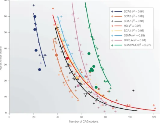
5. Proteostasis, disease and aging... 18
5.1. Small molecules affecting proteostasis ...20
5.2. Chemical genetics efforts to ameliorate proteostasis...23
5.3. C. elegans as a model for understanding proteostasis and disease...24
6. Using HTP and chemical genetics for new treatments of neurodegenerative diseases... 25
7. Aims of the present work ... 26
II – Materials and Methods ... 29
1. Compounds... 29
2. Cell cultures ... 29
3. C. elegans strains ... 30
4. Cell-based determination of small molecule activity ... 30
5. Cytoxicity Assays ... 31
7. C. elegans liquid culture assay for assessment of rescue of aggregation and motility defects... 34
8. Locomotion deficiency assay... 36
9. RNA extraction of nematodes and semi-quantitative RT-PCR... 37
10. Rat brain slice and neuronal co-culture assays (Lo Lab) ... 38
11. Cell-based High-Throughput Assays ... 39
12. Western Blot analysis ... 40 III – Mechanism of the Proteostasis Regulation by the Barbiturate-like Small Molecule F1... 45
1. Background... 45
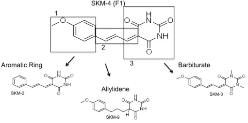
1.1. Structure/function studies of F1 ...45
1.2. SKM-‐4, a newly synthesized F1 ...46
2. Results ... 47
2.1. Activity and toxicity of SKM-‐4 when compared to F1 ...47
2.2. Effect of structural changes on SKM-‐4/F1 function and toxicity...48
2.2.1. Allylidene modifications ... 48 2.2.2. Aromatic ring analogues... 53 2.2.3. Barbiturate substructure substitutions ... 53 2.2.4. Additional analogues... 54
2.3. Endogenous effects on stress pathways ...55
2.3.1. Activation of the endogenous HSR by SKM-‐4/F1 analogues ... 55 2.3.2. Activation of other stress pathways by the barbiturate small molecules... 61
2.4. Effect of SKM-‐4/F1 analogues in the Q35 C. elegans model...70
2.4.1. Aggregation and toxicity effect in Q35-‐YFP C. elegans... 70 2.4.2. Activation of stress pathways by the SKM-‐4/F1 analogues in Q35 worms... 72
2.5. Amelioration of toxicity in C. elegans neuronal models...74
2.6. Protective effect of F1 in rat brain slices and neuronal co-‐culture models
expressing Htt-‐N90Q73...76
2.7. Insights into the molecular mechanism of action of the barbiturate-‐like small molecule SKM-‐4/F1 ...78
3. Discussion: SKM-4/F1 and its class have the potential for more advanced studies ... 81 IV – A High Throughput Screen for new Chemical Regulators of the Heat Shock Response... 85
1. Background... 85
2. Results ... 86
2.1. Optimization of an HTP screen for activators of the HSR ...86
2.2. Optimization of an HTP for inhibitors of the HSR ...89
2.3. Identification of new regulators of the HSR – activators, inhibitors and a new class, co-‐activators...91
2.4. Activity and toxicity profiles of validated screen hits...93
2.4.1. Activators ... 94 2.4.2. Inhibitors... 94 2.4.3. MG132 co-‐activators ... 99
2.6. A731, a new activator of the HSR ...104
2.6.1. Activation of general HSR by A731 ...104 2.6.2. Effect of A731 on proteotoxicity in C. elegans models of protein misfolding ...107
3. Discussion - The triage of the new regulators of the HSR...110 V – General Discussion and Conclusions...115
1. HRS regulation to ameliorate proteostasis ...115
2. Structure, activity and MoA of barbiturate-like activators of the HSR ...116
2.1. Manipulating the chemistry of barbiturate-‐like small molecules for a better understanding of proteostasis disorders...116
2.2. F1 has potential applications in neuronal models of protein misfolding...118
2.3. The HSR is necessary for disease model amelioration in the Q35 worms by F1, but other stress pathways are not ...118
3. Concerted efforts for new HSR regulators of different classes...120
3.1. Refining a screen...120
3.2. The newly identified regulators of the HSR ...120
4. Conclusions and Future Directions...122 Appendix I – Activity and Toxicity Profiles of SKM-4/F1 Analogues...127 Appendix II – HTP Screening at Northwestern University ...137 References...141
I – GENERAL INTRODUCTION
I – General Introduction
1. Protein Folding
Proteins are a major component of all cells and are responsible for most cellular functions. The processes involved in protein biogenesis, namely synthesis, folding, transport and degradation, constitute a constant challenge that must be accomplished efficiently in order to maintain cellular health.
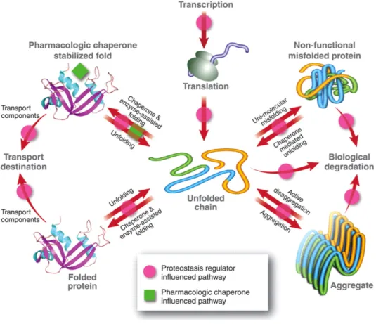
Folding is the process by which a linear polypeptide chain transitions into a three-‐dimensional structure (2), mostly with the information contained by the sequence of amino acids, particularly the attractive or repulsive forces that neighboring amino acids exert on each other. An efficient folding process can be seen as an energy landscape, where the unfolded protein represents the state of maximum energy, and the folded state corresponds to the protein at its lowest energy level, which is also where thermodynamic stability is maximized. In addition, folding intermediates and misfolded peptides have intermediary energy states ((3), (4), (5), (6)). Protein folding is particularly demanding in the crowded intra-‐cellular environment (7) (8). It is commonly accepted that, for the majority of polypeptides in living cells, the intricate folding process is assisted by auxiliary factors that include folding enzymes and molecular chaperones (9).
Figure 1. Hypothetical folding energy landscape for a polypeptide. Energy is
lowered from the unfolded (U) state to the native functional protein (F). Folding intermediates or misfolded peptides (M) have medium energy levels. Adapted from (1)
2. Molecular chaperones and quality control
The main function of molecular chaperones is to maintain proteome integrity, which consists in maintaining proteins in their functional, native conformation. This is regulated by constitutively expressed chaperones that span all cellular compartments and also, under more extreme conditions (like heat shock and other forms of cellular stress), by inducible ones. In addition to their role in protein folding, molecular chaperones also have functions in assembling and regulating macromolecular complexes, in trafficking proteins between different cell compartments or to the cell membrane, and also in targeting proteins for degradation (10), (11), (12), (13). Since molecular chaperones are the main components in pathways ranging from protein biogenesis to protein degradation, they make up the core of the cellular protein quality control machinery, which is essential for maintaining protein homeostasis (proteostasis).
The protein quality control machinery is composed of many molecular chaperones that can act either ubiquitously or at specific cellular locations. Chaperones thus constitute a surveillance system with conformational requisites that proteins must fulfill in order to reach their native functional state and optimal cellular localization. Failure to do so can result in refolding of the polypeptide, or, when refolding fails, in protein degradation, thus avoiding the detrimental effects caused by the accumulation of misfolded and potentially toxic proteins. Hence, protein quality control coordinates gene transcription and protein synthesis with the post-‐translational machineries of folding, trafficking and repair/degradation, and is therefore integral to cellular health (14).
2.1. Cytoplasmic molecular chaperones
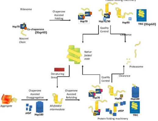
Many of the firstly described molecular chaperones were inducible, i.e., proteins that are up-‐regulated in response to elevated temperatures. As a consequence, these chaperones are often named Heat Shock Proteins (Hsps). Different classes of Hsps were initially separated according to their molecular weights (KDa) and sequence homology: Hsp100, Hsp90, Hsp70, Hsp60, Hsp40 and the small Hsps or sHsps (15) (Figure 2). Each chaperone family differs in the
number of members, substrate specificity, cellular localization and identity of associated co-‐chaperones that assist in their function. Therefore, it is not surprising that chaperone expression differs amongst cell types and correlates with the specific demands of each cell (16).
Figure 2. Different families of chaperones and co-chaperones are involved in protein folding. Adapted from (17).
In the cytosol, two main groups of molecular chaperones facilitate protein folding: ribosome-‐associated chaperones that stabilize nascent chains and allow the initial steps in protein folding, and a second group of chaperones that assist in the later steps of the folding and targeting processes (18). The first group is comprised of the trigger factor in bacteria and by the ribosome-‐associated complex (RAC) and the nascent chain-‐associated complex (NAC) in eukaryotes (3); the second group includes many of the aforementioned Hsps, which we will describe in more detail.
Hsp70s (heat shock proteins of the 70kDa size) are involved in both co-‐ and post-‐translational protein folding in an ATP-‐dependent manner, by cycles of substrate binding and release. The Hsp70 machinery is assisted by co-‐ chaperones and also nucleotide exchange factors (NEFs) like Bag-‐1, which help increase the efficiency of substrate binding and release. These factors also confer
(Hsp40)
specificity, by modulating multiple steps of the ATPase cycle and by regulating the interactions with other components of the quality control network (3) (16). The members of the Hsp40 family are chaperones that contain a signature J domain and are comparable to (and named after) the DnaJ protein in E. coli (19). They are mainly Hsp70 co-‐chaperones that function in stimulating ATP hydrolysis and in stabilizing the binding of Hsp70 to its substrates (20). Some J-‐ domain proteins have the ability to participate in folding independently of Hsp70, binding to unfolded polypeptides and preventing their aggregation. It has been proposed that most unfolded substrates first bind to an Hsp40, which then transfers them to Hsp70 for correct folding (16).
The Hsp60 proteins (also known as Chaperonins) are large multimeric complexes that form a double ring structure. Well-‐known examples are Hsp60, GroEL (from E. coli), and the TRiC (TCP-‐1 ring complex) complex (21). These chaperone machines form cage-‐like structures that encapsulate the protein substrates and accelerate their rate of folding, in an ATP-‐dependent manner (22) (23).
Hsp90 family members are essential for the regulation and maturation of client proteins that are involved in signaling pathways and in control of the cell cycle. Some of these “client” proteins are various transcription factors, hormone receptors and serine-‐threonine kinases (12). The folding intermediates recognized by Hsp90 have near-‐native structures; accordingly, Hsp90 was proposed to act following Hsp70 in the protein folding pathway (24).
Hsp100s are members of a larger family of AAA+ (ATPase associated with diverse activities) proteins with a conserved ATPase unit that contains at least one nucleotide binding domain. Monomers of Hsp100 form functional hexameric rings that provide chaperone activity. Most of these chaperones associate with proteases to participate in protein degradation. However, some Hsp100s like ClpB of E. coli or Hsp104 of S. cerevisiae associate with Hsp70 to promote protein disaggregation. (25). Interestingly, mammals do not possess an Hsp104 homologue. In fact, it was recently found that the mammalian “disaggregase” machinery is the result of a synergetic activity of an Hsp110 (Apg-‐2) with Hsp70 (Hsc70 or Hsp70) and Hsp40 (Hdj1). All of these chaperones were necessary and
sufficient for dissolving disordered aggregates and recovering the native folded state of proteins (26).
Finally, sHsps are chaperones with a conserved α-‐cystallin domain and with low molecular weights between 15 and 40kDa. They are distinct from the other Hsp families in that they do not require ATP for prevention of aggregation. Under stress conditions, sHsps form dimers or larger multimers that bind misfolding proteins in order to prevent them from aggregating (27) (28). These small Hsps cannot promote folding alone, but they act as “holders” that present the client proteins to the Hsp70 machinery for ATP-‐dependent refolding (29).
2.2. Quality control in the endoplasmic reticulum
The ER is the site of folding for proteins that are destined for the secretory pathway. The environment in the ER is unique and differs from the cytoplasm in ion concentrations, redox conditions, and chaperone composition. This creates an optimal environment for formation of disulphide bonds, assembly of multi-‐domain, complex proteins and higher folding efficiencies (30) (31). The most important chaperones with function in the ER include BiP and Grp170, members of the Hsp70 family; Hsp40 family members like ERdj1-‐5; Grp94, belonging to the Hsp90 family; and two important lectin chaperones that aid in the folding of proteins with monoglucosylated N-‐linked glycans -‐ calnexin (CNX) in the ER membrane and calreticulin (CRT) in the ER lumen. Besides chaperones mechanically involved in protein folding, enzymes important for catalyzing isomerization, oxidation and reduction of disulfide bonds such as thiol-‐disulphide oxidoreductases and peptidyl-‐prolyl isomerases (PPIases) also contribute to protein homeostasis in the ER (32) (30).
ER chaperones like BiP, CNX and CRT have the ability to work as “retention anchors”, thereby suppressing the secretion of incompletely folded or misfolded proteins. When an ER resident protein is permanently misfolded or associated with ER chaperones for an extended period, it is targeted for degradation via ER associated degradation (ERAD). During ERAD, the misfolded client is retro-‐translocated to the cytosol where it is degraded by the proteasome (30), (31), (33). While some proteins are ER-‐resident and have an ER retention
signal in their sequence, many others are exported via vesicles to the Golgi complex and the secretion pathway. Alterations in ERAD are particularly problematic for mutant proteins that are partly functional but are still signaled for degradation, leading to diseases such as Cystic Fibrosis (33).
2.3. Protein degradation
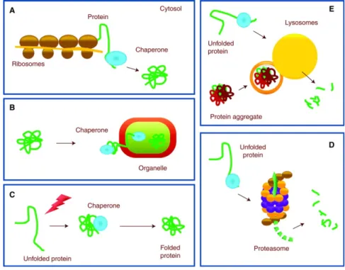
There are two major systems for degradation of proteins in eukaryotic cells: the ubiquitin-‐proteasome system (UPS) and the lysosomal system. These are important not only for clearing misfolded, damaged and mutant proteins, but also for maintaining protein synthesis by renewing the reserves of free amino acids in the cell (34) (35). Both proteolysis systems share a common mechanism: cargo to be degraded is selected and tagged, then recognized and delivered to the proteolytic system, where it undergoes proteolysis.
Figure 3. Chaperones are involved in protein clearance. Chaperones perform functions in
folding new proteins (A), unfolding/refolding for trafficking into cellular compartment (B) and refolding associated with protein damage (C). When these mechanisms fail, unfolded or misfolded proteins can be degraded by a proteolytic system: the ubiquitin-‐proteasome system (UPS) (D) or autophagy (E). Adapted from (35).