ISSN 1517-7076 artigo e-12217, 2018
Autor Responsável: Paola Andrea Forero Sossa Data de envio: 14/07/2017 Data de aceite: 19/02/2018
10.1590/S1517-707620180004.0551
Comparative study between natural and
synthetic Hydroxyapatite: structural,
morphological and bioactivity
properties
Paola Andrea Forero Sossa1,2, Belarmino Segura Giraldo 1,2, Beatriz Clemencia Galviz Garcia1, Elisabeth Restrepo Parra1,3,
Pedro Jose Arango Arango1
1 Laboratorio de Física del Plasma - Universidad Nacional de Colombia Sede Manizales, Laboratorio de Física del Plas-ma, Campus La Nubia, Manizales, Caldas,Colombia.
2 Grupo de investigación en Física y Matemáticas con énfasis en la formación de ingenieros - Universidad Autónoma de Manizales, Carrera 19 A, Manizales, Caldas, Colombia.
3 PCM Computacional Aplicaciones - Universidad Nacional de Colombia Sede Manizales, Laboratorio de Física del Plasma, Campus La Nubia, Manizales, Caldas,Colombia.
e-mail: paaforeroso@unal.edu.co, bsegurag@unal.edu.co, bcgalvizg@unal.edu.co, erestrepop@unal.edu.co, pjaran-goa@unal.edu.co
ABSTRACT
In this work, a study of hydroxyapatite (HAp) powders obtained using both, porcine bones and chemical pre-cursors was carried put. In the case of HAp obtained by means of porcine bones, physical processes as cook-ing, washing and milling were developed, for removing the organic material from the bones; after that, the powders were submitted to a thermal treatment at 800 °C, during 12 h. This procedure was carried out with-out adding chemical alkalines that are harmful for the environment and the human health. On the other hand, HAp powders were also synthetized using the chemical precipitation method widely reported, showing suc-cessful results. Moreover, both kind of powders were characterized using x ray diffraction, Fourier trans-former infrared spectroscopy, scanning electron microscopy and energy dispersive spectroscopy. Further-more, the bioactivity of the materials was determined using the simulated biological fluid (SBF) method. Results showed that the natural HAp exhibited better crystallographic properties. Moreover, according to these results, HAp obtained from porcine bones contains traces of elements as Na and Mg that are favorable for the bioactivity, according to the materials behavior when they are immersed in SBF.
Keywords: Hydroxyapatite, porcine bones, chemical precipitation, simulated biological fluid.
1. INTRODUCTION
Hydroxyapatite is considered as a biomaterial widely employed in many health applications. It is specially used as a source of calcium to produce toothpaste and as an important compound to repair bones. Because of its chemical properties, hydroxyapatite exhibits a great bioactivity and is highly compatible with the adjacent bone and teeth in living beings. Hydroxyapatite is a calcium phosphate ceramic with a high biocompatibility and is nontoxic, being an integral part of bone and teeth tissue in [1]. When HAp is in contact with the osse-ous tissue, it promotes the bone growth toward the implant [2,3]. HAp with chemical formula Ca10(PO4)6(OH)2, has been widely studied since 1926 by Jong et al [4]. They found a relationship between the osseous mineral and HAp [4]; afterwards, Posner et al [5–7] carried out great advances, especially regard-ing the crystallographic structure identification of HAp obtained by means of chemical precipitation. Current-ly, there is a lot of research not only about the production of good quality HAp exhibiting suitable biocom-patible properties, but also in searching economical and not polluting production methods [8,9]. The most used methods for obtaining HAp are: the hydrothermal [10,11], chemical precipitation [12–14], biogenic sources [8,15], among others. The last two methods represent the 30% of the reports in indexed journals from 1999 [9].
bio-genic sources allow the material to keep the native architecture of the animal osseous tissue, producing apa-tite that, under suitable conditions, behaves as an osteoconductive material [16].
Chemical precursor methods to obtain HAp, exhibit several advantages as high crystallinity, purity and suitable Ca/P ratio [9]; nevertheless, the lack of Fe2+, Mg2+, Si2+, Na+, F- ions, that is a specific character-istic of the osseous apatite, is the main disadvantage of HAp produced by this method, compared to that ob-tained using biogenic source methods [3,17]. Furthermore, some of the most biogenic sources used and re-ported in the literature are: egg shells [15], animal bones [18–22], fish spines [8], among others. On the other hand, one of the most used methods for studying the bioactivity was reported by Kokubo et al [23]. In this method, the material is immersed in a simulated biological fluid (SBF) that contains the same ionic concen-tration than the human blood plasma. This characteristic is required, because when the material is implanted into the human body, crystals of apatite, like the bone, are formed on the surface; then, the material can be joined to the bone using the apatite layer. This in vivo formation of apatite can be reproduced on the material surface, when it is immersed in a SBF solution [24]; hence, in an initial approach, the degree of bioactivity can be obtained. In the study performed by Hesaraki et al. [25], HAp was produced using the chemical pre-cipitation method; after that, samples were submerged in a SBF, to obtain carbonated HAp (CHAp). It was determined the behavior of this CHAp, in mesenchymal mother cells (MSC), obtained from mice; in contrast, our work includes, not only the study of synthetic HAp, but also the analysis of biogenic HAp; it is an im-portant advantage, since the natural HAp is more economic and the main raw material is highly accessible. The surface layer of apatite is normally characterized using techniques as x ray diffraction (XRD) and scan-ning electron microscopy (SEM); XRD allows to identify the crystalline phases of this calcium phosphate. Similarly, the SEM micrographs allow us to determine the morphology and the growth of apatite; neverthe-less, few reports of Fourier transformed infrared spectroscopy (FTIR) analysis using the attenuated total re-flectance (ATR) can be found in the literature. The main objective of this analysis is to determine functional groups and vibrational modes of the material molecules; for this reason, this is a suitable method to identify the apatite.
Currently, some researches focused on HAp obtained from porcine bones coincide in using sodium hydroxide [NaOH], to take away the organic part from the bones; nevertheless, NaOH is a highly alkaline chemic that generates important quantities of hazardous waste. It makes that this chemical compound may be used in an especial way, generating an increase in the method cost and environmental damage [19,20,22,26]; moreover, according to the literature, all the works reporting HAp extracted from animal bones use any deri-vate of NaHO. For this reason, it is important to investigate about methods to obtain HAp from biogenic sources trying to protect the environment, avoiding the use of chemical contaminants.
This research presents a method to extract HAp from porcine bones, including physical processes without using hazardous chemical reagents; furthermore, the material obtained will be compared with HAp powders obtained from the chemical precipitation method, to determine the efficiency of the proposed meth-od. Finally, materials obtained were characterized by the x-ray diffraction (XRD), Fourier transformed infra-red spectroscopy (FTIR), scanning electron microscopy (SEM) and energy dispersive spectroscopy (EDS); similarly, the bioactivity of the obtained materials was analyzed using the simulated biological fluid (SBF) technique, applying the Kokubo method [27]; afterwards, the materials were again characterized using FTIR and SEM.
2. EXPERIMENTAL DETAILS
2.1 Synthesis of Natural Hydroxyapatite
Figure 1: Scheme of the methodology used for obtaining HAp from pig bones as a biogenic source (natural method).
2.2 Synthesis of Hydroxyapatite by chemical precipitation
Synthetic powders of HAp were obtained from the next chemical precursors: Calcium nitrate 4-hydrate (Ca(NO3)2˙4H2O) at 98% of purity (Panreac) and ammonium di-hydrogen phosphate((NH4)H2PO4) at 99% of purity (Panreac). The solution of Ca(NO3)2˙4H2O was used at a concentration of 1 mol, while the solution of ((NH4)H2PO4) was employed at 0.6 mol. The nitrate calcium was used as a base solution, adding to it the monobasic phosphate solution drop by drop. With this procedure, pH remained lower than 8 during the
reac-tion by adding ammonia (NH4); similarly, the temperature reacreac-tion was controlled, keeping it constant at 37°C ± 3°C; afterwards, the magnetic shaking was carried out during 3 hours and the aging was maintained
Figure 2: Scheme of the methodology used for obtaining HAp from chemical agents (synthetic method).
2.3 Characterization of the materials
Table 1: List of reagents for the simulated biological fluid preparation and their ion concentration in the SBF and the blood plasma [24]
NO CHEMICAL REAGENTS
ION
CONCENTRATION MMOL BLOOD
PLASMA
SBF
1 Sodium chloride [NaCl]
Na+ 142 142
2 Sodium hydrogen carbonate [NaHCO3]
HCO3- 4,2 27
3 Potassium Chloride [KCl]
K+ 5 5
4 Potassium Hydrogen Phosphate Trihydrate
[K2HPO4·3H2O] HPO42- 1,5 1,5
5 Magnesium Chloride Hexahydrate [MgCl2·6H2O]
Mg2+ 2,5 2,5
6 Hydrogen Chloride [HCl] Cl- 103 147,8
7 Calcium Chloride [CaCl2] Ca2+ 27 4,2
8 Sodium Sulfate [Na2SO4] SO42- 1 1
9 Tris(hydroxymethyl)aminomethane [(HOCH2)3CNH2]
Buffer 0.5 0.5
10 Hydrogen Chloride [HCl] Ph 7.2 – 7.4 7.4
Finally, the powders were analyzed in a simulated biological fluid (SBF) as is reported by Kokubo et al [23]. In order to apply this method, the reagents listed in table 1 were added, one by one, before incorporat-ing the calcium chloride [CaCl2]. It is necessary to establish pH=2 in the solution, usincorporat-ing hydrochloric acid [HCl], to avoid the precipitation of the calcium phosphate in the solution. Finally, reagents 9 and 10 from the list of table 1 were added, in order to obtain a pH=7.42 ±0.02.
3. RESULTS AND DISCUSSION
3.1 FTIR analysis
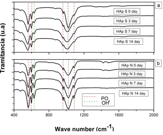
Figure 3: FT-IR spectra of natural and synthetic HAp.
Table 2: Typical vibrational bands of HAp [28]
FUNCTIONAL GROUP VIBRATIONAL MODE WAVE NUMBER (CM-1)
OH
-Stretch 3570
Bending 629
PO4
3-Asymmetric bend 561
598
Asymmetric stretch 1021
1087
Symmetric stretch 961
CO32- Asymmetric stretch 1545
1450
Symetric bend 878
charac-teristic of stoichiometric hydroxyapatite [1]. Moreover, table 2 presents the list of the functional groups, wavelength number and type of vibration mode.
Figure 3 shows the transmittance intensity of the bands at 629 and 357 cm-1 belonging to OH- that is lower in the case of the natural HAp compared to synthetic HAp. This difference can be attributed to the fact that synthetic HAp was obtained by means of reactions between two solutions in an aqueous environment, promoting the formation of a great quantity of this type of functional groups; on the contrary, the natural HAp was obtained using physical reactions. With this method, the organic material of bones was extracted, in order to leave only the inorganic part corresponding to different calcium phosphate, mainly the HAp. During this process, the material did not remain in contact with any aqueous solution, being the possible cause of no
formation of great quantity of hydroxyl groups’ type.
In figure 3, bands at 878, 1450 and 1545 cm-1 corresponding to carbonate groups are identified; moreover, in table 2, the vibrational modes observed in the synthetic Hap are listed. These bands are formed, since with this methodology, a carbonate HAp was obtained. This type of HAp exhibits a substitution of car-bonate ions by hydroxyl ions (A type substitution) and carcar-bonate ions by phosphate ions (B type substitution) [27]. This type of substitutions favors the HAp powders bioactivity, because the negative charge carriers ini-tiate and promote the bone type apatite growth in presence of the SBF [30].
3.2 XRD analysis
Figure 4: Diffraction patterns for natural and synthetic HAp powders
among others, are present in the materials obtained. Therefore, it can be concluded that there is not a mixture of the HAp with other crystalline compounds; moreover, in these diffractograms, a preferential growth in the (211) plane at 2θ = 31.9325° is observed. This is a typical characteristic of the HAp.
The lattice parameters obtained for these patterns are listed in table 3. These values are similar to those reported in the literature [2]; moreover, from the Stokes and Wilson equation, values of micro-stress were obtained as shown in table 3; it is evident that the synthetic structure undergoes a stress 6 times greater than the natural HAp. This behavior is associated to the methods employed; particularly, in the case of the chemical precipitated, the procedure was carried out at low temperatures, being in detriment of the crystalline structure, since the energy is not enough to reach the formation limit of the crystalline HAp that is 11. 2 eV [3]; then, a HAp with poor crystallinity was formed, increasing the lattice stress. The lower crystallinity and higher mocrodeformation exhibited by the synthetic HAp, compared with those observed in the natural HAp is an indicative that the first one presents the production of nanocrystals. These nanocrystals are evidenced by the formation of wide peaks in the diffraction patterns; furthermore, this type of nanocrystals can be formed by nanometric particles which sizes do not favor the bone type HAp growth when they are immersed in SBF.
Table 3: Parámetros cristalográficos obtenidos a partir de los patrones de difracción de la figura 4
HYDROXYAPATITE LATTICE PARAMETER MICRODEFORMATION
a c
SYNTHETIC HAP 9.4455 Å 6.9234 Å 0.6664 ± 0.3114
NATURAL HAP 9.3892 Å 6.8442 Å 0.1585 ± 0.0527
HAP [30] 9.4320 Å 6.8810 Å ---
3.3 SEM Analysis
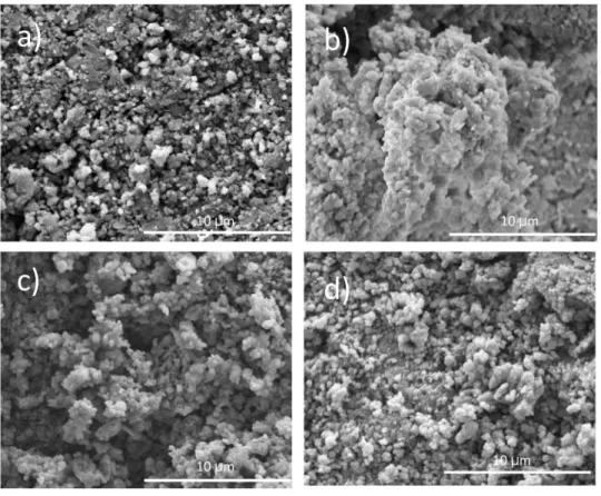
Figure 5: SEM micrographs of a) natural HAp powders at 500X, b) synthetic HAp powders at 500X c) natural HAp at 2500X and d) synthetic HAp at 2500X
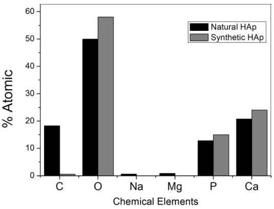
Figure 6: Atomic percentage depending on the chemical element obtained by EDS for both, natural and synthetic HAp.
In figure 6, the atomic percentage depending on the chemical element for both natural and synthetic HAp can be observed. These results were obtained using EDS technique. According to these results, the pre-dominant elements in both samples are P, Ca and O; furthermore, the spectrum of natural HAp reveals the presence of impurity traces of Na and Mg. These impurities are not present in the synthetic HAp. The
prece-dence of these impurities is attributed to the composition of the natural’s Hap precursors (pig bones)
3.4 Bioactivity test using SBF
3.4.1 Fourier transform infrared spectroscopy
Figure 9: SEM micrographs of synthetic HAp submerged in SBF a) 0 days b) 3 days c) 7 days and d) 14 days
The Ca/P relationship was determined from the EDS analyses and values of 1.62 and 1.59 for natu-ral and synthetic Hap, which were obtained respectively. These values are close to the ideal value of 1.67, especially in the case of the natural HAp [3], [8]; the difference between the Ca/P relationship for both natu-ral and synthetic HAp, is due to the obtaining method, since the chemical precipitation was carried out at low temperatures, entailing to produce Ca deficient HAp with lower crystallinity as evidenced in XRD analysis [3].
3.4.2 SEM analyses
Figures 8 and 9 show SEM micrographs for natural and synthetic Hap, respectively submerged in SBF during 0, 3, 7 y 14 days. For the bioactivity analysis, the materials were synthesized and after that, they do not
ex-hibit the same powders’ morphology. The micrograph presented in figure 8(a) was considered the pattern sample, since it was not submerged in SBF. In this image, it is observed uniformity in the grain size and oval morphology. In figures 8(b), (c) and (d), the apatite growth exhibiting spherical shapes, can be observed. These spheres are agglomerated in order to form an interconnected lattice like ordering; the apatite formation was observed 3 days after the immersion in SBF, where the spheres formation increased with the time of immersion [11].
In figure 9(a), a similar morphology to the one presented in figure 8(a), corresponding to natural HAp is observed; nevertheless, the formation of HAp after 3 days of immersion is poor and it increases with the time of immersion; nevertheless, the growth of apatite in samples of synthetic HAp is lower than the one in natural HAp. The greater apatite growth in samples of natural HAp, is attributed to the high intensity of the 630 cm-1 band, corresponding to the flexion vibration type of the OH- functional group, in this set of samples, this growth is observable in IR spectra shown in figures 3 and 7. [10].
The increase in the bioactivity is promoted by the combination of different calcium phosphate phas-es. Powders obtained from pig bones are a combination of hydroxyapatite and tricalcium phosphate. This combination improves the bioactivity with respect to pure HAp [34].
Figure 10 shows SEM micrographs at 50000X for natural and synthetic hydroxyapatite, immersed in SBF during 14 days. Synthetic HAp exhibits grains of nanometric sizes (fig. 10.a); these sizes are similar to those present in the bone. Therefore, natural HAp (fig. 10.b) shows grains of micrometric sizes. The micro-metric grains promoted a greater growth of the apatite bonelike than it promoted by nanomicro-metric grains.
Finally, it can be highlighted that in this work, we performed a comparative study of the bioactivity exhibited by both, natural and synthetic HAp, and this study was carried out using two complementary tech-niques as FTIR and SEM; furthermore, it was observed the great relevance that the particle size exerts on the bioactivity and the chemical response of these materials, by means of the intensity of the functional groups of the hydroxyl phosphates.
4. CONCLUSIONS
Powders of natural and synthetic HAp were obtained by using new methods proposed. It was found that natu-ral HAp exhibited better structunatu-ral properties compared with synthetic HAp. On the other hand, XRD analy-sis allowed us to observe that both, natural and synthetic HAp exhibit a single phase that is in agreement with the reposts; furthermore, the natural HAp presented higher crystallinity than the synthetic HAp. From the FTIR results, typical functional groups of stoichiometric HAp were identified, evidencing that both methods are successfully obtaining crystalline HAp. Finally, SEM results indicated that the morphology of the materi-als is strongly influenced by the method type, since natural HAp exhibited laminar shapes that were no ob-served in synthetic HAp. Similar to the case of bioactivity, it was evidenced a great increase of the OH- and PO43- functional groups for the set of natural HAp samples that indicates the formation of apatite. Using SEM analysis, it was identified a formation of an apatite layer on the surface of HAp samples; furthermore, this layer of apatite exhibited a greater growth in samples obtained by the natural method.
5. ACKNOWLEDGMENTS
The authors gratefully acknowledge financial support from the Dirección Nacional de Investigaciones of the National University.
6. BIBLIOGRAPHY
[1]RAYA I., MAYASARI E., YAHYA A., et al.,
―
Shynthesis and Characterizations of CalciumHydroxy-apatite Derived from Crabs Shells (Portunus pelagicus) and Its Potency in Safeguard against to Dental De-mineralizations
‖,
International Journal of Biomaterials, v. 2015, pp. 469176, Feb. 2015.[2]ZHOU, H., LEE, J., ―Nanoscale hydroxyapatite particles for bone tissue engineering‖,Acta Biomateralia.,
v. 7, n. 7, pp. 2769-2781, Jul. 2011.
[4]DE JONG W.F., ―Le substance minerale dans le os‖
,
Recueil des Travaux Chimiques des Pays-Bas, v. 45,n. 6, pp. 445–448, Apr. 1926
.
[5]POSNER A.S
.
―
Crystal chemistry of bone mineral‖,
Physiological. Reviews., v. 49, n. 4, pp. 760-792,Oct. 1969.
[6]POSNER A.S., PERLOFF A., DIORIO A.F
.,
―Refinement of the hydroxyapatite structure‖,
Acta Crys-tallographica., v. 11, pp. 308-309, Dec. 1958.[7]KAY, M.I., YOUNG R.A., POSNER A.S., ―Crystal structure of hydroxyapatite‖,International journal of science nature, v. 204, pp. 1050-1052, Dec. 1964
.
[8]HUANG Y.-C., HSIAO P.-C., CHAI H.-J.,
―
Hydroxyapatite extracted from fish scale: Effects on MG63 osteoblast-like cells‖,
Ceramics International. v. 37, m. 6, pp. 1825-1831, Aug. 2011.
[9] SADAT-SHOJAI, M., KHORASANI, M.T., DINPANAH-KHOSHDARGI, E., et al., ―Synthesis m eth-ods for nanosized hydroxyapatite with diverse structures‖
,
Acta Biomaterialia, v. 9, n. 8, pp. 7591-7621, Aug.2013.
[10] ZHANG G., CHEN J., YANG S., et al.,―Preparation of amino-acidregulated hydroxyapatite particles by hydrothermal method‖,Materials. Letters, v. 65, n. 3, pp. 572-574, Feb. 2011
.
[11] JOKIC, B., MITRIC, M., RADMILOVIC, V., et al.,
―
Synthesis and characterization of monetite andhydroxyapatite whiskers obtained by a hydrothermal method
‖,
Ceramics International., v. 37, n. 1, pp.167-173, Jan. 2011.
[12] STIPNIECE, L., SALMA-ANCANE, K., BORODAJENKO, N., et al.,
―
Characterization of Mg-substituted hydroxyapatite synthesized by wet chemical method‖,
Ceramics. International., v. 40, n. 2, pp.3261-3267, Mar. 2014
.
[13]KIM D.W., CHO I.-S., KIM J.Y., et al.,
―
Simple large-scale synthesis of hydroxyapatite nanoparticles:in situ observation of crystallization process
‖,
Langmuir, v. 26, n. 1, pp. 384-388, Oct. 2010.
[14]DHANG V., RHEE K.Y., PARK S.-J.,
―
The facile and low temperature synthesis of nanophase hydrox-yapatite crystals using wet chemistry‖,
Materials Science and Engineering: C, v. 36, pp. 152-159, Mar. 2014.[15] KAMALANATHAN P., RAMESH S., MANG L.T., et al.,
―
Synthesis and sintering of hydroxyapatite derived from eggshells as a calcium precursor‖, Ceramics International., v. 40, n. 10B, pp. 16349-16359,Dec. 2014.
[16] MUCALO M.R., 14 – Animal-bone derived hydroxyapatite in biomedical applications in: Hydroxyap-atite Biomed. Appl
.,
The University of Waikato, 1 ed, New Zealand, 2015[17]AKRAM, M., AHMED, R., SHAKIR, I., et al.,―Extracting hydroxyapatite and its precursors from nat u-ral resources‖
,
Journal of Materials Science. v. 49, n. 4, pp. 1461-1475, Feb. 2014.[18]MONDAL, S., MONDAL, B., DEY, A., et al.,
―
Studies on processing and characterization ofhydroxy-apatite biomaterials from different bio wastes
‖,
Journal of Minerals and Materials Characterization and Engineering v. 11, n. 1, pp. 55-67, Jan. 2012.[19] HABERKO, K., BUCKO, M.M., BRZEZINSKA-MIECZNIK, J., et al., ―Natural hydroxyapatite—its behaviour during heat treatment‖,Journal of the European Ceramic Society., v. 26, n. 4-5, pp. 537-542, Aug.
2005
.
[20]JANUS, A.M., FARYNA, M., HABER,KO K., et al., ―Chemical and microstructural characterization of natural hydroxyapatite derived from pig bones‖,Microchimica Acta., v. 161, n. 3-4, pp. 349-353, Jun. 2008.
[21] KIM S.-H., SHIN J.-W., PARK S.-A., et al., ―Chemical, structural properties, and osteoconductive e f-fectiveness of bone block derived from porcine cancellous bone‖, Journal of Biomedical Materials Research Part B: Applied Biomaterials., v. 68, n. 1, pp. 69-74, Nov. 2004
.
[22] SOBCZAK-KUPIEC, A., WZOREK, Z.,―The influence of calcination parameters on free calcium
ox-ide content in natural hydroxyapatite‖,Ceramics International. v. 38, n. 1, pp. 641-647, Jan. 2012.
[23] KOKUBO, T., TAKADAMA, H
.,
―How useful is SBF in predicting in vivo bone bioactivity?‖,
Bio-materials., v. 27, n. 15, pp. 2907-2915, May. 2006.[24] KOKUBO, T., ―Bioactive glass ceramics: properties and applications‖, Biomaterials., v. 12, n. 2, pp.
155-163, Mar. 1991.
[25]HESARAKI, S., NAZARIAN, H., POURBAGUI-MASOULEH, M., et al.,―Comparative study of me
Bi-omater., v. 102, n. 1, pp. 108-118, Jan. 2014
.
[26] BARAKAT, N.A.M., KHIL, M.S., OMRAN, A.M., et al., ―Extraction of pure natural hydroxyapatite from the bovine bones bio waste by three different methods‖, Journal of Materials Processing Technology.,
v. 209, n. 7, pp. 3408-3415, Apr. 2009
.
[27]HENCH L.L, ―Bioceramics: From Concept to Clinic‖, Journal of the American Ceramic Society., v. 74,
n. 7, 1487-1510, Jul. 1991.
[28] BIENENSTOCK A., POSNER A.S., ―Calculation of the x-ray intensities from arrays of small
crystal-lites of hydroxyapatite‖,Archives of Biochemistry and Biophysics. v. 124, pp. 604-607, Dec. 1964.
[29]KOKUBO, T., Bioceramics and their clinical application
s.
Woodhead Pub. and Maney Pub. on behalf of Institute of Materials, 1 ed, Minerals & Mining, 2008.
[30] LIU Y., SHEN Z., ―Dehydroxylation of Hydroxyapatite in Dense Bulk Ceramics Sintered by Spark
Plasma Sintering‖,Journal of the European Ceramic Society., v. 32, pp. 2691-2696, Aug. 2012.
[31] GIRALDO-BETANCUR A. L, ESPINOSA-ARBELAEZ D. G, DEL REAL-LOPEZ A, et al., ―Co
m-parison of physicochemical properties of bio and commercial hydroxyapatite‖, Current Applied Physics., v.
13, n. 7, pp. 1383–1390, Sep. 2013.
[32]HAHN, B. D, LEE, J. M, PARK, D. S, et al.,―Enhanced bioactivity and biocompatibility of nanostru
c-tured hydroxyapatite coating by hydrothermal annealing‖, Thin Solid Films, v. 519, n. 22, pp. 8085–8090,
Sep. 2011.
[33] KONG, L., GAO, Y., LU, G., et al., ―A study on the bioactivity of chitosan/nano-hydroxyapatite
com-posite scaffolds for bone tissue engineering‖,European Polymer Journal., v. 42, n. 12, pp. 3171–3179, Dec.
2006.
[34] FARZADI, A., SOLATI-HASHJIN, M., BAKHSI F, et al., ―Synthesis and characterization of hydrox
y-apatite/β-tricalcium phosphate nanocomposites using microwave irradiation‖, Ceramics International., v. 37,


![Table 1: List of reagents for the simulated biological fluid preparation and their ion concentration in the SBF and the blood plasma [24] NO CHEMICAL REAGENTS ION CONCENTRATION MMOL BLOOD PLASMA SBF 1 Sodium chloride [NaCl] Na + 142 142](https://thumb-eu.123doks.com/thumbv2/123dok_br/16319156.719083/5.892.123.781.155.576/reagents-simulated-biological-preparation-concentration-chemical-reagents-concentration.webp)