Development of
algorithms for
detection and
quantification of
rheumatic diseases
in musculoskeletal
ultrasound
Nelson Costa Martins
Doctoral Programme in Computer Science – MAPi
Department of Computer Science - FCUP 2019 De v e lo p m e n t o f a lg o rit h m s fo r d e te c tio n a n d q u a n tif ic a tio n of rhe um a tic di s e a s e s in m us c ul os k e le ta l ul tr a s ound Nel so n Co st a M ar tin s
D
FCUP DCC 2019 3.º CICLOD
D
D
Development of
algorithms for detection
and quantification of
rheumatic diseases in
musculoskeletal
ultrasound
Nelson Costa Martins
Doctoral Programme in Computer Science - MAPi
Department of Computer Science, Faculty of Sciences University of Porto
2019
Supervisor
Miguel Tavares Coimbra,
Assistant Professor, Department of Computer Science Faculty of Sciences of the University of Porto, Portugal
Co-Supervisor
Manuel João Oliveira Ferreira
Head of Research, Development and Innovation Neadvance, Machine Vision, SA, Braga, Portugal
P
HD T
HESISDevelopment of Algorithms for Detection
and Quantification of Rheumatic Diseases
in Musculoskeletal Ultrasound
Author:
Nelson MARTINS
Supervisors: Doutor Miguel COIMBRA
Doutor Manuel FERREIRA
Submitted in part fulfillment of the requirements for the degree of Doctor of Philosophy in Computer Sciences, for the MAPi doctoral program.
Faculty of Sciences of the University of Porto &
Neadvance, Machine Vision, SA
I, Nelson MARTINS, would like to thank Doctor Manuel Ferreira and Doctor Miguel
Coim-bra as my main supporters. I’m also very thankful to the Neadvance - Machine Vision SA, Faculty of Sciences of the University of Porto and Telecommunications Institute -Porto teams for their help as friends and work partners. To Dra M´onica Bogas and Dra Filipa Teixeira many thanks, their medical experience was very important along this work. And finally, to my family and friends for their unconditional support.
Resumo
Programa Doutoral MAPi
Doutoramento em Inform´atica
Desenvolvimento de Algoritmos de Detecc¸˜ao e Quantificac¸˜ao de Doenc¸as Reum´aticas para Ecografia M ´usculo-Esquel´etica
por Nelson MARTINS
As doenc¸as reum´aticas s˜ao a maior causa de dor e perda de mobilidade nos pa´ıses de-senvolvidos, fazendo delas um grave problema social, econ´omico e de sa´ude p´ublica. Devido `as suas vantagens, a ecografia tem vindo a ser introduzida na pr´atica cl´ınica dos reumatologistas de forma a facilitar o diagn´ostico e o seguimento dos pacientes. Ao contr´ario da radiografia, que ´e atualmente a modalidade imagiol´ogica standard, a ecografia n˜ao utiliza radiac¸˜ao ionizante e permite uma detecc¸˜ao mais precoce e mel-hores seguimentos de algumas doenc¸as. No entanto, a dificuldade na interpretac¸˜ao e aquisic¸˜ao das imagens ecogr´aficas reduz a sua aceitac¸˜ao e, por esse motivo, s˜ao necess´arias soluc¸˜oes inovadoras que potenciem a sua utilizac¸˜ao em contexto cl´ınico. Neste trabalho utilizam-se imagens da segunda articulac¸˜ao metacarpofalˆangica pela sua importˆancia para o diagn´ostico de doenc¸as reum´aticas tal como a Artrite Reumat´oide. Relativamente a estas imagens, foram identificadas problemas em aberto na literatura, os quais se procuraram responder com as abordagens descritas ao longo deste tra-balho.
Segmentac¸˜ao do metacarpo e da falange utilizando contornos ativos locais. Esta tec-nica permitiu identificar 80% das imagens com uma distˆancia m´edia de Hausdorff infe-rior a 3 pixels;
A identificac¸˜ao do tend˜ao extensor foi alcanc¸ada aplicando uma variante do algoritmo de contornos ativos abertos, utilizando simetria de fase como preprocessamento e dados de forma do tend˜ao para reforc¸ar o modelo. Por fim, o tend˜ao foi obtido pela op-timizac˜ao dos valores das energias recorrendo a algoritmos gen´eticos. Os resultados
alcanc¸ados apontam para uma distˆancia m´edia de Hausdorff inferior a 10 pixels (ou 0.5 mm) em 95% das imagens;
Para a identificac¸˜ao da c´apsula articular foram testadas duas abordagens distintas, uma baseada no algoritmo SLIC, e outra baseada em redes neuronais convolucionais, mais propriamente o modelo UNet. Os resultados indicam uma clara superioridade do modelo UNet que tem ainda a vantagem de n˜ao depender de outros m´etodos de segmentac¸˜ao (como ´e o caso do SLIC) no processo de inicializac¸˜ao. Os resultados demonstraram que 90% das imagens foram identificadas com um coeficiente de Dice superior a 0.6.
Por fim, foi testada uma abordagem preliminar de extrac¸˜ao de carater´ısticas e classifica-c¸˜ao para o problema espec´ıfico da sinovite. Esta abordagem partiu das segmentac¸˜oes do metacarpo, falange e tend˜ao extensor para criar um conjunto de m´ascaras para caracterizar localmente a imagem e identificar as alterac¸˜oes criadas na mesma devido a sinovite. Tendo sido obtido um index de Youden’s de 0.84 para a identificac¸˜ao de sinovite e de 0.94 para a classificac¸˜ao.
Palavras-chave: Doenc¸as Reum´aticas; Artrite Reumat´oide; Sinov´ıte; Articulac¸˜ao
Metacar-pofalangica; Ecografia; Contornos ativos; Simetria de Fase; SLIC; Redes Neuronais Convolucionais; M´aquinas de Vectores de Suporte; Extracc¸˜ao de Caracter´ısticas.
Abstract
MAPi Doctoral Program
Doctor of Philosophy in Computer Sciences
Development of Algorithms for Detection and Quantification of Rheumatic Diseases in Musculoskeletal Ultrasound
by Nelson MARTINS
Rheumatic diseases are the main cause of impairment and pain in developed countries, which makes them a critical social, health and economic problem. Due to their main advantages, ultrasounds are now being used in rheumatology to diagnosis and evalu-ate rheumatic diseases in their early stages. Unlike radiography, which is the current standard, ultrasounds are less expensive, do not use ionizing radiation and can lead to better early diagnosis and follow-up outcomes. The difficulties in the interpretation and acquisition of this type of image reduce its acceptance, and thus, new and innovative solutions are needed to help doctors in the diagnostic process.
In this study, images of the second metacarpophalangeal joint will be used because they are of great importance in the diagnosis of rheumatic diseases such as Rheuma-toid Arthritis. Regarding these images, there are image processing problems that are still unsolved, and this work aims to solve them.
The segmentation of the metacarpus and phalangeal bone was achieved using Local-izing Active Contours. This approach allowed the identification of 80% of the images with a Modified Hausdorff Distance below 3 pixels.
The extensor tendon was identified with the proposal of an Open Ended Active Con-tours method using Phase Symmetry pre-processing, prior structure knowledge and Genetic Algorithm based optimization. The results show that the segmentation was achieved with a confidence of 95% for a Modified Hausdorff Distance below 10 pixels;
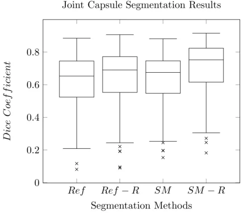
For the segmentation of the joint capsule, two distinct approaches were tested: one using the Simple Linear Iterative Clustering algorithm followed by a special shape con-strained merge strategy, and the other using convolutional neural networks, more pre-cisely the UNet model. The results show that the UNet model outperforms the clus-tering method, without the necessity of other segmentation methods to limit the joint capsule search zone, as is the case for the Simple Linear Iterative Clustering method. The segmentation was achieved with a Dice Coefficient higher than 0.6 in 90% of the images.
Finally, a preliminary feature extraction and classification study was presented, specif-ically addressing the synovitis. The proposed approach started from the segmentation of the metacarpus, phalange and extensor tendon to create a set of masks. These masks were used to locally characterize the images and detect the anatomical changes provoked by the synovitis.
Keywords: Rheumatic Diseases; Rheumatoid Arthritis; Synovitis; Metacarpophalangeal
Joint; Ultrasounds; Active Contours; Phase Symmetry; Simple Linear Iterative Cluster-ing; Convolutional Neural Networks; Support Vector Machines; Feature Extraction.
Acknowledgements iii
Resumo v
Abstract vii
Contents ix
List of Figures xiii
List of Tables xvii
Abbreviations xix 1 Introduction 1 1.1 Motivation . . . 1 1.2 Research Questions . . . 4 1.3 Scientific Contributions. . . 4 1.4 Thesis Outline . . . 7 2 Ultrasound Imaging 9 2.1 Introduction . . . 9 2.2 Acquisition Equipment . . . 10 2.3 Image Characteristics . . . 12 2.4 Doppler Mode. . . 13 3 Rheumatology Background 17 3.1 Introduction . . . 17 3.1.1 Causes . . . 18 3.1.2 Epidemiology . . . 19 3.2 Rheumatoid Arthritis . . . 20 3.2.1 Diagnosis . . . 21 3.2.2 Treatment . . . 24 3.2.3 Costs . . . 25
3.3 Ultrasound for Rheumatoid Arthritis. . . 25
3.3.1 Metacarpophalangeal Joint . . . 26
4 Image Processing and Analysis Background 31
4.1 Ultrasound Imaging Existing Work . . . 32
4.1.1 Pre-Processing . . . 32
4.1.2 Segmentation . . . 33
4.1.3 Feature Extraction and Classification . . . 34
4.2 Active Contours Model . . . 35
4.2.1 Internal Energy . . . 36
4.2.2 External Energy . . . 36
4.2.3 Optimization . . . 37
4.3 Localizing Active Contours. . . 37
4.4 Phase Symmetry . . . 39
4.5 Simple Linear Iterative Clustering . . . 41
4.6 Convolutional Neural Networks . . . 42
4.7 Performance Metrics . . . 45
4.7.1 Modified Hausdorff Distance . . . 45
4.7.2 Dice Similarity Coefficient . . . 47
5 Metacarpus and Phalange Identification 49 5.1 Introduction . . . 49
5.2 Proposed Work . . . 51
5.2.1 Pre-processing . . . 52
5.2.2 Contour Initialization . . . 52
5.2.3 Contour Refinement - LAC . . . 52
5.2.4 Upper Line Extraction . . . 53
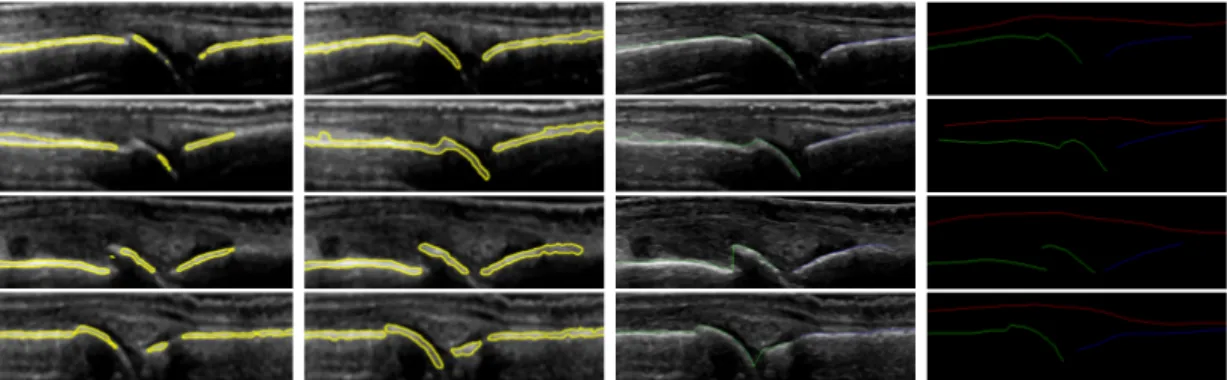
5.3 Results and Discussion . . . 54
5.4 Complementary Results . . . 55
5.5 Conclusions. . . 57
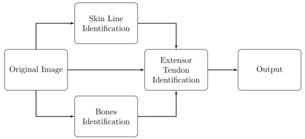
6 Extensor Tendon Identification 59 6.1 Introduction . . . 59 6.2 Segmentation Framework . . . 61 6.2.1 Internal Energy . . . 62 6.2.2 External Energy . . . 63 6.2.3 Area Constraints . . . 65 6.2.4 Additional Constraints . . . 66 6.2.5 Implementation . . . 67
6.3 Results and Discussion . . . 68
6.3.1 Dataset and Metrics . . . 68
6.3.2 Number of Points Optimization . . . 69
6.3.3 Weights Optimization . . . 70
6.3.4 Visual Results . . . 72
6.4 Complementary Results . . . 73
6.4.1 New Data Results . . . 73
6.4.2 Fully Automatic Segmentation . . . 74
6.5 Conclusions. . . 77
7.1 Introduction . . . 81
7.2 Proposed Work . . . 84
7.2.1 Split and Merge. . . 84
7.2.2 Convolutional Neural Networks . . . 88
7.3 Results and Discussion . . . 92
7.3.1 Metrics . . . 93
7.3.2 Results - SM . . . 93
7.3.3 Results - CNN . . . 96
7.3.4 Comparative Results and Discussion. . . 97
7.4 Conclusions. . . 100
8 Synovitis Detection and Grading 103 8.1 Introduction . . . 103
8.2 Proposed Work . . . 105
8.2.1 Feature Extraction . . . 106
8.2.2 Feature Selection . . . 108
8.3 Results and Discussion . . . 109
8.4 Conclusions. . . 112
9 Conclusions and Future work 115 A Database Information and Study 133 A.1 Database Characteristics . . . 133
A.2 Clinical Visual Features . . . 134
A.2.1 Distances Between Bones, Extensor Tendon and Skin Line . . . . 135
A.2.2 Joint Capsule Area . . . 136
1.1 Illustration of the bone anatomy of the hand and respective nomenclature. 3
2.1 Typical acquisition system used in medical ultrasound imaging. . . 10
2.2 Example of ultrasound transducers. . . 11
2.3 Example of image visualization enhancement by speckle noise reduction. 13 2.4 Examples of artifacts found in ultrasound images.. . . 14
2.5 Examples of a B-Mode, Doppler and Power Doppler images of the kidney from different patients. . . 15
3.1 Example of the progression of Rheumatoid Arthritis on the hands. . . 20
3.2 Illustration of a healthy metacarpophalangeal joint. . . 27
3.3 Illustration of a normal joint and a joint with synovitis. . . 28
3.4 Representation of the second metacarpophalangeal joint ultrasound im-age acquisition. . . 29
4.1 Representation of the Localizing Active Contour algorithm . . . 38
4.2 Example of a symmetric and an asymmetric object.. . . 39
4.3 Representation of the odd and even orthogonal filters. . . 40
4.4 Difference between a fully connected layer and a convolutional layer. . . . 43
4.5 Example of receptive field growth in deeper layers. . . 44
4.6 Example of the low level and high-level features learn in shallow and deep layers (from the bottom to the top). . . 44
4.7 Examples of the Hausdorff distance between two lines . . . 46
4.8 Visual representation of the Dice Coefficient in three different cases. . . . 47
5.1 Examples of ultrasound images of the dorsal view of the second metacar-pophalangeal joint. . . 50
5.2 Proposed pipeline for the identification of the metacarpus and phalange. . 51
5.3 Visual results of the metacarpus and phalange segmentation using the Localizing Active Contours algorithm. . . 54
5.4 Segmentation error for the metacarpus and phalange, using the vertical
distance in pixels.. . . 55
5.5 Segmentation error for the metacarpus and phalange using the vertical distance in pixels, in all dataset. . . 56
5.6 Segmentation error for the metacarpus and phalange using the Modified Hausdorff Distance in millimeters, in all dataset.. . . 57
6.1 Example of ultrasound images of the dorsal view of the second metacar-pophalangeal joint. . . 60
6.2 Proposed open ended active contour model framework. . . 62
6.3 Example of the phase symmetry pre-processing results. . . 64
6.4 Box plot of the area measurements. . . 66
6.5 Approximation error using different number of points. . . 69
6.6 Results obtained for different parameter configurations of the proposed method. . . 71
6.7 Visual results of the extensor tendon segmentation. . . 72
6.8 Results obtained for the segmentation of the extensor tendon, in all database. 74 6.9 Diagram of the fully automatic extensor tendon segmentation method. . . 75
6.10 Results obtained in the test data for the different automatic and manual segmentation settings. . . 75
6.11 Visual results of the fully automatic segmentation of the extensor tendon for different configurations.. . . 77
7.1 Example of ultrasound images of the second metacarpophalangeal joint with different joint capsules aspects and synovitis grades. . . 82
7.2 Joint capsule segmentation pipeline using a Split-Merge-Refine approach. 84 7.3 Results of the Simple Linear Iterative Clustering algorithm in the metacar-pophalangeal joint images. . . 86
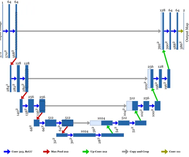
7.4 The UNet model.. . . 89
7.5 Result of adding skip connections. . . 90
7.6 Proposed changes to the UNet model. . . 91
7.7 Boxplot of the DICE values obtained for the different configurations. . . . 94
7.8 Segmentation results obtained for the different configurations.. . . 95
7.9 Visual results obtained for the segmentation of the joint capsule for dif-ferent configurations. . . 95
7.10 Visual results obtained for the segmentation of the joint capsule for dif-ferent UNet parameterizations. . . 97
7.12 Results obtained for the segmentation of the joint capsule using three
different methods. . . 99
7.13 Visual results obtained for the segmentation of the joint capsule using three different methods. . . 100
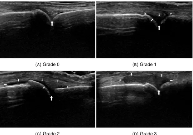
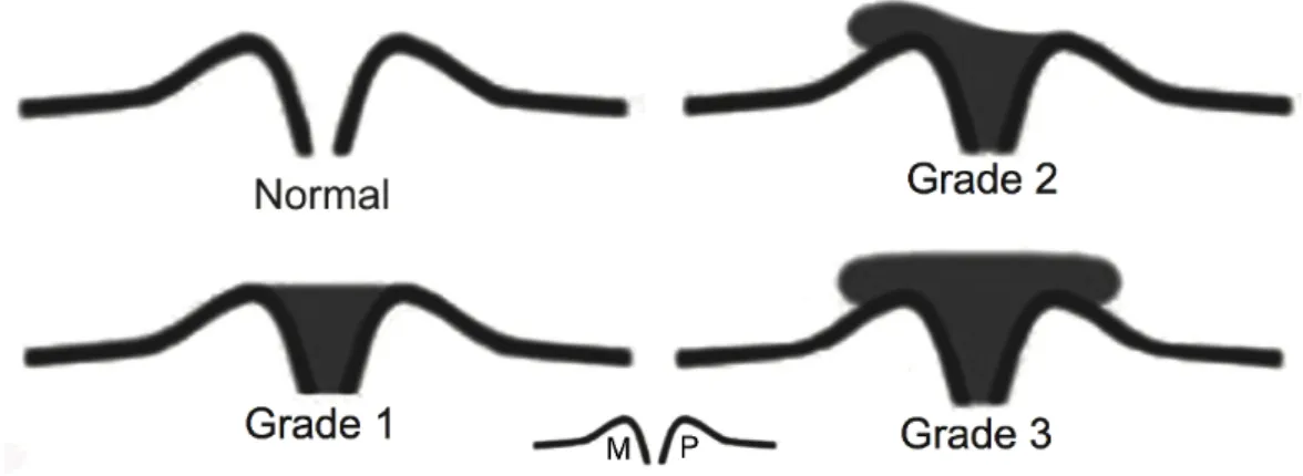
8.1 Representation of the synovitis grades convention. . . 104
8.2 Example of images with different synovitis grades. . . 105
8.3 Manual annotations used in the feature extraction. . . 106
8.4 Proposed masks for the synovitis identification and grading. . . 107
8.5 Selected features for the different synovitis detection and grading tests. . 113
A.1 Manual annotations used to validate the proposed algorithms.. . . 134
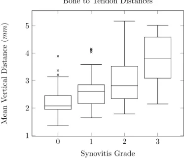
A.2 Box plot of the mean distances between the bones and the extensor tendon. . . 135
A.3 Box plot of the mean distances between the extensor tendon and the skin line. . . 136
A.4 Box plot of the joint capsule area extracted from the manual annotations for each synovitis grade. . . 137
A.5 Plot of the mean intensity extracted from inside and outside the joint capsule manual annotations. . . 139
A.6 Plot of the intensity standard deviation extracted from inside and outside the joint capsule manual annotations. . . 139
3.1 Epidemiology of major rheumatic diseases. . . 20 3.2 2010 ACR/EULAR classification criteria for rheumatoid arthritis.. . . 22 6.1 Confidence obtained for the different configurations and for three MHD. . 76 7.1 p-values resulting from the W elch0s t testof different pairs of methods. 94
7.2 Mean DICE results in the test set. . . 96 7.3 p-values resulting from the W elch0s t testof different pairs of methods. 98
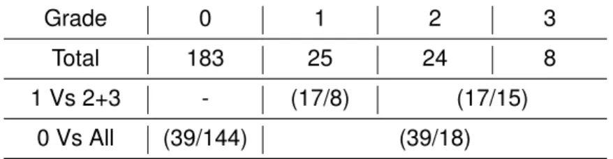
8.1 Database organization and number of images used in the Monte Carlo cross-validation train and validation (Train/Validation) attending to the synovitis grade. . . 110 8.2 Feature selection results in the four tested configurations. . . 111 8.3 Summary of the results obtained before and after the feature selection
phase for each configuration. . . 112 A.1 Final database size according to the synovitis grade. . . 133 A.2 Statistical significance (p-values) between the mean distance
measure-ment and the synovitis degree. . . 136 A.3 Statistical significance of the difference in the joint capsule area in
differ-ent synovitis grades. . . 137 A.4 Correlation between the joint capsule area and the mean distance
ACR American College of Rheumatology AUC Area Under the Curve
bsDMARD biosimilar Disease Modifying AntiRheumatic Drugs CAD Computer Aided Diagnosis
CAT Computed Axial Tomography
DMARD Disease Modifying AntiRheumatic Drugs DNN Deep Neural of Network
EM Expectation Maximization
EULAR EUropean League Against Rheumatism IDC Inverted Dice Coefficient
LAC Localizing Active Contours MCPJ MetaCarpoPhalangeal Joint MHD Modified Hausdorff Distance MRF Markov Random Field
MRI Magnetic Resonance Imaging MTPJ MetaTarsoPhalangeal Joint PIP Proximal InterPhalangeal PCA Pincipal Component Analysis RA Rheumatoid Arthritis
SLIC Simple Linear Iterative Clustering SVM Support Vector Machine
ULSAM Unidade Local Sa´ude Alto Minho US UltraSound
Introduction
Contents 1.1 Motivation . . . 1 1.2 Research Questions . . . 4 1.3 Scientific Contributions . . . 4 1.4 Thesis Outline . . . 71.1 Motivation
Rheumatic diseases are pathologies that affect bones, joints and connective tissues. They are the main cause of impairment and pain in developed countries, making them a serious health problem with high social and economic implications [1]. Due to the in-crease in longevity and negative lifestyles, the number of rheumatic problems is likely to grow in the coming years. This creates pressure for the diagnosis of these diseases be-cause, if not diagnosed and treated properly and in time, they progress faster, causing severe and irreversible physical deformations and, leading to incapacity [2]. An early diagnosis is then fundamental to give the patient a better quality of life and prevent faster deterioration of his/her condition.
The number of rheumatic diseases is very large and covers a broad range of different types of pathologies, but their assessment is usually based on physical exams, imaging techniques and sample based analysis [3]. A physical exam tests the mobility of the joints, pain, and other movement characteristics, such as morning stiffness and lack of
strength. Sample based analysis tests the presence and concentration of specific anti-bodies and other substances in the body [4]. The imaging techniques allow visualization of the internal body structures. In rheumatology, the most common imaging techniques are magnetic resonance imaging (MRI), radiography and ultrasound imaging. Due to their characteristics, ultrasounds have seen their use increase in the last few years. These characteristics include their low cost, ease of operation, ease of acceptance by patients, possibility of acquiring several images in different positions, and comparable
results to the other imaging techniques [5,6]. One limitation of the imaging techniques
is the subjectivity of the diagnosis and the difficulty of image interpretation. This is es-pecially true in ultrasound images, because the images tend to be more noisy and their acquisition more operator dependent. Moved by this problem and its potential impact, the research, development and testing of new advanced image processing algorithms are proposed in this work. Due to the previously mentioned advantages, ultrasound imaging was selected for use in this study. The aim is to contribute to the creation of a computer aided diagnosis (CAD) system to automatically extract meaningful informa-tion from the images that complements the informainforma-tion that the rheumatologists already extract from them empirically. In this manner, it is also expected to help improve the diagnosis and follow up for rheumatic diseases.
In medicine, and rheumatology in particular, the use of quantitative information is es-sential to obtain an evidence based diagnosis. Consequently, a CAD system can be an added value for doctors and their patients. In fact, such systems have been used to help visualize and extract information from medical information. The introduction of digital images and videos created the opportunity to use digital manipulation. From simple contrast enhancement, zooming and panning to more complex tools, such as statistical analysis and even artificial intelligence, they all help handle and enhance the information present in the images.
To achieve the proposed CAD system, work on automatic detection of physiologi-cal structures will be conducted, as well as quantification and classification of rele-vant physiological parameters. In this manner, it is expected that the assessment of rheumatic diseases will be improved, making it less subjective and reducing the vari-ability intra and inter observer. Moreover, with more precise measures, the follow-up for these diseases could be more accurate and the treatment adjustment more assertive,
allowing a direct comparative analysis of the treatment responses for each patient. Ide-ally, this could also be used to create gold standards, improving the diagnosis even further.
Given that the number of possible acquisition protocols is very broad, it was decided to narrow the scope and focus the work on a single acquisition. The longitudinal-dorsal view of the metacarpophalangeal joint (MCPJ) was selected because it provides clues regarding the hand small joint, which is often the first structure to be affected by
rheumatic diseases, such as rheumatoid arthritis. In Fig. 1.1an illustration of the hand
bone structure can be seen.
FIGURE 1.1: Illustration of the bone anatomy of the hand and respective
nomencla-ture.1
The metacarpals are the bones in the palmar region of the hand; they are directly connected to the phalanges and carpal bones, which are, respectively, the fingers and
wrist bones. The numbers above each finger in Fig.1.1indicates the convention in the
naming of each finger, which is used to facilitate communication between medical staff. The rule is that the thumb is the first finger, the index finger is the second, and so on, until the pinky, which is the fifth finger. This is used for both hands and feet.
1Adapted from: Blausen.com staff. Medical gallery of blausen medical 2014. WikiJournal of Medicine
The selection of the longitudinal-dorsal view was based on a literature review (explained
in more detail in Chapter3) and the experience of the rheumatologist that contributed
in the creation of this work. Nevertheless, the proposed algorithms and metrics might be useful for other ultrasound images, such as metatarsophalangeal joints and inter-phalangeal joints, because the image characteristics are similar.
1.2 Research Questions
Three main research questions will be addressed during this PhD:
• Is it possible to, accurately and automatically, identify the structures present in ultrasound images of the MCPJ (bones, tendon and joint capsule)?
• Using feature extraction techniques, is it possible to classify an image regarding the presence or absence of pathology?
• Can this approach improve diagnosis and/or follow up for rheumatic diseases? Regarding the last question we assume that, given its complexity and necessary re-sources, we may not fully answer it. It involves several patients and Rheumatologists and a long period of time to track the evolution of the disease. Because of that we will aim for a preliminary study and move later to a larger study.
1.3 Scientific Contributions
Throughout the PhD work several contributions were proposed; next, a summary of all contributions is presented:
Journal publications
• N. Martins, S. Sultan, D. Veiga, M. Ferreira, F. Teixeira, and M. Coimbra. A new active contours approach for finger extensor tendon segmentation in ultrasound images using prior knowledge and phase symmetry. IEEE Journal of Biomedical and Health Informatics, pages 1–1, 2018. ISSN 2168-2194. doi: 10.1109/JBHI.
Conference publications
• N. Martins, M. S. Sultan, D. Veiga, M. Ferreira, and M. Coimbra. Segmentation of the metacarpus and phalange in musculoskeletal ultrasound images using local active contours. In 2016 38th Annual International Conference of the IEEE Engi-neering in Medicine and Biology Society (EMBC), pages 4097–4100, Aug 2016. doi: 10.1109/EMBC.2016.7591627
• N. Martins, M. S. Sultan, D. Veiga, M. Ferreira, and M. Coimbra. Joint capsule segmentation in ultrasound images of the metacarpophalangeal joint using a split and merge approach. In 2018 IEEE EMBS International Conference on Biomed-ical Health Informatics (BHI), pages 243–246, March 2018. doi: 10.1109/BHI. 2018.8333414
• Martins, N., et al. ”Fully Automatic Finger Extensor Tendon Segmentation in Ul-trasound Images of the Metacarpophalangeal Joint.” Engineering in Medicine and Biology Society (EMBC), 2018 IEEE 40th Annual International Conference of the. IEEE, 2018.
Other Contributions
• M. S. Sultan, N. Martins, M. J. Ferreira, and M. T. Coimbra. Segmentation of bones mcp joint region of the hand from ultrasound images. In 2015 37th An-nual International Conference of the IEEE Engineering in Medicine and Biology Society (EMBC), pages 3001–3004, Aug 2015. doi: 10.1109/EMBC.2015.7319023 • Malik Saad Sultan, Nelson Martins, Diana Veiga, Manuel Ferreira, and Miguel Coimbra. Automatic segmentation of extensor tendon of the mcp joint in ultra-sound images. In Proceedings of the International Joint Conference on Biomed-ical Engineering Systems and Technologies, BIOSTEC 2016, pages 71–76, Por-tugal, 2016. SCITEPRESS - Science and Technology Publications, Lda. ISBN 978-989-758-170-0. doi: 10.5220/0005692500710076
• M. S. Sultan, N. Martins, D. Veiga, M. J. Ferreira, and M. T. Coimbra. Tracking of the anterior mitral leaflet in echocardiographic sequences using active con-tours. In 2016 38th Annual International Conference of the IEEE Engineering
in Medicine and Biology Society (EMBC), pages 1074–1077, Aug 2016. doi: 10.1109/EMBC.2016.7590889
• Malik Saad Sultan, Nelson Martins, Eva Costa, Diana Veiga, Manuel Jo˜ao Fer-reira, Sandra da Silva Mattos, and Miguel Tavares Coimbra. Real-time ante-rior mitral leaflet tracking using morphological operators and active contours. In Proceedings of the 10th International Joint Conference on Biomedical En-gineering Systems and Technologies (BIOSTEC 2017) - Volume 2: BIOIMAG-ING, Porto, Portugal, February 21-23, 2017., pages 39–46, 2017. doi: 10.5220/ 0006244700390046
• Eva Costa, Nelson Martins, Malik Saad Sultan, Diana Veiga, Manuel Jo˜ao Fer-reira, Sandra da Silva Mattos, and Miguel Tavares Coimbra. A preliminary ap-proach for the segmentation of mitral valve regurgitation jet in doppler ecocar-diography images. In Proceedings of the 10th International Joint Conference on Biomedical Engineering Systems and Technologies (BIOSTEC 2017) - Volume 2: BIOIMAGING, Porto, Portugal, February 21-23, 2017., pages 47–54, 2017. doi: 10.5220/0006248900470054
• M. S. Sultan, N. Martins, E. Costa, D. Veiga, M. J. Ferreira, S. Mattos, and M. T. Coimbra. Tracking large anterior mitral leaflet displacements by incorporating optical flow in an active contours framework. In 2017 39th Annual International Conference of the IEEE Engineering in Medicine and Biology Society (EMBC), pages 3244–3247, July 2017. doi: 10.1109/EMBC.2017.8037548
• M. S. Sultan, N. C. Martins, E. Costa, D. Veiga, M. J. Ferreira, S. Mattos, and M. T. Coimbra. Virtual m-mode for echocardiography: A new approach for the segmentation of the anterior mitral leaflet. IEEE Journal of Biomedical and Health Informatics, pages 1–1, 2018. ISSN 2168-2194. doi: 10.1109/JBHI.2018.2799738 • M.S. Sultan, N. Martins, E. Costa, D. Veiga, M.J. Ferreira, S. Mattos, and M. Coimbra, “A New Method for the Anterior Mitral Leaflet Segmentation in Echocar-diography Videos by Using the Virtual M-mode Space”, in Proc. of IEEE EMBC 2018, Honolulu, USA, Jul 2018.
• L. Pires, M.S. Sultan, N. Martins, E. Costa, D. Veiga, M.J. Ferreira, S. Mattos, and M. Coimbra, “Extracting Thickness Profiles of Anterior Mitral Leaflets in Echocar-diography Videos”, in Proc. of IEEE EMBC 2018, Honolulu, USA, Jul 2018.
1.4 Thesis Outline
This PhD thesis is divided into nine chapters, each as independent as possible from the others to facilitate the reading. There was care to separate the background fun-damentals from the contributions, and the structure of this thesis is thus organized as follows:
Chapter1-Introductioncreates the basis for the entire thesis; it presents the general problem and addresses the motivation and expected contributions.
Chapter2 -Ultrasound Imaging introduces the acquisition system and the resultant images and justifies its choice over other imaging techniques, as well as its advantages and disadvantages.
Chapter 3 - Rheumatology Background introduces the clinical point of view of the problem; it presents a brief overview of rheumatic diseases with a higher focus on rheumatoid arthritis, the MCPJ and the ultrasound imaging, which are the main topics in the study.
Chapter4-Image Processing and Analysis Backgroundpresents the main frame-works used in this work. This chapter is composed of existing frame-works, which are used as bases for the proposals in the following chapters.
Chapter5- Metacarpus and Phalange Identificationaddresses the automatic seg-mentation of the metacarpus and phalange in the ultrasound images of the MCPJ using Localizing Active Contours.
Chapter 6- Extensor Tendon Identification addresses the segmentation of the ex-tensor tendon in the ultrasound images of the second MCPJ using a new open-ended active contours framework with phase symmetry pre-processing and anatomical prior knowledge constraints.
Chapter7-Joint Capsule Identificationaddresses the segmentation of the joint cap-sule in the ultrasound images of the second MCPJ using two approaches. One is using a split and merge approach, where the Simple Linear Iterative Clustering algorithm is used, followed by region growing with shape constraints. The other is based on convo-lutional neural networks, or more precisely, the UNet model.
Chapter8-Synovitis Detection and Gradingaddresses the detection and quantifica-tion of synovitis in the ultrasound images of the second MCPJ using a set of proposed masks and features in an SVM model.
Chapter9 - Conclusions and Future workis the final chapter and the one that ag-gregates all of the main conclusions achieved in this work. It points out the main ideas that can be extracted from the work, and based on that, it proposes possible paths for future work.
Ultrasound Imaging
Contents 2.1 Introduction . . . 9 2.2 Acquisition Equipment . . . 10 2.3 Image Characteristics . . . 12 2.4 Doppler Mode . . . 132.1 Introduction
Ultrasound Imaging is a diagnostic tool used to visualize the internal structure of the hu-man body using sound waves with high frequency (above 20 kHz). The main principle is that a portion of the sound waves’ energy is reflected when they hit an acoustic tran-sition, while the remaining energy continues on its path. An acoustic transition exists when materials with different acoustic impedances are in contact, for example, blood and cardiac muscle or muscle and bone. The percentage of reflected energy is depen-dent on the characteristics of the acoustic transition and can be used to characterize tissues or other structures [18]. It is one of the most used medical imaging systems due to its advantages when compared with other techniques. The low cost, highly informa-tive outputs and real-time visualization and interaction with the patient are among them. Additionally, ultrasounds do not use ionizing radiation, while the results are comparable with the remaining techniques [19]. Looking at ultrasound disadvantages, the images are difficult to read because of several artifacts that the user needs to be aware of. The
depth to which the ultrasound can penetrate is limited, and the spatial resolution of the image is inversely proportional to the depth. It is also impossible to visualize bones’ interiors, because they are acoustic barriers. Finally, the lack of agreement regarding the metrics and parameters to be used in specific cases is problematic. With research and technology progression, most of these limitations are expected to be reduced.
2.2 Acquisition Equipment
The ultrasound equipment has a specific set-up that is needed to acquire the ultrasound
images. In Fig. 2.1, the typical apparatus used in medical ultrasound imaging is shown.
FIGURE2.1: Typical acquisition system used in medical ultrasound imaging.
It consists of: a processing unit (a computer or embedded device), which is also respon-sible for the data storage; a visualization unit (screen); and a transducer. Regarding the processing unit, it should be able to handle and store all of the information, and thus sufficient processing power and data space need to be available for these tasks. The vi-sualization unit should have high contrast so that small changes in the gray-scale image are easily detected by the human eye. Finally, the transducer is a critical component
and should be chosen according to the structure we want to visualize. In Fig.2.2, some ultrasound transducers are shown.
FIGURE 2.2: Example of ultrasound transducers, adapted from [20]. 1 and 2 - 2D
linear probes used to visualize small body parts; 3, 4 and 5 - 2D convex probes used to visualize larger body parts; 6 - 4D probe used to visualize larger structures in 3D + time; 7 and 8 - 4D and 2D endocavity probes used to visualize internal structures
using the patient’s cavities.
The transducers have different shapes, characteristics and purposes, which need to be taken into account during the diagnosis. The first feature that needs to be selected is the probe shape, with the most common being linear, curvilinear and endocavity. The first is used for smaller parts and typically has higher frequencies values, providing higher details and lower depths. The curvilinear probes are used in obstetrics and gen-eral abdominal applications, and they have lower frequency values and higher depths compared to linear probes. The endocavity is a special type used for vaginal, rectal and transesophageal applications, where the probe needs to be positioned inside the cavities of patients. For each probe type, it is also necessary to select the most suitable probe size and other characteristics, such as Doppler support. One additional and fun-damental component is the ultrasound gel. It is used to reduce the acoustic impedance between the transducer and the skin, allowing the sound waves to travel from the trans-ducer to the interior of the patients without significant signal loss. In rheumatology, the most commonly used probes are linear ones with high frequencies so that higher detail is obtained. For the case of small joints, it is preferable to use smaller probes so that contact with the surface of the skin is easier.
2.3 Image Characteristics
Ultrasound images have a characteristically noisy appearance, which is known as speckle. It is considered a multiplicative noise and, therefore, proportional to the lo-cal gray-level intensity of the signal. Moreover, there are several artifacts that may occur during the image acquisition [21]. Both are considered limitations of ultrasound acquisition systems and will be discussed in more detail next.
Speckle noise:
Every acquisition system is susceptible to noise interference, and in ultrasound imaging devices, this problem is especially relevant. The speckle noise appears as a granular structure and results from the interaction between scattered sound waves and signal sound waves. It is typically found in other acquisition systems, such as sonar, synthetic aperture radar and optical coherence tomography. Objects smaller than the wavelength of the transmitted wave are also promoters of speckle noise, resulting in the cancellation or enhancement of the signal [22]. There are some ways to reduce the speckle noise. One possible method is to increase the transducer frequency, which decreases the wavelength of the transmitted signal, making it less prone to these effects. The problem, when using ultrasounds is that higher frequencies result in lower penetration depths, which can be problematic in some applications. Additionally, the frequency is limited by the equipment and the technology development. Another method is to use filters to
remove or minimize the noise. In Fig. 2.3, the effect of a digital speckle reduction filter
is shown.
Artifacts:
One additional characteristic of ultrasound imaging systems is the presence of artifacts. Artifacts differ from noise in that they are not caused by the random interference of the sound waves but rather are due to interactions that result in visible structures in the image that do not exist in the real world. Artifacts can be divided into two main groups: those that carry information and are useful in the diagnosis and those that do not carry meaningful information and only make the diagnosis more difficult. Both can be minimized with proper selection of the acquisition parameters, but the former are
FIGURE2.3: Example of image visualization enhancement by speckle noise reduction.
Image courtesy of ContextVision.
In Fig. 2.4- A, a shadow cast by the patella can be seen. This artifact occurs when
the ultrasound waves find a transversal high acoustic impedance transition, such as a bone, calcification or gas collection. In these cases, the structures beyond the high
transitions are not visible because no ultrasound waves can reach them. In Fig. 2.4
-B, a lateral acoustic shadowing can be seen due to the presence of a tangential high acoustic impedance transition; it works in a similar manner to the acoustic shadowing, but because the acoustic transition is tangential, the shadowing is only visible in the
laterals, parallel to the sound waves. In Fig. 2.4- C, a posterior acoustic enhancement
can be seen due to the presence of an anechoic region (liquid). These structures create a signal response that is over-enhanced by the ultrasound device, resulting in a bright
structure after the anechoic region. Finally, in Fig. 2.4 - D, a reverberation artifact
is shown. It occurs when the sound waves travel back and forth reflecting several times between two structures. As the sound wave goes back and forth, the transducer receives several echoes that are seen as sequential acoustic transitions parallel to each other.
2.4 Doppler Mode
The Doppler Mode is a special ultrasound acquisition mode that uses the Doppler effect to capture motion patterns. The Doppler effect is observed when a wave hits a moving
(A) (B)
(C) (D)
FIGURE2.4: Examples of artifacts found in ultrasound images; A) acoustic shadowing on a patella - dark region; B) lateral acoustic shadowing on a finger extensor tendon - darker regions pointed by the arrows; C) posterior acoustic enhancement - brighter region pointed by the arrows; D) reverberation - successive parallel lines. Adapted
from [19].
object. When that happens, the reflected wave has its frequency changed proportion-ally to the velocity of the object. For instance, if the object is moving towards the wave source, the echo will have a higher frequency than the sent wave. For the opposite case of an object moving away from the wave, the frequency of the echo will be lower. This mode is commonly used to capture the blood direction and velocity, as well as inflammatory activity. The power Doppler mode is a variation of the Doppler mode in which the direction of the object is discarded and only the magnitude is used to capture the presence of small movements. This is especially interesting in inflammatory activity,
where the direction is not as important as the presence of movement. Fig.2.5presents
FIGURE2.5: Examples of a B-Mode, Doppler and Power Doppler images of the kidney
from different patients.
From Fig. 2.5, it is possible to gain an idea of the output of the Doppler and power
Doppler modes. On the right, a B-Mode image is presented in which different tissues are visible. In the middle, the Doppler signal is visualized over the B-Mode; the pseudo-color presented in the Doppler mode represents the direction, where blue is moving away and red is moving towards the probe. The power Doppler is on the right and only has one color, representing the presence or absence of movement.
Rheumatology Background
Contents 3.1 Introduction . . . 17 3.1.1 Causes . . . 18 3.1.2 Epidemiology . . . 19 3.2 Rheumatoid Arthritis . . . 20 3.2.1 Diagnosis . . . 21 3.2.2 Treatment . . . 24 3.2.3 Costs . . . 253.3 Ultrasound for Rheumatoid Arthritis . . . 25
3.3.1 Metacarpophalangeal Joint . . . 26
3.3.2 Selected Acquisition Protocol . . . 28
3.1 Introduction
Rheumatology is a sub-specialty of internal medicine that focuses on the diagnosis and therapy of rheumatic diseases. These diseases involve clinical problems in the mus-culoskeletal system, such as bones, muscles and connective tissues. They can affect individuals of all ages, with greater incidence in older people [23]. If not diagnosed and treated properly and in a timely manner, they can cause severe and irreversible phys-ical deformations that considerably reduce the quality of life of those who suffer from these diseases. In fact, they are the main cause of impairment and pain in developed
countries, which makes them a serious health problem with high social and economic implications [2].
The most common manifestations of rheumatic diseases are pain, inflammation, swelling, movement stiffness and fatigue, but other symptoms can be present in the eyes, mouth and skin, such as dryness and rashes. The location and distribution of the symptoms will dictate the first diagnosis, and the complementary exams will confirm or disprove the prognosis. A correct early diagnosis is crucial to the treatment, significantly reduc-ing the long-term consequences and possible functional limitations [2]. To achieve this, diagnostic tools are essential to achieve evidence-based decisions. The follow-up is as important as the initial diagnosis, ensuring that the condition of the patient is improved or kept as stable as possible over his/her life. Here, the diagnosis tools also play an important role and will be discussed in more detail later in this document.
3.1.1 Causes
Gout, ankylosing spondylitis, osteoarthritis, Sjogren’s syndrome and rheumatoid arthri-tis are some examples of rheumatic diseases. Their causes are related to lifestyle, age, genetics, autoimmunity, trauma, biochemical abnormalities, inflammation and other
fac-tors [23,24]. Lifestyle is a cause also associated with the age, since a negative lifestyle
will accelerate the natural aging processes of the body. For instance, smoking, drinking alcohol and a sedentary lifestyle are potential promoters of early rheumatic problems since they create additional unwanted stress in the body. A patient’s profession may also be a risk factor. Whenever a worker needs to perform repetitive tasks or when they have bad posture in the work-place, it is common to have higher incidences of some rheumatic conditions. The previous causes are time-related, and the first signs can take years to manifest; because of that, they are often overlooked. Genetic causes are associated with the individual predisposition to these problems. Here, the patient’s fam-ily history is important because genetic causes can be traceable to the same disease in other family members. The autoimmune causes are not yet fully understood and are often associated with genetics, where an abnormal response of the immune system triggers an attack on healthy body structures. The trigger for this abnormal response is still to be discovered and is an open research topic. Trauma concerns the conditions
associated with accidents, falls, hits, and their severity is variable. They can be tem-porary or cause permanent disability or even death. Finally, biochemical abnormalities may trigger rheumatic diseases due to the lack or excess of a given component in the body. Lifestyle can also play an important role here, but it is not the only contributor. One example is that the presence of abnormal quantities of uric acid in the blood may trigger joint paint because of crystal deposits or gout [24].
3.1.2 Epidemiology
Although the associated mortality is low, the mid- and long-term effects of rheumatic diseases are huge. A study [25] concluded that, in the United States of America, rheumatic diseases affect approximately 21.6% of the population, with higher incidence in women than men (25.4% vs. 17.6%). An incidence of 50% was observed in the pop-ulation over 65 years old. The authors also presented the incidence results for two age groups, those 18-44 years old and 45-64 years old, with incidence values of 7.9% and 29.3%, respectively. Finally, it was also concluded that, of these cases, 34% and 41% experience some physical limitation, which is especially problematic because it affects the active population. In Portugal, there are some projects that have studied the
population, such as RheumaPT [26] and EpiReumaPt [27,28]. These studies suggest
that 30% of the Portuguese population have some type of musculoskeletal rheumatic manifestation. Of these, 20% have some significant problem, which means that they are classified as ill, 7% have some degree of incapacity, and 0.5% are dependent on others. The study of Monjardino et. al. [29] concluded that the prevalence of rheumatic diseases in Portugal is between 16% and 24%, affecting more women than men and increasing with age. According to the Second National Program for the Rheumatic Dis-eases [2], these disDis-eases represent approximately 16%-23% of all General Practice consultations, are the first cause of temporary incapacity, are responsible for 30% of all domicile mobility limitations and 40%-60% of prolonged limitation for daily activities and
are the cause of most early retirements (35%-41%). In Table3.1, the epidemiology of
TABLE3.1: Epidemiology of major rheumatic diseases [30].
Point prevalence Incidence Age Ration Gender ratio Disease /1000 /1000 (65:25yr) (F:M) Rheumatoid arthritis 8.0 0.5 6:1 2.5:1 Juvenile chronic arthritisa 0.7 0.1 N/A 2:1-7:1
Osteoarthritis (knee)b 100 N/A 0 2:1
Ankylosing spondylitis 2.0 0.07 0 1:3 Systemic lupus erythematosus 0.4 0.05 1.5:1 3:1-9:1 Systemic sclerosis 0.1 0.01 3:1 4:1 Gout N/A 1.0 2:1 1:6
a Children <15yr
b Prevalence among persons aged 35-74 yr.
3.2 Rheumatoid Arthritis
Rheumatoid arthritis (RA) is a chronic, systemic, autoimmune disorder that manifests it-self as a disabling inflammatory activity, mostly in the synovial joints. This inflammatory activity has the long-term effect of joint destruction, which, in later stages, creates high
disability (Fig. 3.1). The cause of this disease is not yet fully understood, but it is known
that it involves the immune system targeting healthy joints, resulting in the thickening of the joint capsule and the destruction of the cartilage and bone. It affects from 0.5% to 1.0% of the population, with a higher incidence in women than in men (approximately 3 to 5 times more often) [31]. The first manifestations are mostly between 40 and 50 years of age in women and later in men. Given its progressive nature, it is crucial to achieve early detection so that the inflammatory activity is reduced and the damage to the joints minimized.
Other complications may also appear due to RA [32]. Because some of the medications used target the immune system, it gets less effective, and the patients are more prone to infections, which are among the main causes of death in people with RA. A higher risk of heart attacks and strokes is also associated with the pericardium being attacked by the RA. Anemia, osteoporosis, leg ulcers, and pleural effusions are also possible complications [32]. They all lead to a loss in a patient’s quality of life, which is also a trigger for depression, which is increased in these patients.
3.2.1 Diagnosis
Medical diagnosis is a difficult task involving the analysis of different sources of in-formation. The rheumatology field is no different, and the gathering of information is fundamental for a good diagnosis and follow-up for the diseases. Regarding RA, early diagnosis is crucial for a proper follow-up to slow the disease progression. The chal-lenge lies in the identification of the subtle changes that the RA causes in the patient. To help the rheumatologist in this task, the American College of Rheumatology (ACR) and the European League against Rheumatism (EULAR) created a set of guidelines in
2010. In Table3.2, a short version of the criteria is presented:
From Table3.2different sources of information can be seen. Some are objective, such
as the serology, and others are more subjective, such as the duration of symptoms. The joint involvement is dependent on the rheumatologist’s experience and the patient deployment. Given its importance, the use of imaging techniques is advised so that a more precise diagnosis can be obtained. Note that the joint involvement can add up to 5 to the score, which is especially important since 6 is the threshold for a decision. Among all joints, it is also possible to verify that the small joints are more important than the large ones. An example is that a large joint adds 0 to the score, while a small joint can add 2.
The criteria presented in Table3.2 are enabled by the diagnosis tools available to the
rheumatologist. In general, they can be divided into three categories, each with a specific role and importance: physical exams, imaging techniques and sample-based analysis. Next, each one will be addressed in more detail:
TABLE3.2: 2010 ACR/EULAR classification criteria for RA [3].
Feature Details Score
Joint Involvement
1 large* joint 0
2-10 large joints 1
1-3 small** joints (+/- large joints) 2
4-10 small joints(+/- large joints) 3
>10 joints (at least 1 small joint) 5
Serology
Negative RF and anti-CCP antibodies 0
Low# positive RF or anti-CCP antibodies 2 High## positive RF or anti-CCP antibodies 3
Acute Phase Reactants Normal CRP and ESR 0
Abnormal CRP or ESR 1
Duration of Symptoms <6 weeks 0
>6 weeks 1
Classification criteria are applicable to patients with synovitis in at least one joint without an alternative clear explanation. Joint involvement includes any tender or swollen joint.
* Large joints refer to shoulders, elbows, hips, knees and ankles.
**Small joints refer to metacarpophalangeal, proximal interphalangeal, second to fifth metatarsophalangeal, thumb interphalangeal joints and wrists.
# Low positive refers to values higher than and up to three times the upper limit of normal.
## High positive refers to values three times above the upper limit of normal. A score of at least 6/10 is needed for classification of a patient as having definite RA.
CCP cyclic citrullinated peptide, CRP C-reactive protein, ESR erythrocyte sed-imentation rate, RF rheumatoid factor
Physical exams test the mobility of the joints and the presence of swelling, pain, and
other movement characteristics, such as morning stiffness. The rheumatologist also takes into account the patient’s history, such as professional or family disease history, since it carries important information. It is the first line of examination and of major importance considering that it creates the first prognosis and will dictate which steps to take next. Its limitations are the subjectivity of the exam since it is heavily dependent on the experience of the rheumatologist. Another limitation is the patient’s complaints, since some of the indicators are dependent on their opinions. Pain, discomfort and other sensorial aspects are subjective concepts and hard to quantify for different pa-tients. Nevertheless, it is crucial for the diagnosis and is not replaceable by any other diagnostic tool.
The laboratory tests look for deviations from the normal homeostasis and, the
pres-ence and concentration of specific antibodies and other substances in the body fluids, such as blood, urine and synovial fluid. These tests include, for example, the presence of immunoglobulin M-rheumatoid factor, in rheumatoid arthritis and inflammatory analy-sis, such as the erythrocyte sedimentation rate and the C-reactive protein [4]. The main advantage is that quantitative results are obtained, independent from the patient and medical experience. The disadvantages are that the results are not obtained during the consultation time. This may require the patient to visit several locations, wait for the results and book additional consultations. This not only disrupts their normal personal and professional routines but creates anxiety in the patients since these results can have a huge impact on their lives. Another limitation is the fact that some results are not specific, and conclusions can only be taken if the results are, for example, positive or supported by other evidence [3].
The imaging techniques test the visual aspect of the anatomical structures based
on their expected normality pattern [33]. The typical modalities used in rheumatology are radiography, magnetic resonance imaging (MRI) and ultrasound. Radiography is helpful for understanding the internal structure of the patient’s body to identify bone pathology and some soft-tissue problems. Its limitations are the use of ionizing radia-tion and that some indicators, such as synovitis and bone erosion, are only visible in more advanced stages, which makes it more interesting for monitoring than for early diagnosis. The MRI is used to study the presence of pathology in tendons, ligaments and cartilage. It is especially important to study the spine since it can create a 3D model of the anatomical structure. Its disadvantages are the cost of the equipment and the exam, which make it harder to perform in smaller medical institutions or as a mass diagnosis tool. Unlike radiography or CT, it does not use ionizing radiation, but in some cases, intravenous contrast is used, which is prone to other unwanted reactions. Ultrasound imaging is useful for studying soft tissues, specifically the more superficial ones. It is used to identify early synovitis, effusions and bone erosions and abnormal changes in the tendons, muscles and ligaments. The power Doppler mode can also be used to identify blood flow and obtain further information regarding the presence of active inflammation. Further advantages are the low cost of the equipment and exams, which is lower than those of other imaging modalities, such as MRI. It can be performed during the consultation, it is well accepted by patients, and some studies hint that the
results are comparable to MRI and better than radiography [5,6]. These advantages have increased its acceptance to the point that it is now an important tool for
rheumatol-ogists [34,35], for example, in the assessment of rheumatoid arthritis (RA)[5,36]. The
limitation of an ultrasound exam is that it is complex to perform, both its gesture and image understanding, require intensive training and experience [35]. Computer sys-tems could help doctors in this interpretation by using image processing and analysis techniques, enhancing the visualization, and helping in the identification of structures and extraction of objective clinical measures.
3.2.2 Treatment
RA has no known cure, but some treatments are helpful in slowing the disease pro-gression and stabilizing it. There are different treatment possibilities that need to be discussed with the rheumatologist. They differ considerably in their efficiency, side ef-fects and cost, and the idea is to get to a point of remission or a stable condition with low symptoms so that the patients can perform their daily activities. The ACR and EU-LAR provide guidelines to help with the treatment planning because a single treatment that works for all patients does not exist. The drug groups used in the management of RA are:
• Analgesics
• Non-steroidal anti-inflammatory drugs • Disease-modifying anti-inflammatory drugs • Corticosteroids
• Biologic agents
Disease-modifying antirheumatic drugs (DMARDs) are the most effective, and it is rec-ommended that the treatments start early and aggressively [37]. DMARD is an umbrella term for a somewhat unrelated set of drugs that are known to improve the condition of patients with rheumatoid arthritis. The most-used DMARD is methotrexate, but other alternatives exist, which are normally used when methotrexate does not produce the
used and combined with analgesics to help with pain management. Same patients ben-efit from the use of non-steroidal anti-inflammatory drugs to reduce the inflammatory activity, but due to their side effects, some precautions must be taken, such as gastro protectants. Corticosteroids are also used to decrease the inflammatory activity, acting quickly and improving the condition of the patients. Some side effects are attributed to this treatment, and oral administration is not advised over long periods of time. It is often used as a primary drug in the beginning of the treatment and as a bridging agent, when a therapy change occurs [38]. An alternative to DMARDs are biologic agents, also known as biosimilar DMARDs, bsDMARDs or simply Biologics. They are protein-based drugs engineered to inhibit or augment part of the immune system [38]. They have a higher probability of secondary effects, such as infections and are more expensive, which justifies their use only in special cases, for which methotrexate and other DMARDs do not produce the desired effects.
3.2.3 Costs
The cost of RA treatment is an important aspect to take into consideration when choos-ing a treatment. It is estimated that the medication costs can range from $1,500 to $30,000 annually for a single patient in the USA. Other expenses might also be asso-ciated, such as movement helpers, hospital expenses and missing work days for the patient and family. A study from GBI Research estimated that, in the USA, the RA mar-ket might grow from $6.4 billion in 2013 to $9.3 billion in 2020. This clearly indicates that the burden of such diseases is not negligible, and because of that, measures to reduce their impact need to be taken. This growth is associated with new and more expensive treatments and with the increase in longevity and negative lifestyles, which promote the occurrence of such diseases.
3.3 Ultrasound for Rheumatoid Arthritis
The use of ultrasounds in rheumatology, and more precisely in the assessment of RA, is a relatively recent topic when compared with other well-established protocols, such as neonatal and cardiovascular assessment. Advances in equipment capabilities, with
the spread of probes with higher frequencies (above 10 MHz), have allowed the ac-quisition of images with better spatial resolution. These high frequency probes allow
the differentiation of the small structures present in the joints [34,35,39]. Note that, in
the case of Rheumatoid Arthritis assessment, the loss in depth penetration due to the increase in frequency is not a problem since the structures to be analyzed are mostly superficial.
Regarding the use of ultrasounds to infer the presence of RA, some studies were found in the literature. In [40], a scoring system was proposed where the implication of the proximal interphalangeal (PIP) and metacarpophalangeal joints (MCPJ) was studied on the palmar and dorsal sides. The ”3 finger method” achieved the best results (using the index, middle and ring fingers), and the palmar acquisitions lead to better sensitivities, while no differences were found between the PIP and MCPJ. Alternately, [41] studied the second MCPJ on 8 different planes and concluded that the longitudinal dorsal was more important than the palmar view. These articles are somehow contradictory and show that the assessment of RA is not yet fully understood. Another finding in MCPJ US images related to RA is bone erosion. The authors in [42] studied the specificity of US to detect bone erosions. The results showed that, although bone erosions are not specific to RA, large erosions in the second and fifth MCPJ, fifth metatarsophalangeal joint (MTPJ) and distal ulna were highly specific to RA assessment.
Based on the literature review and the advice of expert rheumatologists, the longitudinal view of the second MCPJ was selected. Nevertheless, the remaining MCPJ, as well as the PIP joints, are very similar, and thus most of the work proposed here might be easily applicable to them.
3.3.1 Metacarpophalangeal Joint
As previously shown, the small joints play a vital role in the assessment of RA. Among all small joints, the MCPJ was also shown to be an important structure, and thus it
was selected for this work as the object of study. In Fig. 3.2, a healthy MCPJ can be
visualized.
1Adapted from: Blausen.com staff. Medical gallery of blausen medical 2014. WikiJournal of Medicine
FIGURE3.2: Illustration of a healthy metacarpophalangeal joint.1
From Fig. 3.2, it is possible to visualize the metacarpus in the proximal region and the
phalange in the distal. Their main function is to give mechanical support to the hand and finger. The extensor and flexor muscles, together with the tendons, are responsible for the movement of the finger, where the first generates force and the second transfers this force from the muscles to the bones. The ligaments are responsible for limiting the movements so that unwanted movements are prevented. The bursa is a structure that reduces the friction between the tendon and the bones. Finally, the articular cartilage, joint capsule, synovial membrane and synovial fluid all work together to ensure that the bones do not deteriorate with movement by reducing the friction between them. The synovial membrane is responsible for keeping the synovial fluid in good condition, i.e., responsible for its renewal, so that the lubrication between the bones is proper maintained.
One of the indicators of RA is the presence of inflammatory activity in the synovial
membrane, known as synovitis [4]. In Fig. 3.3, it is possible to see a representation of
FIGURE3.3: Illustration of a normal joint and a joint with synovitis.2
In RA, the immune system attacks the synovial membrane, making it thicker and com-promising the normal lubrication process. The first effect is that the joint movement is less efficient due to the increase of the friction between the bones, which promotes cartilage deterioration. The other effect is the joint capsule swelling due to the accu-mulation of synovial fluid, which creates a distention of the joint capsule. [4]. These problems have long-term effects as already discussed in this chapter.
3.3.2 Selected Acquisition Protocol
The study of the MCPJ using ultrasound images can follow different acquisition pro-tocols. In this work, the dorsal longitudinal view was selected, because it allows the visualization of the joint capsule, cartilage and possible bone erosions and other
ten-don problems, in a single acquisition. In Fig. 3.4, the probe positioning and the resultant
image are shown.
The metacarpus are the bones of the hand that are connected to the first bone of the fingers, called phalanges and have high brightness in the ultrasound images. The skin line is the most external organ and the first protection layer of the hand, and it also has a bright appearance with a darker region below it due to the presence of a fat layer. The extensor tendon, responsible for the opening of the hand, is situated between the bones and the skin and is darker than its surroundings. Between the metacarpus, phalange and extensor tendon is the joint capsule, responsible for the friction reduction of the 2Adapted from: February 2016. URL https://www.niams.nih.gov/Health_Info/Rheumatic_
FIGURE 3.4: Representation of the second MCPJ ultrasound image acquisition.
Adapted from [12]
fingers due to their natural movement. In the ultrasound images, it is not always visible, but when synovitis is present, it is usually visible as a darker structure. In the head of the metacarpus, it is also possible to see a small darker region, which is the cartilage. The use of the power Doppler mode is also important in the analyses of synovitis. A high Doppler signal is indicative of inflammation activity since more blood movement is involved in these cases. Our choice in this work was to focus on the B-mode ultrasound images, but power Doppler should be considered by future computer vision researchers given its usefulness to rheumatologists.
Image Processing and Analysis
Background
Contents
4.1 Ultrasound Imaging Existing Work. . . 32
4.1.1 Pre-Processing . . . 32
4.1.2 Segmentation . . . 33
4.1.3 Feature Extraction and Classification . . . 34
4.2 Active Contours Model . . . 35
4.2.1 Internal Energy . . . 36
4.2.2 External Energy . . . 36
4.2.3 Optimization . . . 37
4.3 Localizing Active Contours . . . 37
4.4 Phase Symmetry . . . 39
4.5 Simple Linear Iterative Clustering . . . 41
4.6 Convolutional Neural Networks . . . 42
4.7 Performance Metrics . . . 45
4.7.1 Modified Hausdor↵ Distance . . . 45
4.1 Ultrasound Imaging Existing Work
As stated in Chapter 2, ultrasound images are usually corrupted by noise and
arti-facts. This characteristic increases the interest in image processing techniques that allow better image interpretation [44]. Several areas already use image processing techniques with success, such as echocardiography, breast ultrasound, vascular
ul-trasound and gynecological and obstetric ulul-trasound [21, 45–48]. The typical image
processing pipeline can be divided into four main steps: pre-processing; segmentation; feature extraction and classification. The literature review, which will be presented next, follows this division.
4.1.1 Pre-Processing
Image pre-processing can be defined in several ways, but the main idea is to start from an image and obtain a new image with less variability and greater information availability with the aim to improve and simplify the overall processing pipeline. This includes crop-ping, color/gray-scale rearrangements, noise removal, and others. Because ultrasound images are frequently corrupted by noise, it will be the focus of this subsection. The main type of noise found in ultrasound images is speckle. Some studies suggest that it
can be modeled as a Rayleigh distribution [44,49], while others suggest that Gamma
or Fisher-Tippett distributions are good approximations [21, 50, 51], or even a
multi-plicative model [52]. Several methods to remove speckle have been proposed: some use the wavelet transform [53–56], others are based on anisotropic diffusion [57–59], and others are based on partial differential equations [60]. In [61], three pre-processing methods were tested in the classification of ultrasound kidney images: the median filter, wiener filter and histogram equalization. The results showed that the median filter was the best among these three.
In [62], a modeling system is proposed to recover the radio frequency original signal from the log-compressed ultrasound images outputted by the equipment systems. The authors argue that the post-processing, used for display, results in better looking images but compromise the use of realistic models in image analysis. It is reported that, with the proposed framework, the observed data is better represented for modeling purposes
![Fig. 5.5 shows the results for all 240 images, expanded from the original 164 published in [9].](https://thumb-eu.123doks.com/thumbv2/123dok_br/15713168.1069424/77.893.176.770.134.502/fig-shows-results-images-expanded-original-published.webp)