Anti-inflammatory effects of intravenous methotrexate
associated with lipid nanoemulsions on antigen-induced
arthritis
Suzana B.V. Mello,IElaine R. Tavares,II Maria Carolina Guido,IIEloisa Bonfa´,IRaul C. Maranha˜oII,III,*
Instituto do Corac¸a˜o (InCor), Faculdade de Medicina da Universidade de Sa˜o Paulo,
IDivisa˜o de Reumatologia,IILaborato´rio de Metabolismo de Lipı´dios,
Sa˜o Paulo/SP, Brazil.IIIUniversidade de Sa˜o Paulo, Faculdade de Cieˆncias Farmaceˆuticas, Sa˜o Paulo/SP, Brazil.
OBJECTIVE:To test the hypothesis that intravenous use of methotrexate associated with lipid nanoemulsions can achieve superior anti-inflammatory effects in the joints of rabbits with antigen-induced arthritis compared with commercial methotrexate.
METHODS:Arthritis was induced in New Zealand rabbits sensitized with methylated bovine serum albumin and subsequently intra-articularly injected with the antigen. A nanoemulsion of methotrexate labeled with
3H-cholesteryl ether (4 mg/kg methotrexate) was then intravenously injected into four rabbits to determine the
plasma decaying curves and the biodistribution of the methotrexate nanoemulsion by radioactive counting. Additionally, the pharmacokinetics of the methotrexate nanoemulsion were determined by high-pressure liquid chromatography. Twenty-four hours after arthritis induction, the animals were allocated into three groups, with intravenous injection with saline solution (n=9), methotrexate nanoemulsion (0.5mmol/kg methotrexate, n=7), or commercial methotrexate (0.5mmol/kg, n=4). The rabbits were sacrificed 24 h afterward. Synovial fluid was then collected for protein leakage and cell content analyses and synovial membranes were collected for histopathological analysis.
RESULTS:The methotrexate nanoemulsion was taken up mainly by the liver and the uptake by arthritic joints was two-fold greater than that by control joints. The methotrexate nanoemulsion treatment reduced leukocyte influx into the synovial fluid by nearly 65%; in particular, mononuclear and polymorphonuclear cells were reduced by 47 and 72%, respectively. In contrast, cell influx was unaffected following treatment with commercial methotrexate. Protein leakage into the arthritic knees of the rabbits was also more limited following methotrexate nanoemulsion treatment than following commercial methotrexate treatment.
CONCLUSIONS:The intravenous methotrexate nanoemulsion showed anti-inflammatory effects on the synovia of arthritic joints that were clearly superior to the effects of a commercial methotrexate preparation. This result is conceivably due to greater methotrexate uptake by the joints when the drug is associated with a nanoemulsion.
KEYWORDS: Nanoparticles; Methotrexate; Rheumatoid Arthritis; Experimental Arthritis; Solid Lipid Particles. Mello SB, Tavares ER, Guido MC, Bonfa´ E, Maranha˜o RC. Anti-inflammatory effects of intravenous methotrexate associated with lipid nanoemulsions on antigen-induced arthritis. Clinics. 2016;71(1):54-58
Received for publication onSeptember 17, 2015;First review completed onNovember 12, 2015;Accepted for publication onNovember 12, 2015
E-mail: [email protected]
*Corresponding author
’ INTRODUCTION
In a recent study, we showed that a preparation consisting of a methotrexate (MTX) derivative, namely, didodecyl MTX, associated with a lipid nanoemulsion (LDE) had the ability to markedly reduce inflammation when injected into rabbits with antigen-induced arthritis (AIA) via the intra-articular
route. This effect was not attained when a commercial MTX preparation at the same dose was intra-articularly injected into rabbits (1). In fact, it is well known that MTX is not effective when administered directly into a joint.
As shown by Maranhão et al. (2,3), the targeting capabilities of the LDE system are attained by constructing LDEs with a composition and structure that resemble those of low-density lipoprotein (LDL), but without proteins. LDEs have affinity for the different exchangeable apolipoproteins (apo) contained in the plasma. One of the acquired apo molecules, or apo E, is recognized by the LDL receptor and is thereby internalized into cells via a receptor-mediated pathway. In cancer, atherosclerosis, organ graft rejection, arthritis, and other diseases that include proliferative and inflammatory processes, the lipoprotein recep-tors are up-regulated, so LDEs and drugs associated with the
DOI:10.6061/clinics/2016(01)09
Copyright&2016CLINICS–This is an Open Access article distributed under the terms of the Creative Commons License (http://creativecommons.org/licenses/by/ 4.0/) which permits unrestricted use, distribution, and reproduction in any medium or format, provided the original work is properly cited.
nanoemulsions can be concentrated at targeted sites. The reduc-tion in the toxicity of chemotherapeutic agents is remarkable, as shown in experimental animals (4-6) and in clinical trials enrolling patients with advanced cancer (7-9). Moreover, the pharmacological action of the agents can be increased using this drug targeting system (10,11).
MTX used in monotherapy or in combination with other drugs is the drug of choice for the treatment of rheumatoid arthritis (RA). RA is an autoimmune disease and one of the principal causes of joint pain. RA is specifically characterized by symmetric, peripheral polyarthritis. Inflammation of the cells lining the synovium produces progressive erosion of the synovial joints and often results in joint damage and disability, along with several other systemic manifestations, such as an increased incidence of ischemic heart disease.
MTX is a folic acid antagonist that has both anti-proliferative and immunosuppressive actions. This drug also has an extensive toxicity range and, in addition to its relatively high toxicity, the variability and unpredictability of its pharmacological action are also drawbacks to the use of MTX in RA treatment. Due to poor response, high toxicity, or both, approximately 26% of patients are expected to dis-continue MTX treatment.
MTX is hydrophilic and cell uptake of this compound, which depends on the folate receptor pathway, is poor, constituting a limiting factor for the therapeutic effectiveness of the drug and favoring the development of resistance to treatment. Association with an LDE increases the uptake of MTX by cultured neoplastic cells (K562 and HL60 cell lineages) up to ninety-fold as the drug is internalized via LDL-receptor-mediated endocytic pathways, instead of the folate receptors (12). When injected into the inflamed joints of rabbits with AIA, the uptake of a radioactive LDE by arthritic joints is 2.5-fold greater than uptake by normal joints. To achieve an optimum yield for MTX incorporation into an LDE and to improve the stability of the preparation of MTX associated with an LDE (LDE-MTX), MTX was previously esterified with dodecyl bromide.
In view of the positive outcome of the experiments with intra-articular LDE-MTX, experiments were designed to test the hypothesis that the systemic use of LDE-MTX can also achieve superior anti-inflammatory effects in comparison with commercial MTX. The pharmacokinetics and biodis-tribution of the novel formulation were determined in control rabbits and the anti-inflammatory effects were investigated in an antigen-induced RA rabbit model.
’ METHODS
The experiments were approved by the Animal Ethics Committee of the Brazilian College of Experimental Animals (CAPPesq 1093/07). Preparation of LDE-MTX was per-formed as described in our previous study (1).
The pharmacokinetics of LDE-MTX were determined using high-pressure liquid chromatography (HPLC) equipment (Model SPD-10AV, Shimadzu Corporation, Kyoto, Japan using a UV detector at 300 nm). Chromatographic separation was achieved with a ShimPack C18(2) (5mm, 15 cm x 6 mm) analytical column (Phenomenex, Torrance, CA) and pro-tected by a Luna C18(2) guard cartridge. The mobile phase was methanol at a flow rate of 1 mL/min for MTX. Stock solutions of MTX were prepared at 0.1 mg/mL in methanol and stored at 4o
C. A stock solution of N-oleyl daunorubicin, used as an internal standard (IS), was diluted in methanol
and stored at 4o
C. Thawed blank plasma was spiked with the 0.1 mg/mL MTX stock solution to prepare a 5000 ng/mL primary plasma stock, which was diluted further with blank plasma to prepare plasma standards of 78.125, 156.25, 312.5, 625, 1250, 2500 and 5000 ng/mL. To each tube, 50mL (5000 ng) of the IS was added and evaporated under nitrogen flow, after which 300mL of plasma standard and 600mL of ethyl acetoacetate:isopropyl alcohol (1:1) were added. The tubes were vortexed for approximately 1 min and then centrifuged at 3500 rpm for 15 min, after which 500mL of supernatant was transferred into a clean tube and evaporated at 36o
C under nitrogen. The residue was reconstituted with 50mL of methanol and 20mL was injected onto the HPLC system to set up the calibration curve.
Extraction of the MTX by precipitation with ethyl aceto-acetate:isopropyl alcohol (1:1) was used for rabbit plasma sample preparation. In particular, rabbit plasma standards or samples (300mL) containing MTX were aliquoted into 5 mL tubes, spiked with 50mL (5000 ng) of the IS and then mixed with ethyl acetoacetate:isopropyl alcohol (1:1). The tubes were vortexed for approximately 1 min and then centrifuged at 3500 rpm for 15 min, after which 500mL of supernatant was transferred into a clean tube and evaporated at 36o
C under nitrogen. The residue was reconstituted with 50 mL of the mobile phase (methanol) and 20 mL was injected onto the HPLC system at 1 mL/min. All calculations were performed using the corrected peak area (peak area/migration time). Calibration graphs were calculated by analyzing seven different standard solutions of MTX using the IS method and weighted linear regression (1/x).
The pharmacokinetics of MTX were determined in control rabbits weighing approximately 3 kg. For this purpose, LDE-MTX (2 mL total volume; 60 mg total lipids and 12 mg drug) was injected as a single bolus into the marginal ear vein of four rabbits for determination of the plasma decaying curves. Blood samples were collected at pre-established intervals over a 24 h period (5 and 15 min and 1, 2, 4, 8 and 24 h). The plasma was separated by a 15 min centrifugation (3,000 g) and the concentration of MTX was then determined by HPLC. The pharmacokinetic parameters of MTX were calculated using a multicompartmental model via computer software (PK Solutions, Ashland, OH).
A 100mL volume of LDE-MTX labeled with14C-cholesteryl ether (Perkin Elmer, Boston, MA) was intravenously injected into the ear vein of 3 rabbits. The animals were kept in individual cages, and 24 h after injection, they were sacrificed. Fragments of liver, spleen, lung, heart, kidney, muscle, pancreas and synovial tissue were excised and kept in cold saline solution prior to lipid extraction with chloro-form:methanol (2:1 v/v) (13). After lipid extraction, the sol-vent was evaporated under nitrogen flow and resuspended with 500 mL of chloroform:methanol (2:1 v/v) and half the suspension was placed into vials with 5 mL of scintillation solution (Packard BioScience, Groeningen, Netherlands). The radioactivity was then measured with a Packard 1600 TR liquid scintillation spectrometer (Palo Alto, CA).
The animals were sacrificed 48 h after arthritis induction. Immediately after sacrifice, 2 mL of saline containing EDTA (1 mg/mL) was injected into each knee joint. Synovial fluid was collected with a needle, and the joint was then opened to recover the remaining fluid. The collected synovial fluid was diluted, and the total cell count was determined by light microscopy. A differential cell count was also obtained using smears prepared from cell pellets and stained with Giemsa. To assess the vascular permeability, before the induction of arthritis, the animals had been intravenously injected with 20 mg/kg Evans Blue in 2.5% saline solution. This dye binds to plasma proteins, tagging macromolecule that cross the endothelial barrier only when tissues are inflamed. The joint fluid was centrifuged and the density was assessed using a colorimetric method at 630 nm. The dye concentration in the joint was estimated from a standard curve of Evans Blue serial dilutions in saline solution. The results are expressed as mg of protein/mL of synovial fluid.
’ RESULTS AND DISCUSSION
The standard and calibration curve equations showed that the concentrations of MTX had a good linear correlation (n=7, r2=0.999) for the standard solution of the drug in serum samples in the concentration range of 78.125 to 5000 ng/mL. Figure 1 shows the plasma kinetics of MTX associated with the nanoemulsion after a bolus intravenous injection into the rabbits, as followed over 24 h and determined by HPLC. The kinetic parameters derived from the curve are displayed in Figure 1.
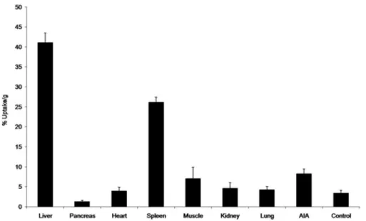
Regarding the biodistribution of the LDE 3H-cholesterol oleate ether in rabbits with AIA, the liver was the main tissue with uptake, followed by the spleen. Uptake by arthritic joints was two-fold greater than uptake in control saline-injected joints, representing the tissue with the third highest uptake (Figure 2).
Figure 3 shows the total and differential leukocyte counts in the synovial fluid of the arthritic joints of the rabbits with AIA treated with saline, commercial MTX or LDE-MTX. Commercial MTX was ineffective in reducing the leukocyte influx into the articular cavity. In contrast, the LDE-MTX preparation reduced the leukocyte influx by nearly 65%; specifically, LDE-MTX treatment reduced the mononuclear (47%) and polymorphonuclear (72%) cell counts. The latter are the predominant cell lineage in this phase of the inflammatory process. The superior anti-inflammatory effi-cacy of LDE-MTX was also shown by the data on the protein leakage into the arthritic knees of the rabbits with AIA, which was more limited in this treatment group compared with both controls and commercial MTX-treated rabbits with AIA (Figure 4).
Several drug delivery systems have been proposed to improve the pharmacological index of MTX. Those systems include liposomes, dendrimers, human serum albumin, solid lipid nanoparticles, polymeric nanoparticles and micelles, carbon nanotubes, and magnetic and gold nanoparticles. At this stage, it is difficult to evaluate the advantages of one system over the others because most of studies have been pre-clinical testing reports. With respect to the MTX preparations tested in subjects, namely, liposomes and human serum albumin, there is still a lack of clinical evidence to support their overall superiority (14,15). Being a low-cost oral medication with widespread and long-standing clinical use in RA and other rheumatic diseases, it is unlikely that commercial MTX could be replaced by parenteral drug delivery systems in the setting of chronic treatment of those diseases. However, periods of treatment with drug delivery systems that can increase the anti-inflammatory action of MTX could reverse the stages of disease aggravation demanding prompt intervention, especially when orally administered medications are not effective.
The potential advantages of LDE-MTX are its lack of immunogenicity and toxicity related to nanotechnological
products, which have become major issues in the field. The fact that the LDE constituent materials are only lipids, which are present in the organism and are furnished by the chemical industry; the low cost; and the ease of large-scale production are also chief advantages.
It is noteworthy that the anti-inflammatory effects of intravenous LDE-MTX injection, as illustrated in Figures 3 and 4, closely resemble those obtained following injection by the intra-articular route (1). The overwhelming superiority of the LDL-receptor-mediated endocytic pathway over the
folate receptor in promoting cell uptake of MTX (16) may have been responsible for the marked improvement of the drug’s anti-inflammatory properties when the LDE was used as a carrier. In commercial MTX treatment, low cell uptake via the folate receptor can conceivably facilitate drug resistance. In view of the current results in the AIA rabbit model, the possibility exists that use of an LDE as an MTX carrier may also have the potential to disrupt drug resistance mechanisms to allow the continuation of MTX treatment.
Figure 2 -Tissue uptake of LDE labeled with3H-cholesteryl oleyl ether. The tissue samples were excised for radioactivity counting 24 h after intravenous injection of the labeled LDE into the rabbits with AIA. Contralateral synovia without arthritis induction were used as controls.
Figure 3 - Leukocyte count (monocytes: white column; poly-morphonuclear cells: gray column) in the synovial fluid of rabbits 48 h after AIA induction. The animals received commercial MTX (0.5mmol/kg), LDE-MTX (0.5mmol/kg) or saline solution intrave-nously 24 h after AIA induction.*po0.05 compared with the
control saline-treated rabbits with AIA. The results are expressed as the mean±SEM. The data have been subjected to repeated analysis of variance. Post-analysis was conducted using Newman-Keuls multiple comparison tests. In all analyses, po0.05 was
considered statistically significant.
Figure 4 -Vascular permeability, as assessed by the Evans Blue method, in joint fluid collected from the joints of rabbits 48 h after AIA induction. The animals received intravenous commer-cial MTX (0.5mmol/kg), LDE-MTX (0.5mmol/kg) or saline solution 24 h after the induction of arthritis.*po0.05 compared with the
control saline-treated rabbits with AIA. The results are expressed as the mean±SEM. The data have been subjected to repeated
analysis of variance. Post-analysis was conducted using Newman-Keuls multiple comparison tests. In all analyses, po0.05 was
’ ACKNOWLEDGMENTS
This work was supported by the São Paulo State Foundation for Research Support (FAPESP, São Paulo, Brazil) and the National Council for Scientific and Technological Development (CNPq, Brasilia, Brazil). Dr. Maranhão is a Research Awardee of CNPq.
’ AUTHOR CONTRIBUTIONS
Melo SB was responsible for the design of the study and the data analysis. Tavares ER was responsible for the technical execution and the manuscript writing. Guido MC helped with the manuscript writing. Bonfá E and Maranhão RC participated in the study design and the manuscript writing and providedfinancial support.
’ REFERENCES
1. Mello SB, Tavares ER, Bulgarelli A, Bonfá E, Maranhão RC. Intra-articular methotrexate associated to lipid nanoemulsions: anti-inflammatory effect upon antigen-induced arthritis. Int J Nanomedicine. 2013;8:443-9. 2. Maranhão RC, Cesar TB, Pedroso-Mariani SR, Hirata MH, Mesquita CH.
Metabolic behavior in rats of a nonprotein microemulsion resembling low-density lipoprotein. Lipids. 1993;28(8):691-6, http://dx.doi.org/10.1007/ BF02535988.
3. Maranhão RC, Roland IA, Toffoletto O, Ramires JA, Gonc¸alves RP,
Mesquita CH, et al. Plasma kinetic behavior in hyperlipidemic subjects of a lipidic microemulsion that binds to low density lipoprotein receptors. Lipids. 1997;32(6):627-33, http://dx.doi.org/10.1007/s11745-997-0080-6. 4. Kretzer IF, Maria DA, Maranhão RC. Drug-targeting in combined cancer
chemotherapy: tumor growth inhibition in mice by association of pacli-taxel and etoposide with a cholesterol-rich nanoemulsion. Cell Oncol (Dordr). 2012;35(6):451-60, http://dx.doi.org/10.1007/s13402-012-0104-6. 5. Rodrigues DG, Maria DA, Fernandes DC, Valduga CJ, Couto RD, Ibanez OC, et al. Improvement of paclitaxel therapeutic index by derivatization and association to a cholesterol-rich microemulsion: in vitro and in vivo studies. Cancer Chemother Pharmacol. 2005;55(6):565-76, http://dx.doi. org/10.1007/s00280-004-0930-y.
6. Valduga CJ, Fernandes DC, Lo Prete AC, Azevedo CH, Rodrigues DG, Maranhão RC. Use of a cholesterol-rich microemulsion that binds to
low-density lipoprotein receptors as vehicle for etoposide. J Pharm Phar-macol. 2003;55(12):1615-22, http://dx.doi.org/10.1211/0022357022232. 7. Pinheiro KV, Hungria VT, Ficker ES, Valduga CJ, Mesquita CH, Maranhão
RC. Plasma kinetics of a cholesterol-rich microemulsion (LDE) in patients with Hodgkin’s and non-Hodgkin’s lymphoma and a preliminary study on the toxicity of etoposide associated with LDE. Cancer Chemother Pharma-col. 2006;57(5):624-30, http://dx.doi.org/10.1007/s00280-005-0090-8. 8. Pires LA, Hegg R, Valduga CJ, Graziani SR, Rodrigues DG, Maranhão RC.
Use of cholesterol-rich nanoparticles that bind to lipoprotein receptors as a vehicle to paclitaxel in the treatment of breast cancer: pharmacokinetics, tumor uptake and a pilot clinical study. Cancer Chemother Pharmacol. 2009;63(2):281-7, http://dx.doi.org/10.1007/s00280-008-0738-2. 9. Dias ML, Carvalho JP, Rodrigues DG, Graziani SR, Maranhão RC.
Pharma-cokinetics and tumor uptake of a derivatized form of paclitaxel associated to a cholesterol-rich nanoemulsion (LDE) in patients with gynecologic cancers. Cancer Chemother Pharmacol. 2007;59(1):105-11, http://dx.doi.org/ 10.1007/s00280-006-0252-3.
10. Teixeira RS, Curi R, Maranhão RC. Effects on Walker 256 tumour of car-mustine associated with a cholesterol-rich microemulsion (LDE). J Pharm Pharmacol. 2004;56(7):909-14, http://dx.doi.org/10.1211/0022357023826. 11. Maranhão RC, Graziani SR, Yamaguchi N, Melo RF, Latrilha MC,
Rodrigues DG, et al. Association of carmustine with a lipid emulsion: in vitro, in vivo and preliminary studies in cancer patients. Cancer Che-mother Pharmacol. 2002;49(6):487-98, http://dx.doi.org/10.1007/s00280-002-0437-3.
12. Moura JA, Valduga CJ, Tavares ER, Kretzer IF, Maria DA, Maranhão RC. Novel formulation of a methotrexate derivative with a lipid nanoemul-sion. Int J Nanomedicine. 2011;6:2285-95.
13. Folch J, Lees M, Sloane Stanley GH. A simple method for the isolation and purification of total lipids from animal tissues. J Biol Chem. 1957;226(1): 497-509.
14. Wong PT, Choi SK. Mechanisms and implications of dual-acting metho-trexate in folate-targeted nanotherapeutic delivery. Int J Mol Sci. 2015; 13;16(1):1772-90.
15. Abolmaali SS, Tamaddon AM, Dinarvand R. A review of therapeutic challenges and achievements of methotrexate delivery systems for treat-ment of cancer and rheumatoid arthritis. Cancer Chemother Pharmacol. 2013;71(5):1115-30, http://dx.doi.org/10.1007/s00280-012-2062-0. 16. Bulgarelli A, Leite AC Jr, Dias AA, Maranhão RC. Anti-atherogenic effects