1
Arquivos Brasileiros de Cardiologia - Volume 83, Nº Especial, Dezembro 2004
Original Article
The Use of Intravascular Ultrasound in Deciding
on the Treatment of Moderate Coronary Lesions
Andrea Claudia Leão de Sousa Abizaid, Leopoldo Soares Piegas, Alexandre Antonio Cunha
Abizaid, Luiz Fernando Leite Tanajura, Aurea Jacob Chaves, Marinella Patrizia Centemero,
Ana Cristina C. Seixas, Luiz Alberto Piva Mattos, Ibraim Pinto, Amanda Guerra Morais Rego
Sousa, J. Eduardo Morais Rego Sousa
São Paulo, SP - Brazil
Individuals with moderate angiographic lesions (vascular lumen obstruction between 40 and 70%) account for a significant number of patients in our daily practice. However, the choice of a criterion that may differentiate the patients who should from those who should not undergo coronary revascularization procedures is yet to be established.
In that subgroup of patients, anginal symptoms are not discri-minatory, because they may or may not be present. In some situa-tions, noninvasive functional tests may be normal or inconclusive, underestimating the functional meaning of those lesions 1-3.
Coronary angiography has become the most used procedure in the field of invasive cardiology. However, despite the qualities that made it universally accepted, the technique has some limi-tations: 1) angiography shows the lumen as a simple planar si-lhouette, being, therefore, also called a luminogram 4; 2) we
should consider the phenomenon of positive arterial remodeling, which is nothing more than vessel enlargement in an attempt to preserve the lumen in the presence of a great amount of atheros-clerotic plaque 5; and 3) the ultrasound has already shown that,
most of the time, the disease is diffuse, without a completely disease-free segment 6. Therefore, those 3 factors cited limit the
applicability of the method in that subgroup of patients. Intravascular ultrasound is also an invasive modality, based on acquisition of tomographic images, from the interior of the coronary artery, by using a catheter that has a single transducer in its extremity. That method may provide 2-dimensional tomographic assessments of the coronary arteries in vivo, allowing the identi-fication of the components of the atherosclerotic plaques and of the vascular wall, in addition to the highly accurate measurement of the arterial lumen 7-9.
Currently, one of the major indications for its use is the as-sessment of moderate angiographic lesions. Therefore, the choice of a criterion that may differentiate those lesions from the prog-nostic point of view is required.
We assessed the use of the ultrasound criterion of minimum luminal area (MLA) > 4.0, which showed a linear relation with the coronary flow reserve > 2.0, ie, not causing ischemia 10.
Therefore, we wanted to test the hypothesis that the use of intravascular ultrasound, using the cutoff value of the minimum lu-minal area, in patients with moderate lesions, according to coronary angiography, is an adequate assessment for differentiating patients who should (MLA < 4.0 mm2) or should not (MLA ≥ 4.0 mm2)
Instituto Dante Pazzanese de Cardiologia
Mailing address - Andrea Claudia Leão de Sousa Abizaid - Rua Dom Paulo Pedrosa, 673/72B - Cep 05687-001 – São Paulo, SP, Brazil E-mail: diretoriaidpc@uol.com.br
Received for publication: 06/10/2003 Accepted for publication: 08/03/2004 English version by Stela Maris Costalonga
Objective
To investigate the ultrasound criterion of minimum luminal area (MLA) with a cutoff value of 4.0 mm2 in an attempt to differentiate
the lesions that should be treated from those that should not.
Methods
The study comprised 173 consecutive patients with moderate angiographic lesions (percentage of stenosis between 40 and 70) who underwent ultrasound imaging. They were divided into 2 groups as follows: group 1 or clinical group (MLA ≥ 4.0 mm2);
and group 2 or revascularization group (MLA < 4.0 mm2). The
patients were followed up to determine the rate of major cardiac events (MCE) in 2 years, the need for revascularization of the target lesion, and to identify the clinical, angiographic, and ultrasound predictors of events.
Results
Seventy-five (43%) patients had MLA ≥ 4.0 mm2 and were
clinically treated; 98 (57%) patients had MLA < 4.0 mm2 and
were treated with coronary stents. On quantitative coronary an-giography, no significant difference was observed between the percentage of vessel stenosis [group 1: 48% vs group 2: 53%; P=0.06]. By the other hard ultrasound measurements, as MLA was significantly greater in group 1 compared with that in group 2 [4.54 mm2 vs 2.45 mm2; P < 0.001)]. The clinical impact of
the decision was favorable, and no difference was observed in regard to the occurrence of major cardiac events: [group 1: 5 (7%) vs group 2: 14 (15%); P = 0.09]. The need for revasculari-zation of the target lesion also did not differ (group 1: 3 (4%) vs group 2: 11 (12%); P = 0.07). The predictive variables for MCE were diabetes, functional class III (FC III) angina before hospitali-zation, and MLA assessed on ultrasound.
Conclusion
This strategy for deciding on appropriate treatment guarantees low rates of MCE in both groups in the 24-month follow-up, with reduced revascularization rates. The predictive variables of major cardiac events were as follows: diabetes mellitus, FC III angina, and MLA on intravascular ultrasound.
Keywords
2
Arquivos Brasileiros de Cardiologia - Volume 83, Nº Especial, Dezembro 2004
The Use of Intravascular Ultrasound in Deciding on the Treatment of Moderate Coronary Lesions
undergo treatment. This assures that those cases will evolve with low rates of major cardiac events during late clinical follow-up.
Therefore, the primary objectives of this study were as follows: 1) to detect the rates of major cardiac events (death, myocardial infarction, and the need for revascularization of the target lesion) in a 2-year follow-up; 2) to detect the rates of revascularization of the target lesion during the same observation period; and 3) to identify the prognostic clinical, angiographic, and ultrasound va-riables for cardiac events in the follow-up.
Methods
From August 2000 to March 2001, 173 patients were pros-pectively and consecutively included in the study to undergo intra-vascular ultrasound in the Section of Coronary Angioplasty of the Instituto Dante Pazzanese de Cardiologia. They had angiographically moderate lesions, ie, luminal obstruction between 40 and 70% on visual evaluation.
The inclusion criteria were as follows: presence of typical or atypical precordial pain or confirmation of silent ischemia, or both; angiographic evidence of single-vessel coronary disease; target vessel with a reference diameter > 3.0 mm, primary target lesion located in the natural coronary web; and coronary anatomy favo-rable to the passage of the catheter of intravascular ultrasound through the lesion to be investigated.
All ultrasound examinations were performed with the Clear View device (Boston Scientific, USA). The catheter was placed inside the coronary artery at least 10 mm distally from the target lesion, and automate traction was initiated at the constant velocity of 0.5 mm/s, which allowed a careful scanning of the entire segment to be studied, identifying the point of minimum luminal area. Once the minimum luminal area was measured, the cutoff value of 4.0 mm2 was used for decision making, and the patients
were then assigned to the 2 groups.
Patients with a minimum luminal area ≥ 4.0 were assigned to group 1 and clinically followed up. Those in group 2 had a mini-mum luminal area < 4.0 and had to undergo coronary revascula-rization. Major cardiac events were computed over 24 months.
Other quantitative ultrasound data were assessed: the area of the external elastic membrane (EEM), the area of the plaque plus the media layer (area of the external elastic membrane – minimum luminal area), the maximum and minimum diameters, the per-centage of obstruction of the plaque (ratio between the area of the plaque plus the media layer and the area of the EEM multiplied by 100), and the percentage of obstruction of the MLA (difference between the areas of the reference lumen and the MLA multiplied by 100 and divided by the area of the reference lumen).
The following qualitative ultrasound data were also assessed: the morphology of the plaque (lipid, fibrotic, and calcified), the aspects of vulnerability (thin fibrous cap or lipid deposits, or both), and the presence of arterial remodeling (positive or negative).
The statistical analysis was performed using SPSS for Windows, version 10.0. The categorical variables were compared between the 2 groups by using the chi-square test, and the continuous variables were compared using the Student t test. The correlations between the measurements of intravascular ultrasound and quantitative angio-graphy were performed by using the Pearson correlation. The accu-mulated probabilities of major cardiac events were estimated by using
the Kaplan-Meier curves. The Cox regression analysis (multivariate approach) was performed to determine the variables with independent prognostic value for the occurrence of cardiac events.
Results
According to the ultrasound criterion of MLA, 75 (43%) patients had nonimportant lesions and were clinically followed up (group 1), and 98 (57%) patients had severe lesions treated with stent implantation (group 2).
No statistically significant difference in regard to the clinical characteristics was observed between the 2 groups (tab. I), only the presence of functional class III angina was significantly greater in group 2 patients than in group 1 patients (22% vs 2%; P = 0.03). Patients in both groups had significant differences neither in the percentage of luminal stenosis [group 1: 48% (SD 7%) vs group 2: 53% (SD 6%); P = 0.06], nor in the minimum luminal diameter [group 1: 1.60 mm (SD 0.33) vs group 2: 1.52 mm (SD 0.36); P = 0.08]. This method did not allow differentiating the severity of the lesions between both groups.
The major ultrasound results in the site of the target lesion are shown in table II.
By the end of 24 months, 19 (11%) patients of our case series had major cardiac events as follows: 5 (7%) patients in group 1 and 4 (15%) patients in group 2; P = 0.09. The results in group 1 were as follows: no patient died; 2 patients had acute myocardial infarction due to progression of the target lesion, which already had aspects of vulnerability; and 3 patients required re-vascularization of the target lesion. On the other hand, the results in group 2 were as follows: 1 patient died due to a cardiac cause (acute myocardial infarction); 2 had nonfatal infarction; and 11 patients required new revascularization of the target lesion due to angiographic restenosis.
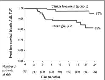
The Kaplan-Meier curve showed that by the end of 24 months of evolution, 91% of the group 1 patients were free of major cardiac events, and, in 83% of the group 2 patients that decrease in the curve in the first year was mainly due to the need for new revascu-larization of the target lesion because of angiographic restenosis, a problem inherent to the coronary endoprosthesis (fig. 1).
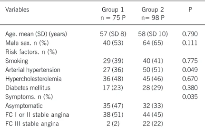
Table I – Clinical data of 173 patients with moderate coronary lesions divided into 2 groups, according to the minimum luminal area (MLA)
on ultrasound [group 1 (MLA ≥≥≥≥≥ 4.0 mm2; clinical follow-up) and
group 2 (MLA < 4.0 mm2; percutaneous intervention)].
Variables Group 1 Group 2 P
n = 75 P n= 98 P
Age. mean (SD) (years) 57 (SD 8) 58 (SD 10) 0.790 Male sex. n (%) 40 (53) 64 (65) 0.111 Risk factors. n (%)
Smoking 29 (39) 40 (41) 0.775
Arterial hypertension 27 (36) 50 (51) 0.049 Hypercholesterolemia 36 (48) 45 (46) 0.670 Diabetes mellitus 17 (23) 28 (29) 0.380
Symptoms. n (%) 0.035
Asymptomatic 35 (47) 32 (33) FC I or II stable angina 38 (51) 44 (45) FC III stable angina 2 (2) 22 (22)
3
Arquivos Brasileiros de Cardiologia - Volume 83, Nº Especial, Dezembro 2004
The Use of Intravascular Ultrasound in Deciding on the Treatment of Moderate Coronary Lesions
Fifteen clinical, angiographic, and ultrasound variables were chosen for inclusion in the univariate model, such as hypertension, diabetes, dyslipidemia, smoking, previous myocardial infarction, angina before hospitalization, vessel approached, site of the target lesion, minimum luminal diameter, percentage of vessel obstruction, lipid plaque, arterial remodeling, minimum luminal area, percentage of plaque obstruction, and percentage of obstruction of the minimum luminal area. When a multivariate model of logistic regression was used, the major variables independently responsible for the prognosis were as follows: presence of diabetes (OR = 3.17; 95% CI = 2.17-10.49; P = 0.006), functional class III angina (OR = 2.18; 95% CI = 1.02-8.41; P = 0.04), and ultrasound assessment of the severity of the minimum luminal area > 4.0 mm2 (OR = 0.59;
95% CI = 0.39-0.89; P = 0.0123).
Discussion
The quantitative analysis of the angiogram lacked discriminatory power to accurately define moderate lesions, which was only obtained after ultrasound assessment. Therefore, by using the
ultrasound criterion of minimum luminal area ≥ 4.0 mm2, more
than half (57%) of our patients with angiographically depicted moderate lesions had, in fact, significant lesions. At any rate, the information obtained by using intravascular ultrasound is purely anatomical and, particularly in the presence of moderate lesions, the pathophysiological repercussion needs to be known so that the most appropriate therapeutic approach may be chosen, based on the presence of ischemia. Therefore, the reason for choosing an anatomical or morphological criterion to manage those patients has to be clarified.
The recent literature has shown that the physiological criteria of coronary flow reserve and flow reserve fraction may be safely used in deciding whether intervening or not in patients with mode-rate lesions, assuring a good late clinical result 11-14. Therefore, the
functional criteria proved to be useful for the clinical decision ma-king process, although they are of difficult application in daily routine. In parallel, in a retrospective study including 300 patients with moderate lesions, revascularization was not indicated, based, mostly, on the verification of minimum luminal area ≥ 4.0 mm2.
This demonstrates that that criterion is safe and has excellent late results, with 8% of overall cardiac events in the 13-month follow-up with an event-free survival of 92% 15.
Considering that the anatomical and functional criteria have a high degree of concordance and that the detection of the degree of stenosis is much simpler and accurate with the use of ultraso-nography, the latter became the method chosen in our service for managing lesions that were of concern.
Our results corroborate the correctness of that option. In the present prospective study, revascularization was also not chosen when the minimum luminal area was greater than 4.0 mm2 (group
1). That strategy was safe, because only 5 (7%) patients in the group evolved with major cardiac events in a longer follow-up (24 months). On the other hand, patients with minimum luminal area < 4.0 mm2 (group 2) had a safe evolution, with 14 (15%)
cardiac events by the end of 2 years, considering that a group at greater risk, in which all patients had undergone implantation of coronary stents. Event-free survival was 82% in 2 years, compa-rable to that of patients undergoing stent implantation in the BENESTENT II study 16 (event-free survival of 84.3%), and, more
recently, greater than that in the control group of the RAVEL study 17 (event-free survival of 70.9%). It is worth noting that
most events in our case series (9/14 equal to 64%) were due to restenosis and revascularization of the target lesion. Currently, when interventional cardiology enters the era of drug-eluting stents, in which restenosis has been drastically controlled, the perspective of those patients is much more favorable, with an event-free survival probably greater than 90% 18,19.
The current guidelines recommend that percutaneous coronary interventions should be performed preceded by the objective evidence of myocardial ischemia. However, sometimes this does not occur due to multiple reasons. In the cases in which coronary angiography shows a moderate coronary lesion, and a noninvasive assessment of its functional meaning is not available, additional exploration of its severity or repercussion is essential for clinical decision making. We recommend, for the profile of patients included in this investigation, the use of the ultrasound criterion with as-sessment of the minimum luminal area, with a cutoff value of 4.0 mm2, which has guaranteed these patients, after the choice Time (months)
Fig. 1 - Kaplan-Meier curve showing the event-free survival by the end of 24 months according to minimum luminal area (MLA) on ultrasound: [group 1 (MLA
≥ 4.0 mm2; clinical follow-up) and group 2 (MLA < 4.0 mm2; percutaneous
intervention)]. TLR = target lesion revascularization
Table II - Quantitative ultrasound analysis in the site of the target lesion of 173 cases investigated, divided into 2 groups according to the minimum luminal area (MLA) on ultrasound: [group 1 (MLA ≥≥≥≥≥ 4.0
mm2; clinical follow-up) and group 2 (MLA < 4.0 mm2; percutaneous
intervention)].
Variables Group 1 (75 P) Group 2 (98 P) P mean (SD) mean (SD)
Area of the EEM (mm2) 13.25 (3.59) 13.83 (4.41) 0.35
MLA (mm2) 4.54 (0.71) 2.46 (0.85) < 0.001
Area of P + M (mm2) 8.70 (3.51) 11.37 (4.21) < 0.001
Maximum luminal diameter (mm) 2.75 (0.35) 1.99 (0.61) < 0.001 MLD (mm) 2.25 (0.30) 1.58 (0.45) < 0.001 % plaque (%) 63 (10) 81 (7) < 0.001 % area obstruction (%) 49 (10) 73 (9) < 0.001 Negative remodeling (%) 51 (68) 69 (70) 0.744 Positive remodeling (%) 24 (32) 29 (30) 0.862
P- patients; SD- standard deviation; %- percentage; EEM- external elastic membrane; MLA- minimum luminal area; P + M- plaque plus media; MLD- minimum luminal diameter.
Event-free survival (death, AMI, TLR)
Number of patients at risk
Clinical treatment (group 1)
4
Arquivos Brasileiros de Cardiologia - Volume 83, Nº Especial, Dezembro 2004
The Use of Intravascular Ultrasound in Deciding on the Treatment of Moderate Coronary Lesions
1. Schulman SP, Losorda D, Farah T, et al. Correlations between coronary flow reser-ve measured with a Doppler guide wire and treadmill exercise testing. Am Heart J.1997; 134:99-104.
2. Picano E, Parodi O, Lattanzi F, et al. Assessment of anatomic and physiological severity of single-vessel coronary artery lesions by dipiridamol echocardiography: comparison with positron emission tomography and quantitative arteriography. Circulation.1994; 89:753-61.
3. Naqvi TZ, Hachamovitch R, Berman D, Buchbinder N, Kiat H, Shah PK. Does the presence and site of myocardial ischemia on perfusion scintigraphy predict the oc-currence and site of future myocardial infarction in patients with stable coronary artery disease? Am J Cardiol. 1997; 79:1521-24.
4. Nissen SE, Gurley JC, Demaria AN. Assessment of vascular disease by intravascular ultrasound. Cardiology. 1990; 77: 398-410.
5. Glagov S, Weisenberg E, Zarins CK, Stankunavicius R, Kolettis GJ. Compensatory enlargement of human coronary arteries. N Engl J Med.1987; 316:1371-5. 6. Mintz GS, Painter JA, Pichard AD, et al. Atherosclerosis in angiographically
“nor-mal” coronary artery reference segments: an intravascular ultrasound study with clinical correlations. J Am Coll Cardiol. 1995; 25:1479-85.
7. Gussenhoven EJ, Essed CE, Lancee CT, et al. Arterial wall characteristics determi-ned by intravascular ultrasound imaging: an in vitro study. J Am Coll Car-diol.1989; 14:947-52.
8. Mintz GS, Nisses SE, Anderson WD, et al. American College of Cardiology clinical expert consensus document on standards for acquisition, measurement and repor-ting of intravascular ultrasound studies (IVUS): A report of the American College of Cardiology Task Force on clinical expert consensus documents developed in colla-boration with the European Society of Cardiology Endorsed by the Society of Car-diac Angiography and Interventions. J Am Coll Cardiol. 2001; 37:1478-92. 9. Nishimura RA, Edwards WD, Warnes CA, et al. Intravascular ultrasound imaging:
in vitro validation and pathologic correlation. J Am Coll Cardiol. 1990; 16:145-54.
References
10. Abizaid A, Mintz GS, Pichard AD, et al. Clinical, intravascular ultrasound, and quan-titative angiographic determinants of the coronary flow reserve: before and after percutaneous transluminal coronary angioplasty. Am J Cardiol. 1998; 82: 423-8. 11. Kern MJ, Donohue TJ, Aguirre FV, et al. Clinical outcome of deferring angioplasty
in patients with normal translesional pressure-flow velocity measurements. J Am Coll Cardiol. 1995; 25:178-87.
12. Ferrari M, Schnell B, Werner GS, Figulla HR. Safety of deferring angioplasty in pa-tients with normal coronary flow velocity reserve. J Am Coll Cardiol. 1999; 33:83-7. 13. Gruberg L, Kapeliovich M, Roguin A, Grenadier E, Markiewicz W, Beyar R. Defer-ring angioplasty in intermediate coronary lesions based on coronary flow criteria is safe: comparison of a deferred group to an intervention group. Int J Cardiovasc Interv. 1999; 2:35-40.
14. Pijls NHJ, Van Gelder B, Van Der Voort P, et al. Fractional flow reserve: a useful index to evaluate the influence of an epicardial coronary stenosis on myocardial blood flow. Circulation. 1995; 92:3183-93.
15. Abizaid AS, Mintz GS, Mehran R, et al. Long-term follow-up after percutaneous transluminal coronary angioplasty was not performed based on intravascular ultra-sound findings: importance of lumen dimensions. Circulation. 1999; 100:245-61. 16. Serruys PW, Van Hout B, Bonnier H, et al. Randomised comparison of implanta-tion of heparin-coated stents with balloon angioplasty in selected patients with coronary artery disease (BENESTENT II). Lancet.1998; 352:673-81. 17. Morice MC, Serruys PW, Sousa JE, et al. Randomized study with sirolimus –coated
BX Velocity balloon expandable stent in the treatment of patients with de novo native coronary lesions. N Engl J Med.2002; 346:1773-80.
18. Sousa JE, Costa MA, Abizaid A, et al. Sustained suppression of neointimal proli-feration by sirolimus-eluting stens. One –year angiographic and intravascular ul-trasound follow-up. Circulation. 2001; 104:2007-11.
19. Sousa JE, Costa MA, Sousa AG, et al. Two-year angiographic and intravascular ul-trasound follow-up after implantation of sirolimus-eluting stents in human coronary.
of appropriate treatment, a favorable long-term prognosis. In addition, if the patient with an intermediate lesion has symptoms or is diabetic, or both, a more strict assessment should be pro-grammed, as well as a more careful late control.
In conclusion, the strategy for deciding on treatment by using the ultrasound criterion of minimum luminal area, with a cutoff
value of 4.0 mm2, in patients with moderate lesions on coronary