Larissa Suzuki*, André Wilson Machado**, Marcos Alan Vieira Bittencourt***
INTRODUCTION AND LITERATURE REVIEW The smile is a key factor in the composition of an individual’s overall beauty. Hence, it’s noticed the modern society’s growing demand for beauti-ful, healthy smiles. Within this context, orthodon-tics plays a role of paramount importance.
Several parameters are available to assess smile esthetics, such as amount of gingival display, midline,
buccal corridor, incisor width/height ratio, incisor crown inclination, gingival contour and smile arch ap-pearance, among others.13,19 Among these parameters,
one should highlight the importance of assessing the amount of gingival display on smiling as it may be related to several factors, such as vertical maxillary excess, upper lip hyperactivity and length, and height of the clinical crowns of maxillary incisors.16
An evaluation of the influence of gingival
display level in the smile esthetics
Objective: The aim of this study was to evaluate the influence of the amount of gingival display on
smile esthetics. Methods: Two extraoral photographs were used: One of the close-up smile and one frontal view of the smiling face of four individuals (one Caucasian and one Afro-Brazilian man, and one Caucasian and one Afro-Brazilian woman). The photographs were manipulated in a computer and five images were created for each original photograph with different degrees of gingival display: 0 mm, 1 mm, 3 mm, 5 mm and 7 mm. Then the images were evaluated by 60 indi-viduals who assigned a score from zero to ten to each image on a visual analogue scale. Results and Conclusions: Statistical analysis and results showed that levels of gingival display equivalent to 0 mm and 1 mm received the highest mean scores, i.e., 6.6 and 6.2, respectively, and showed no statistical difference between them (p>0.05). Gingival displays of 3 mm, 5 mm and 7 mm received lower, decreasing scores of 5.0, 3.5 and 2.9, respectively, without any statistical difference between levels 5 mm and 7 mm (p>0.05). Furthermore, the use of close-up photographs of the smile or frontal view photographs of the smiling face showed no statistical difference (p>0.05).
Abstract
Keywords: Esthetic dentistry. Gingiva. Orthodontics. Smile.
* Student Specialty Course in Orthodontics, Federal University of Bahia UFBA.
** MSc in Orthodontics, PUC/Minas. PhD in Orthodontics, UNESP/Araraquara - UCLA/USA. Assistant Professor, UCLA/USA. Professor, Specialty Course in Orthodontics, UFBA.
*** PhD in Orthodontics, UFRJ. Professor of Orthodontics, UFBA. How to cite this article: Suzuki L, Machado AW, Bittencourt MAV. An
evalua-tion of the inluence of gingival display level in the smile esthetics. Dental Press
J Orthod. 2011 Sept-Oct;16(5):37.e1-10.
» The authors report no commercial, proprietary, or inancial interest in the
The literature comprises classifications for different types of smiles based on the relation-ship between the upper lip and the anterosuperi-or teeth. Smiles fall into five different categanterosuperi-ories: Class I, when the edge of the lip lies above the cervical portion of the incisor crowns (“gummy” smile); class II, when the edge of the lip is located at the cervical third of the incisor surfaces; class III, when the edge of the lip lies in the middle third of the incisor surfaces; class IV, when the edge of the lip is located at the incisal third of the incisors; and class V, when the edge of the lip covers the entire incisor surfaces. The authors concluded that more than 98% of the sample was in classes I and II.24
Another method of smile classification em-ploys degrees of dental crown exposure and gingi-val tissue display, which fall into three categories: High, medium and low. In the high smile there is total exposure of the clinical crowns of anterosu-perior teeth and a continuous strip of gingival tis-sue. The medium smile reveals most (75%) or all (100%) of the clinical crowns of anterosuperior teeth and the interdental or interproximal papil-lae, only. The low smile shows less than 75% of the clinical crowns of anterosuperior teeth and no display of gingival tissue.17,25
Photographic models and individuals re-garded as having good facial esthetics exhibit, on smiling, the entire length of the anterosupe-rior teeth and often a narrow strip of gingival margin.6 This is precisely the key issue discussed
in the clinical and scientific literature. Can the display of gingival tissue on smiling be consid-ered esthetic? If yes, what is the ideal amount of gingival display? Or else, how much exposure would be acceptable?
According to the literature, the appropriate relationship is one in which the upper lip rests on the gingival margin of the central maxillary incisors.2,11,14 Likewise, in the so-called ideal
smile the upper lip should be positioned so as to expose the entire crown of the maxillary
in-cisors and up to 1 mm of gingiva.1,4,9,10 On the
other hand, up to 3 mm of gingiva display can be considered esthetically acceptable.5,12,13
The literature also reports the difference in the amount of gingival display on smiling between genders. Several authors agree that women have a higher smile line with greater gingiva display, while men have a lower smile line.3,4,12,18,20,21,25
The “gummy” smile is not necessarily un-aesthetic to the public’s eye. Some movie stars and models, especially women, display some gingival tissue on smiling but their smile is nev-ertheless still considered pleasant.16 Moreover,
the smile pattern varies with patient age, with children displaying a greater amount of gingiva than adults. It is noteworthy that with advanc-ing age, loss of tissue tone causes the upper lip to stretch and upper teeth to overlap, thereby reducing gingival display.3
Another influencing factor is ethnicity, as Afro-descendants tend to display less of their up-per teeth and gingiva, probably due to the shape and volume of their lip muscles.2 In contrast, a
study that investigated six different clinical vari-ables, including amount of gingival display in 253 patients from six different ethnic groups, found that the Afro-descendant group had the greatest amount of gingival display.18
Another recently raised question suggests that the method used to assess the smile can potentially influence the results. In a research study using different images, i.e., a frontal view photograph of the face, a photograph depicting the lower third of the face and a close-up dental view, the authors found that after the images had been evaluated by a group of laypersons the esthetic impact was lower in the facial pictures. In other words, the influence of global facial es-thetic factors had masked how the smile was perceived and evaluated.8
According to Sarver22 in the last decade
their smile esthetics. However, although the liter-ature cites a variety of clinical opinions regarding what would be an ideal or acceptable degree of gingival display, most lack scientific evidence. In-deed, few studies have researched, evaluated and compared the different degrees of gingival display. Kokich Jr et al13 evaluated, among other
es-thetic criteria, the perceived amount of gingival display using photographs of smiles intentionally altered on a computer. Variations in the distance between the upper lip and the gingival margin of maxillary incisors were created, generating five types of close-up smile images: 2 mm of the in-cisors overlapped by the lips, lips touching the gingival margin of the incisors (0 mm of gingival display), 2 mm, 4 mm and 6 mm of gingival dis-play. The images were subjected to three groups of raters consisting of orthodontists, laypersons and general practice (GP) dentists. The smiles with the upper lip touching the gingival margins of the incisors (0 mm) were generally assigned the highest scores. When the groups of raters were separated, laypersons and GP dentists considered displays of up to 4 mm acceptable while ortho-dontists rated displays above 2 mm as unpleasant.
Hunt et al,12 in a similar study,
manipulat-ed two photographs (one of a man and one of a woman) and created seven types of relations between lips and teeth, ranging from 2 mm to 4 mm, i.e., in the first, the upper lips overlapped the crowns by 2 mm and the last had a gingival tissue display of 4 mm. Then the images were evaluated by 120 laypersons. The results showed that gingival display in the 0 mm group received the highest scores while displays above 2 mm re-ceived progressively lower scores.
The literature has discussed the use of anatom-ic references for characterizing the smile. How-ever, many esthetic parameters are based on the clinical perception of some authors or on subjec-tive assessments whereas the standards supported by scientific research have not yet been estab-lished in Brazil. Therefore, it should be stressed
that there is a need for studies aimed at determin-ing the ideal gdetermin-ingival display on smildetermin-ing accorddetermin-ing to the esthetics of the Brazilian population, whose composition is rather heterogeneous.
In view of the above, the aim of this study was to assess and compare the degree of esthetic ac-ceptance of five levels of gingival display on smiling (0 mm, 1 mm, 3 mm, 5 mm and 7 mm) using ma-nipulated photographs, and to investigate whether or not there are any differences, in this evaluation, between frontal view extraoral photographs of the smile and close-up smile photographs.
MATERIAL AND METHODS
Two extraoral photographs were used (one frontal view of the smile, one of the close-up smile) and intraoral frontal views of four indi-viduals, two Afro-Brazilians (one man and one woman) and two Caucasians (one man and one woman) aged between 20 and 30 years, totaling eight photographs altogether.
The photographs were taken by the same op-erator with a digital Canon Rebel camera, Can-on MR-14 ring flash and CanCan-on macro lens 100 (Canon Inc., Taiwan). The eight photographs of the smiles of all individuals were manipulated in Adobe Photoshop® 7.0 (Seattle, WA, USA).
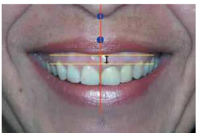
In manipulating the images, the teeth and gin-giva were erased from the smile photographs as depicted in Figure 1A. Subsequently, the image from the previously taken frontal view intraoral photograph was inserted into the smile photo and then manipulated (upwards or downwards) to cre-ate different levels of gingival display (Fig 1B, C). With this purpose, an adaptation of the method described by Peck et al20 was performed, as shown
A B C
midsagittal plane. Two horizontal lines were then drawn, one tangent to the superior most gingival margin of the central incisors and one tangent to the inferior most contour of the upper lip, both perpendicular to the vertical line. Finally, as illus-trated in Figure 1C, the central image was moved upwards or downwards according to the horizon-tal reference lines, and the distances in millimeters were recorded to create the images.
In the images of close-up smiles, measure-ments made in millimeters were used at a 100% ratio, i.e., 1 mm on the image was equivalent to 1 mm in reality. Thus, five images were generated according to the following criteria:
» 0 mm gingival display: Gingival margin of maxillary central incisors positioned in the lower contour of the upper lip.
» 1 mm gingival display: Gingival margin of maxillary central incisors positioned 1 mm below the lower contour of the upper lip. » 3 mm gingival display: Gingival margin of
maxillary central incisors positioned 3 mm below the lower contour of the upper lip. » 5 mm gingival display: Gingival margin of
maxillary central incisors positioned 5 mm below the lower contour of the upper lip. » 7 mm gingival display: Gingival margin of
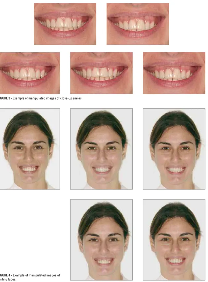
maxillary central incisors positioned 7 mm below the lower contour of the upper lip. The five photographs of each individual — generated from JPEG format files with 300 dpi resolution and 25 cm x 38 cm size — were ran-domly distributed on the same page (Fig 3).
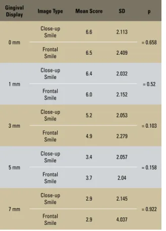
For facial photographs, measurements in mil-limeters were calculated at a 25% ratio, i.e., 1 mm on the image was equivalent to 4 mm in reality. Thus, five images were generated for each existing photograph, adopting the same criteria described before. The images were also randomly distribut-ed and savdistribut-ed in files with the same features of the close-up smile photographs (Fig 4).
Subsequently, these files were processed in a specialized digital lab using professional equipment, model Noritsu 2901 (Noritsu Bra-zil S/A, Manaus, AM), on Kodak Edge Genera-tions paper (Kodak Brazil, Manaus, AM) with photographic quality on standard A3 size paper (29.7 cm x 42 cm). A photographic album with eight pages containing all images was then made and the four pages containing the facial images were then randomly ordered and followed by the four pages with the close-up smiles.
FIGURE 1 - Illustration of the method to standardize the creation of manipulated images.
FIGURE 3 - Example of manipulated images of close-up smiles.
2 3 4 5 6 7
After the album was ready, 60 individuals, among them orthodontists, oromaxillofacial sur-geons and laypersons were asked to rate the images.
Along with the album, each examiner re-ceived a printed form containing a printed simulation of a ruler (visual analogue scale) for each image (5 rulers per page, totaling 40 rul-ers). On these rulers they were asked to mark with an “X” the quality degree associated with each image. The scale was designed to show an ascending order of quality from right to left. It was explained to each rater that it was pos-sible to place a mark anywhere on the ruler. The visual analogue scale15,17,26 had 10 cm, and
a dash was drawn at its center, giving raters the perception of regular quality. The distance (in mm) between the mark made by the rater and the point on the far left served as an estimation of the degree of quality determined for each
image rated.23 At the end of the evaluation
process, a total of 40 images were examined by each rater.
The data were statistically analyzed, central tendency and dispersion were calculated, and normal distribution tested (KS test). ANOVA and Tukey’s test were also applied with signifi-cance level of 5% in order to identify differences among the groups.
RESULTS
Table 1 and Figure 5 show that, regardless of the photograph type, smiles with 0 mm, 1 mm, 3 mm, 5 mm and 7 mm gingival display had mean scores of 6.6, 6.2, 5.0, 3.5 and 2.9, respec-tively. Between the scores assigned to the smiles with 0 mm and 1 mm display, no statistically significant difference was found. Likewise, no statistically significant differences were found between smiles with 5 mm and 7 mm gingival display. A gingival display of 3 mm, on the other hand, differed from the other levels, as did the gingival displays of 5 mm and 7 mm, to the det-riment of the others.
In comparing the assessments made of the close-up smile images with those of frontal view smiles it becomes clear that no statistically sig-nificant difference exists between the scores as-signed to any degree of gingival display (p>0.05). For close-up smiles and frontal view smiles, re-spectively, the top scores were once again given to the 0 mm degree of display, namely, 6.6 and 6.5, while the 7 mm display received the lowest scores, i.e., 2.9 and 2.9 (Table 2).
To test the influence of ethnicity and gender the data were submitted to ANOVA (p<0.05). Table 3 and Figure 6 show the means and con-fidence intervals associated with the degree of gingival display in all images evaluated accord-ing to ethnicity and gender of individuals.
For the smiles with 0 mm gingival display, the overall mean scores assigned to the Caucasian
Gingival Display Mean Score SD
0 mm 6.6* 1.976
1 mm 6.2* 1.819
3 mm 5.0 1.926
5 mm 3.5** 1.764
7 mm 2.9** 2.590
TABLE 1 - Mean and overall standard deviation of scores of different types of smiles.
FIGURE 5 - Mean and confidence interval of scores of different types of smiles.
* No statistical difference found between 0 mm and 1 mm displays (p>0.05).
** No statistical difference found between 5 mm and 7 mm displays (p>0.05).
6.84788
6.40397
5.27304
3.74227
3.23146 6.34438
5.94124
4.7833
0 mm display 1 mm display 3 mm display 5 mm display 7 mm display 3.29356
8 7 6 5 4 3 2 1
man, Afro-Brazilian man, Caucasian woman and Afro-Brazilian woman were 5.7, 6.7, 6.7 and 7.3, respectively. For the smiles with 1 mm gingival display, the overall mean scores assigned to the same groups were 5.7, 5.9, 5.9 and 6.9, respec-tively. For the 3 mm display, the mean scores were 4.6, 4.8, 4.9 and 5.7, respectively. For the 5 mm display, 3.3, 3.0, 3.6 and 4.2, respectively. Finally, for the 3 mm display, the mean scores were 2.5, 2.2, 3.2 and 3.8, respectively.
At all levels of gingival display the scores assigned to the Afro-Brazilian woman were higher than all others. However, this result was only statistically significant for the 1 mm gin-gival display. For the 0 mm and 3 mm displays, the Afro-Brazilian woman received signifi-cantly higher scores than the Caucasian man. For the 5 mm and 7 mm displays, however,
this difference was seen among Caucasian and Afro-Brazilian men (p<0.05). The results were similar in all other scenarios.
Gingival
Display Image Type Mean Score SD p
0 mm
Close-up
Smile 6.6 2.113
= 0.658 Frontal
Smile 6.5 2.409
1 mm
Close-up
Smile 6.4 2.032
= 0.52 Frontal
Smile 6.0 2.152
3 mm
Close-up
Smile 5.2 2.053
= 0.103 Frontal
Smile 4.9 2.279
5 mm
Close-up
Smile 3.4 2.057
= 0.158 Frontal
Smile 3.7 2.04
7 mm
Close-up
Smile 2.9 2.145
= 0.922 Frontal
Smile 2.9 4.037
TABLE 2 - Mean scores and standard deviations in assessments of close-up smiles and frontal view smiles.
TABLE 3 - Mean and standard deviation of scores in different groups of individuals.
Gingival
Display Groups
Mean
Score SD Conclusions
0 mm
1 - Caucasian man 5.7 2.018
2 - Afro-Brazilian man 6.7 1.978 (2 = 3)
3 - Caucasian woman 6.7 1.677 (4 > 1)
4 - Afro-Brazilian woman 7.3 1.893
1 mm
1 - Caucasian man 5.9 1.770
2 - Afro-Brazilian man 5.9 1.891 (1 = 2 = 3)
3 - Caucasian woman 5.9 1.764 (4 > 1, 2, 3)
4 - Afro-Brazilian woman 6.9 1.692
3 mm
1 - Caucasian man 4.6 1.941
2 - Afro-Brazilian man 4.8 1.900 (1 = 2 = 3)
3 - Caucasian woman 4.9 2.113 (4 > 1)
4 - Afro-Brazilian woman 5.7 1.571
5 mm
1 - Caucasian man 3.3 1.651
2 - Afro-Brazilian man 3.0 1.662 (1 = 2 = 3)
3 - Caucasian woman 3.6 4.2 (4 > 1, 2)
4 - Afro-Brazilian woman 1.878 1.681
7 mm
1 - Caucasian man 2.5 1.613
2 - Afro-Brazilian man 2.2 1.605 (1 = 2 = 3)
3 - Caucasian woman 3.2 4.102 (4 > 1, 2)
4 - Afro-Brazilian woman 3.8 1.889 0 mm display
Caucasian man Caucasian man Caucasian man Caucasian man Caucasian man Afro-Brazilian manCaucasian woman Afro-Brazilian manCaucasian woman Afro-Brazilian manCaucasian woman Afro-Brazilian manCaucasian woman Afro-Brazilian manCaucasian woman
Afro-Brazilian woman Afro-Brazilian woman Afro-Brazilian woman Afro-Brazilian woman Afro-Brazilian woman
1 mm display 3 mm display 5 mm display 7 mm display
DISCUSSION
Evaluation of all images showed that among the levels of gingival display researched, the highest scores were assigned to the group with no gingival display (0 mm) and 1 mm display, i.e., 6.6 and 6.2, respectively. These two types showed no statistically significant difference between them, in agreement with the litera-ture, which also assigns the highest scores to a 0 mm gingival display.12,13 The argument found
in the literature that a variation of up to 1 mm gingival display is imperceptible further cor-roborates these findings.10,20 This result also
confirms the idea that, on smiling, the proper relationship is one where the upper lip rests on the gingival margin of the maxillary central in-cisors, which is represented by the group with 0 mm gingival display.2,11,14
The literature also argues that gingival dis-play of up to 2 mm is esthetically acceptable.3,12
Although this study did not include a group with 2 mm gingival display, but only 1 mm and 3 mm, it can still be inferred, given the proxim-ity of these values and the results achieved, that a 2 mm gingival display would be acceptable.
For the smiles with 3 mm, 5 mm and 7 mm gingival display, the overall mean scores as-signed were 5.0, 3.5, and 2.9, respectively. All these values were noticeably below the 0 mm and 1 mm gingival display levels (p<0.05). Moreover, these values slowly decreased, i.e., the larger the display, the less esthetic the smile became. Furthermore, given the similarities be-tween the 5 mm and 7 mm display groups, one could suggest that from a certain point onwards, perception of unsightliness becomes a constant. One striking feature that raised some doubt was the low scores assigned by the raters. The highest scores found in this investigation were 6.6 and 6.2, for levels 0 mm and 1 mm, respec-tively. On a scale of 0 to 10, one realizes that these values were not high, thereby demonstrat-ing that the images were not up to standard from
an esthetic point of view. Moreover, the main goal was not to evaluate the quality of images individually, but rather to compare the different levels of gingival display on smiling. Among the factors responsible for the low scores, one could highlight (a) the individual esthetics of the sub-jects, who had different smile patterns, and (b) image manipulation, which can produce lower quality images than the original photos.
Some adjectives used in the literature, such as “ideal,” “acceptable” and “pleasant” are diffi-cult to interpret. As an example, the 3 mm gin-gival display received an average score of 5.028, i.e., 50%. It is obvious that, as mentioned earlier, an absolute value such as a 5.0 score can hardly qualify a 3 mm gingival display. However, Cas-tro5 found that a gingival exposure of up to
3 mm is considered pleasant. The question then is whether or not a 5.0 score may be considered esthetically acceptable or even if this degree of exposure may or may not be considered un-pleasant. Moreover, due to differences between the average scores of 0 mm and 1 mm, and the 3 mm score, and between the latter and the 5 mm and 7 mm scores, one could well argue that a 3 mm gingival display occupies an inter-mediate position, with the first groups achieving higher scores than the last groups.
Therefore, it is a moot question whether or not a 3 mm gingival display, or even a 5 mm or 7 mm display, are unpleasant, since qualify-ing a smile as pleasant or unpleasant depends on many other factors. This explains why certain Brazilian and international beauty models dis-play their gingiva on smiling but even so their smiles are not considered unpleasant.
close-up view, highlighting only the smile, affords the same degree of perception, suggesting that the face has no bearing on the esthetic evaluation of different levels of gingival display. This result is in disagreement with the literature, whose assess-ments of the facial photographs showed a decrease in the level of perception.8 This study, however,
did not assessed the influence of gingival display on smiling using manipulated photographs, but rather the esthetic impact of three photographs types of 18 smiling individuals.
On the other hand, a more detailed evalua-tion of Table 2 shows that the 3 mm display, de-spite statistical similarities with the values found, points to a different tendency. In other words, it appears that since the 3 mm display constitutes a boundary or turning-point between “esthetic” and unaesthetic, the photographs types may have influenced the evaluation. A more detailed inter-pretation of these results would require further studies with more numerous images and raters.
Although this was not among the aims of this study, the potential influence of gender and ethnicity on the evaluations of different degrees of gingival display was also investigated. At all gingival levels investigated, the Afro-Brazilian woman received the highest scores. From a sta-tistical point of view the differences did not follow a pattern, as the scores assigned to the Afro-Brazilian woman were higher than those of the Caucasian man with 0 mm and 3 mm
gingival display, and higher than the scores assigned to the Caucasian and Afro-Brazilian man with 5 mm and 7 mm gingival display. These results should be analyzed with caution, as the findings do not indicate that the Afro-Brazilian woman’s smile is more beautiful than the others or that the smile of the Caucasian or Afro-Brazilian man is less attractive. It is worth mentioning again that, since this study utilized a sample comprising four individuals and their manipulated images, intrinsic variables com-plicate the analysis of some absolute values in-volved, such as individual esthetic and image manipulation technique.
Thus, the study underscores the need for further research with the inclusion of a larger sample and different ethnic groups.
CONCLUSIONS
It can be concluded that the 0 mm and 1 mm degrees of gingival display exhibited the high-est mean scores, and no statistical difference was found between them (p>0.05). Degrees of gingival display 3 mm, 5 mm and 7 mm were considered less esthetic and received lower, de-creasing scores.
1. Ackerman MB, Brensinger C, Landis JR. An evaluation of dynamic lip-tooth characteristics during speech and smile in adolescents. Angle Orthod. 2004;74(1):43-50.
2. Ahmad I. Geometric considerations in anterior dental aesthetics: restorative principles. Pract Periodontics Aesthet Dent. 1998;10(7):813-22; quiz 824.
3. Arnett GW, Bergman RT. Facial keys to orthodontic diagnosis and treatment planning: Part II. Am J Orthod Dentofacial Orthop. 1993;103(5):395-411.
4. Câmara CALP. Estética em Ortodontia. Parte I: diagrama de referências estéticas dentais (DRED). Rev Dental Press Estét. 2004;1(1):40-57.
5. Castro MVM. Aferição da proporção áurea em sorrisos agradáveis [dissertação]. São Paulo (SP): Universidade de Taubaté; 2005.
6. Crawford EC. The face: an orthodontic perspective. Aust Orthod J. 1991;12(1):13-22.
7. Dong JK, Jin TH, Cho HW, Oh SC. The esthetics of the smile: a review of some recent studies. Int J Prosthodont. 1999;12(1):9-19.
8. Flores-Mir C, Silva E, Barriga MI, Lagravere MO, Major PW. Lay person’s perception of smile aesthetics in dental and facial views. J Orthod. 2004;31(3):204-9; discussion 201.
9. Geron S, Atalia W. Inluence of sex on the perception of oral and
smile esthetics with different gingival display and incisal plane inclination. Angle Orthod. 2005;75(5):778-84.
10. Graber TM, Vanarsdall JR. Ortodontia: princípios e técnicas atuais. 3ª ed. Rio de Janeiro: Guanabara Koogan; 2000. 11. Hulsey CM. An esthetic evaluation of lip-teeth relationships
present in the smile. Am J Orthod. 1970 Feb;57(2):132-44. 12 Hunt O, Johnston C, Hepper P, Burden D, Stevenson M. The
inluence of maxillary gingival exposure on dental attractiveness
ratings. Eur J Orthod. 2002 Apr;24(2):199-204.
13. Kokich VO Jr, Kiyak HA, Shapiro PA. Comparing the perception of dentists and lay people to altered dental esthetics. J Esthet Dent. 1999;11(6):311-24.
REfERENCES
14. Mackley RJ. An evaluation of smiles before and after orthodontic treatment. Angle Orthod. 1993;63(3):183-9; discussion 190. 15. Maple JR, Vig KW, Beck FM, Larsen PE, Shanker S. A
comparison of providers’ and consumers’ perceptions of
facial-proile attractiveness. Am J Orthod Dentofacial Orthop.
2005;128(6):690-6.
16. Mondelli J. Estética e cosmética em clínica integrada restauradora. 1ª ed. São Paulo: Ed. Santos; 2003.
17. Montini RW, McGorray SP, Wheeler TT, Dolce C. Perceptions
of orthognathic surgery patient’s change in proile. A ive-year
follow-up. Angle Orthod. 2007;77(1):5-11.
18. Owens EG, Goodacre CJ, Loh PL, Hanke G, Okamura M, Jo KH, et al. A multicenter interracial study of facial appearance. Part 2: A comparison of intraoral parameters. Int J Prosthodont. 2002;15(3):283-8.
19. Patnaik VVG, Rajan S, Sanju B. Anatomy of a beautiful face and smile. J Anat Soc India. 2003;52(1):74-80.
20. Peck S, Peck L, Kataja M. The gingival smile line. Angle Orthod. 1992;62(2):91-100; discussion 101-2.
21. Rigsbee OH 3rd, Sperry TP, BeGole EA. The inluence of facial
animation on smile characteristics. Int J Adult Orthodon Orthognath Surg. 1988;3(4):233-9.
22. Sarver DM. Principles of cosmetic dentistry in orthodontics: Part 1. Shape and proportionality of anterior teeth. Am J Orthod Dentofacial Orthop. 2004;126(6):749-53.
23. Scott SH, Johnston LE Jr. The perceived impact of extraction and nonextraction treatments on matched samples of African
American patients. Am J Orthod Dentofacial Orthop. 1999 Sep;116(3):352-60.
24. Teo CS. An evaluation of the smiling lip-line. Singapore Dent J. 1981;6(1):27-30.
25. Tjan AH, Miller GD, The JG. Some esthetic factors in a smile. J Prosthet Dent. 1984;51(1):24-8.
26. Wilmot JJ, Barber HD, Chou DG, Vig KW. Associations between severity of dentofacial deformity and motivation for orthodontic-orthognathic surgery treatment. Angle Orthod. 1993;63(4):283-8.
Contact address
André Wilson Machado
Rua Eduardo José dos Santos, 147, salas 810/811, Ed. Fernando Filgueiras, Garibaldi – Salvador/BA - Brazil CEP: 41.940-455 – E-mail: [email protected]
Submitted: August 21, 2008